

Malaria protection in beta 2-microglobulin-deficient mice lacking major histocompatibility complex class I antigens: essential role of innate immunity, including gammadelta T cells
- PMID: 17916163
- PMCID: PMC2266043
- DOI: 10.1111/j.1365-2567.2007.02661.x
Malaria protection in beta 2-microglobulin-deficient mice lacking major histocompatibility complex class I antigens: essential role of innate immunity, including gammadelta T cells
Abstract
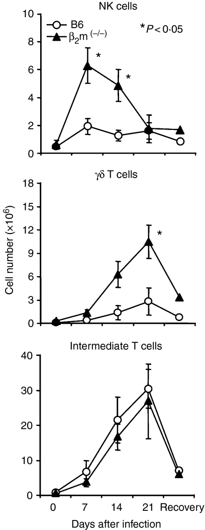
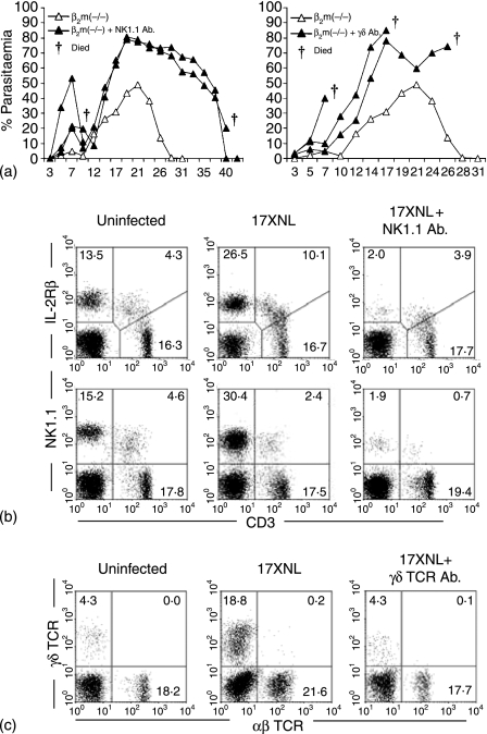
It is still controversial whether malaria protection is mediated by conventional immunity associated with T and B cells or by innate immunity associated with extrathymic T cells and autoantibody-producing B cells. Given this situation, it is important to examine the mechanism of malaria protection in beta(2)-microglobulin-deficient (beta(2)m(-/-)) mice. These mice lack major histocompatibility complex class I and CD1d antigens, which results in the absence of CD8(+) T cells and natural killer T (NKT) cells. When C57BL/6 and beta(2)m(-/-) mice were injected with parasitized (Plasmodium yoelii 17XNL) erythrocytes, both survived from the infection and showed a similar level of parasitaemia. The major expanding T cells were NK1.1(-) alphabeta T-cell receptor(int) cells in both mice. The difference was a compensatory expansion of NK and gammadelta T cells in beta(2)m(-/-) mice, and an elimination experiment showed that these lymphocytes were critical for protection in these mice. These results suggest that malaria protection might be events of the innate immunity associated with multiple subsets with autoreactivity. CD8(+) T and NKT cells may be partially related to this protection.
Figures


Similar articles
-
Resistance to malarial infection is achieved by the cooperation of NK1.1(+) and NK1.1(-) subsets of intermediate TCR cells which are constituents of innate immunity.Cell Immunol. 2001 Aug 1;211(2):96-104. doi: 10.1006/cimm.2001.1833. Cell Immunol. 2001. PMID: 11591113
-
Reasons why DBA/2 mice are resistant to malarial infection: expansion of CD3int B220+ gammadelta T cells with double-negative CD4- CD8- phenotype in the liver.Immunology. 2006 Jan;117(1):127-35. doi: 10.1111/j.1365-2567.2005.02273.x. Immunology. 2006. PMID: 16423048 Free PMC article.
-
Essential role of extrathymic T cells in protection against malaria.J Immunol. 2002 Jul 1;169(1):301-6. doi: 10.4049/jimmunol.169.1.301. J Immunol. 2002. PMID: 12077258
-
T cells as mediators of protective immunity against liver stages of Plasmodium.Trends Parasitol. 2003 Feb;19(2):88-93. doi: 10.1016/s1471-4922(02)00053-3. Trends Parasitol. 2003. PMID: 12586477 Review.
-
Role of apolipoproteins in gammadelta and NKT cell-mediated innate immunity.Immunol Res. 2005;33(3):241-55. doi: 10.1385/ir:33:3:241. Immunol Res. 2005. PMID: 16462001 Review.
Cited by
-
Gammadelta T cells but not NK cells are essential for cell-mediated immunity against Plasmodium chabaudi malaria.Infect Immun. 2010 Oct;78(10):4331-40. doi: 10.1128/IAI.00539-10. Epub 2010 Jul 26. Infect Immun. 2010. PMID: 20660608 Free PMC article.
-
The spleen CD4+ T cell response to blood-stage Plasmodium chabaudi malaria develops in two phases characterized by different properties.PLoS One. 2011;6(7):e22434. doi: 10.1371/journal.pone.0022434. Epub 2011 Jul 21. PLoS One. 2011. PMID: 21814579 Free PMC article.
-
Characterization of γδT cells in lung of Plasmodium yoelii-infected C57BL/6 mice.Malar J. 2021 Feb 15;20(1):89. doi: 10.1186/s12936-021-03619-z. Malar J. 2021. PMID: 33588839 Free PMC article.
-
Gamma/Delta T Cells and Their Role in Protection Against Malaria.Front Immunol. 2018 Dec 20;9:2973. doi: 10.3389/fimmu.2018.02973. eCollection 2018. Front Immunol. 2018. PMID: 30619330 Free PMC article. Review.
-
Aspecific binding of anti-NK1.1 antibodies on myeloid cells in an experimental model for malaria-associated acute respiratory distress syndrome.Malar J. 2024 Apr 18;23(1):110. doi: 10.1186/s12936-024-04944-9. Malar J. 2024. PMID: 38637828 Free PMC article.
References
-
- Guebre-Xabier M, Schwenk R, Krzych U. Memory phenotype CD8+ T cells persist in livers of mice protected against malaria by immunization with attenuated Plasmodium berghei sporozoites. Eur J Immunol. 1999;29:3978–86. - PubMed
-
- Morrot A, Zavala F. Effector and memory CD8+ T cells as seen in immunity to malaria. Immunol Rev. 2004;201:291–303. - PubMed
-
- Morrot A, Zavala F. Regulation of the CD8+ T cell responses against Plasmodium liver stages in mice. Int J Parasitol. 2004;34:1529–34. - PubMed
-
- Plebanski M, Hannan CM, Behboudi S, Flanagan KL, Apostolopoulos V, Sinden RE, Hill AV. Direct processing and presentation of antigen from malaria sporozoites by professional antigen-presenting cells in the induction of CD8 T-cell responses. Immunol Cell Biol. 2005;83:307–12. - PubMed
Publication types
MeSH terms
Substances
LinkOut - more resources
Full Text Sources
Other Literature Sources
Medical
Molecular Biology Databases
Research Materials
