. 2007 Dec;8(12):1829-1840.
doi: 10.1111/j.1600-0854.2007.00658.x. Epub 2007 Oct 17.

Dominant-negative behavior of mammalian Vps35 in yeast requires a conserved PRLYL motif involved in retromer assembly

Affiliations

Dominant-negative behavior of mammalian Vps35 in yeast requires a conserved PRLYL motif involved in retromer assembly

Xiang Zhao et al. Traffic. 2007 Dec.

Abstract

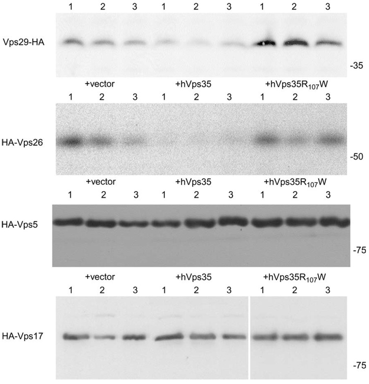
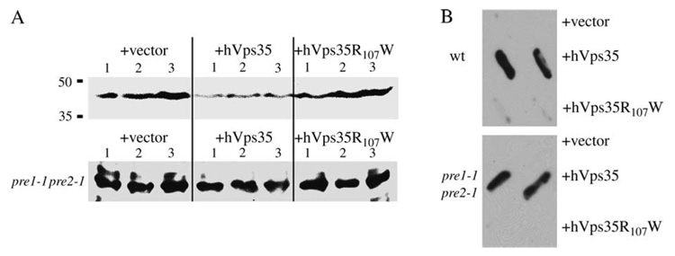
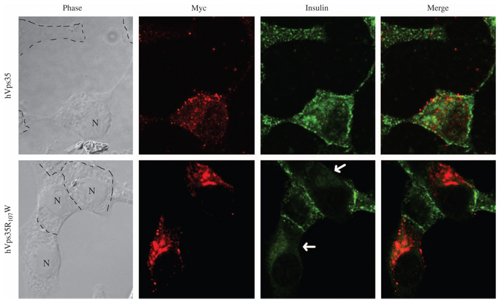
The retromer protein complex assists in recycling selected integral membrane proteins from endosomes to the trans Golgi network. One protein subcomplex (Vps35p, Vps26p and Vps29p) combines with a second (Vps17p and Vps5p) to form a coat involved in sorting and budding of endosomal vesicles. Yeast Vps35p (yVps35) exhibits similarity to human Vps35 (hVps35), especially in a completely conserved PRLYL motif contained within an amino-terminal domain. Companion studies indicate that an R(98)W mutation in yVps35 causes defective retromer assembly in Saccharomyces cerevisiae. Herein, we find that the expression of hVps35 in yeast confers dominant-negative vacuolar proenzyme secretion and defective secretory proprotein processing. The mutant phenotype appears to be driven by hVps35 competing with endogenous yVps35, becoming incorporated into defective retromer complexes and causing proteasomal degradation of endogenous Vps26 and Vps29. Increased expression of yVps35 displaces some hVps35 to a 100 000 x g supernatant and suppresses the dominant-negative phenotype. Remarkably, mutation of the conserved R(107)W of hVps35 displaces some of the protein to the 100 000 x g supernatant, slows protein turnover and restores stability of Vps26p and Vps29p and completely abrogates dominant-negative trafficking behavior. We show that hVps35 coprecipitates Vps26, whereas the R(107)W mutant does not. In pancreatic beta cells, the R(107)W mutant shifts hVps35 from peripheral endosomes to a juxtanuclear compartment, affecting both mannose phosphate receptors and insulin. These data underscore importance of the Vps35 PRLYL motif in retromer subcomplex interactions and function.


Figures

Figure 1
Figure 1. Mutation of the conserved R107 residue induces a dominant-negative phenotype in the context of a chimera of hVps35–yVps35
A) Within a conserved domain extending to residue 288 of yVps35, a sequence of near identity surrounds yVps35-R98, corresponding to hVps35-R107 (enlarged font). The ‘nhcy1’ chimera was constructed to contain the N-terminal 263 residues of hVps35 linked to C-terminal residues 245–944 of yVps35. Panels B–E each employ low-copy YCp plasmids with expression driven by the TDH3 promoter. B) Wild-type (WT) (W303) and vps35Δ mutant strains are shown in duplicate above, while nhcy1 (in quadruplicate) exhibits partial complementation of vacuolar protein sorting in a vps35Δ background. The R107W mutant of nhcy1 exhibits no complementation. C) vps35Δ mutant cells transformed with empty vector (control) or plasmids encoding yVps35, nhcy1 or nhcy1-R107W were pulse labeled for 10 min with 35S-amino acids and chased for 45 min before immunoprecipitation of CPY from intracellular (I) and extracellular (E) fractions. Quantification of these data is summarized beneath the panel. D) Wild-type control (W303) and vps35Δ mutant strains are shown above, while the nhcy1 mutant (in quadruplicate) produces a milder dominant-negative phenotype than does the nhcy1-R107W mutant. Not shown in panels B + D: expression of yVps35 in vps35Δ cells resulted in CPY secretion comparable to controls. E) Wild-type SNY36-9A cells transformed with empty vector (control) or plasmids encoding expression of yVps35, nhcy1 or nhcy1-R107W were analyzed as in panel C with quantification of the data shown below. ‘p2’ form = uncleaved Golgi-modified precursor; ‘m’ form = mature vacuolar species.
Figure 2
Figure 2. Untagged or tagged hVps35 induces a dominant-negative vacuolar sorting phenotype in yeast
Panels A–C each employ low-copy YCp plasmids with expression driven by the TDH3 promoter. A) Unlike wild-type yVps35 (not shown), none of the constructs shown can complement vacuolar protein sorting in a vps35Δ mutant. B) Transformation of wild-type (wt) cells to express each of the constructs shown induces CPY secretion beyond that observed for cells transformed with an empty vector. C) Cells expressing C-terminally myc-tagged or N-terminally HA-tagged hVps35 secrete the post Golgi (P2) form of CPY to the extracellular medium (E) with decreased mature CPY retained intracellularly (I).
Figure 3
Figure 3. Proalpha factor (proαF) secretion from cells expressing hVps35
Wild-type yeast bearing plasmids expressing the indicated proteins (yVps35 constructs expressed from multi-copy Yep plasmids driven by the VPS35 promoter; hVps35myc from a YCp plasmid with expression driven by the TDH3 promoter) were metabolically labeled with 35S-amino acids for 50 min as described in Materials and Methods. The media from each strain was immunoprecipitated with an antibody recognizing alpha factor-containing peptides. The positive control (Pos. control) comes from the secretion from a kex2Δ strain. In this SDS–PAGE system, a number of partially processed alpha factor peptides run with mature alpha factor at the dye front (arrowhead).
Figure 4
Figure 4. The dominant-negative phenotype induced by hVps35 is rescued by overexpression of yVps35
Panels A and B each employ yVps35 expressed from multicopy Yep plasmid driven by the VPS35 promoter and hVps35myc from a YCp plasmid with expression driven by the TDH3 promoter. A) The CPY filter overlay. Each wild-type strain is transformed with a centromeric plasmid listed first and a 2µ plasmid listed second with the protein (or lack of protein) encoded by these plasmids described (at right). B) Sedimentation behavior of hVps35-myc in the presence or absence of overexpressed yVps35. At left, hVps35 is nearly quantitatively associated with the pellet after centrifugation at 100 000 × g for 1 h (P100). At right, upon overexpression of yVps35, a fraction of hVps35 is displaced into the supernatant (S100). The hVps35 protein was identified by Western blotting with anti-myc.
Figure 5
Figure 5. The dominant-negative phenotype induced by hVps35 is blocked by the R107W mutation
Panels A–C each employ constructs expressed from a YCp plasmid with expression driven by the TDH3 promoter. A) Western blot establishing that the R107W mutant produces an expressed protein. B) Five independent transformants expressing the hVps35R107 mutant show loss of CPY secretion by filter blot compared with five transformants expressing hVps35. One colony of W303 cells transformed with empty vector (Vector) is shown as a negative control. C) Analysis of proalpha factor (pαF) processing by the strains shown in panels A and B was carried out as described in the legend to Figure 3. In this SDS–PAGE system, a number of partially processed alpha factor peptides run with mature alpha factor (αF) at the dye front.
Figure 6
Figure 6. Turnover, sedimentation and Vps26 association of hVps35 with and without the R107W mutation
Panels A–C each employ constructs expressed from a YCp plasmid with expression driven by the TDH3 promoter. A) Wild-type (W303) cells expressing hVps35-myc or hVps35R107W-myc were metabolically labeled for 5 min with 35S-amino acids and chased for the times indicated. At each chase time, cells were lysed and immunoprecipitated with anti-myc, normalized to trichloroacetic acid-precipitable counts. The immunoprecipitates were analyzed by SDS–PAGE and fluorography. B) Wild-type (W303) cells transformed to express the proteins shown or transformed with empty vector (Control) were lysed and the lysates were sedimented at 100 000 × g for 60 min as described in Materials and Methods. The pellet was resuspended in a volume identical to the supernatant volume, and equal volumes of the pellet and supernatant fractions were analyzed by SDS–PAGE and Western blotting with anti-myc. C) Yeast strain SNY217 carrying plasmid pVps26-HA (9) and either plasmid pGTH12 (hVps35-myc) or pGTH12-R107W[hVps35(R107W)-myc, also called simply R107W] was lysed under native conditions. To confirm the presence of comparable starting amounts of yVps26-HA in each strain, 2% of each lysate was analyzed by SDS–PAGE and immunoblotting with mouse anti-HA antibody (upper gel). From the remaining lysates, immunoprecipitation was performed with rabbit anti-HA antibody using protein A–Sepharose. Fifty percent of the immunoprecipitate (I) and 3.2% of the supernatant (S) were analyzed by SDS–PAGE gel and immunoblotting with mouse anti-c-myc antibody (lower gel) to detect coprecipitated hVps35-myc and hVps35(R107W)-myc. Densitometry of the bands, with correction for the percent loaded, back-calculated to the same total myc-tagged protein levels in each strain, and this was also directly confirmed by Western blotting (not shown). P, pellet; S, supernatant.
Figure 7
Figure 7. Immunoblotting to show steady-state levels of Vps29, Vps26, Vps5 and Vps17 in cells expressing hVps35, hVps35R107W or empty vector (+vector)
Constructs were expressed from a YCp plasmid with expression driven by the TDH3 promoter. Each of the retromer components contains an HA-epitope tag as described in Materials and Methods. Triplicate independent transformants are shown. Note that only the components of the large retromer subcomplex (Vps29 and Vps26) exhibit reduced levels in the presence of hVps35. Quantification from these data shows that in cells expressing hVps35, there is at least a 60% reduction of Vps29 and a 75% reduction of Vps26 compared with the empty vector control. By contrast, cells expressing hVps35R107W show no reduction in Vps29 or Vps26 levels.
Figure 8
Figure 8. Expression of hVps35 in yeast induces proteasomal turnover of Vps26
Constructs were expressed from a YCp plasmid with expression driven by the TDH3 promoter. A) Transformants (in triplicate) expressing hVps35, hVps35R107W or empty vector (+vector). The pre1-1 pre2-1 cells were grown, and all experiments were performed at 30°C. The Vps26 Western blot in the upper panel shows the results from control cells, while the lower panel (pre1-1 pre2-1) shows the results from proteasome-deficient cells. B) The CPY filter overlay from the strains shown in panel A. wt, wild type.
Figure 9
Figure 9. Expression of myc-tagged hVps35 or hVps35R107W in INS-1 cells
By Western blotting (not shown), INS-1 cells stably expressing hVps35 have equal protein levels to those of the cells expressing hVps35R107W. Cells expressing hVps35 show anti-myc labeling in small vesicles throughout the cytoplasm, whereas the R107W mutant is concentrated in juxtanuclear structures. Scale bar = 10 µm.
Figure 10
Figure 10. Double immunofluorescence labeling of myc-tagged hVps35 or hVps35R107W with the CI-MPR (MPR) in INS-1 cells
The hVps35R107W mutant tends to concentrate in juxtanuclear structures, and MPR exhibits decreased overall immunofluorescence intensity in these cells compared with cells that do not express hVps35R107W (see text). Scale bar = 10 µm. The arrow highlights decreased MPR immunofluorescence intensity in cells expressing the mutant hVps35.
Figure 11
Figure 11. Double immunofluorescence labeling of myc-tagged hVps35 or hVps35R107W with anti-insulin in INS-832/13 cells
Transient expression of hVps35 (upper panels) does not impair accumulation of subplasmalemmal insulin secretory granules. By contrast, expression of the R107W mutant in these cells inhibits granule accumulation (lower panels and see text). Dashed lines drawn on phase-contrast images outline the cell borders. ‘N’ highlights selected nuclei. Scale bar = 10 µm. The arrows highlight decreased immunofluorescent insulin secretory granules in cells expressing the mutant hVps35.

Similar articles

Cited by

References

    1. Zhang B-y, Chang A, Kjeldsen TB, Arvan P. Intracellular retention of newly-synthesized insulin in yeast is caused by endoproteolytic processing in the Golgi complex. J Cell Biol. 2001;153:1187–1197. - PMC - PubMed
    1. Seaman MN. Recycle your receptors with retromer. Trends Cell Biol. 2005;15:68–75. - PubMed
    1. Hettema EH, Lewis MJ, Black MW, Pelham HR. Retromer and the sorting nexins Snx4/41/42 mediate distinct retrieval pathways from yeast endosomes. EMBO J. 2003;22:548–557. - PMC - PubMed
    1. Griffin CT, Trejo J, Magnuson T. Genetic evidence for a mammalian retromer complex containing sorting nexins 1 and 2. Proc Natl Acad Sci U S A. 2005;102:15173–15177. - PMC - PubMed
    1. Seaman MN, McCaffery JM, Emr SD. A membrane coat complex essential for endosome-to-Golgi retrograde transport in yeast. J Cell Biol. 1998;142:665–681. - PMC - PubMed

Publication types

MeSH terms