. 2007 Nov;208(1):145-58.
doi: 10.1016/j.expneurol.2007.08.011. Epub 2007 Aug 29.

Modulation of the cAMP signaling pathway after traumatic brain injury

Affiliations

Modulation of the cAMP signaling pathway after traumatic brain injury

Coleen M Atkins et al. Exp Neurol. 2007 Nov.

Abstract

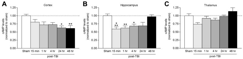
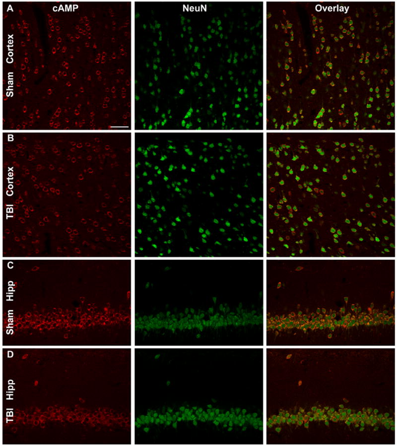
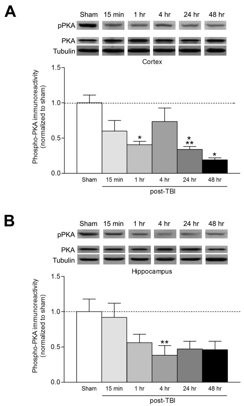
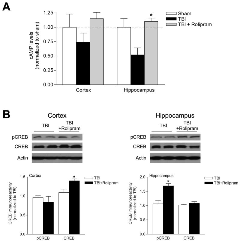
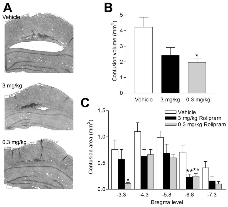
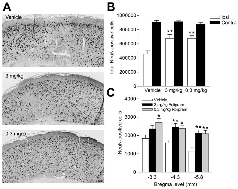
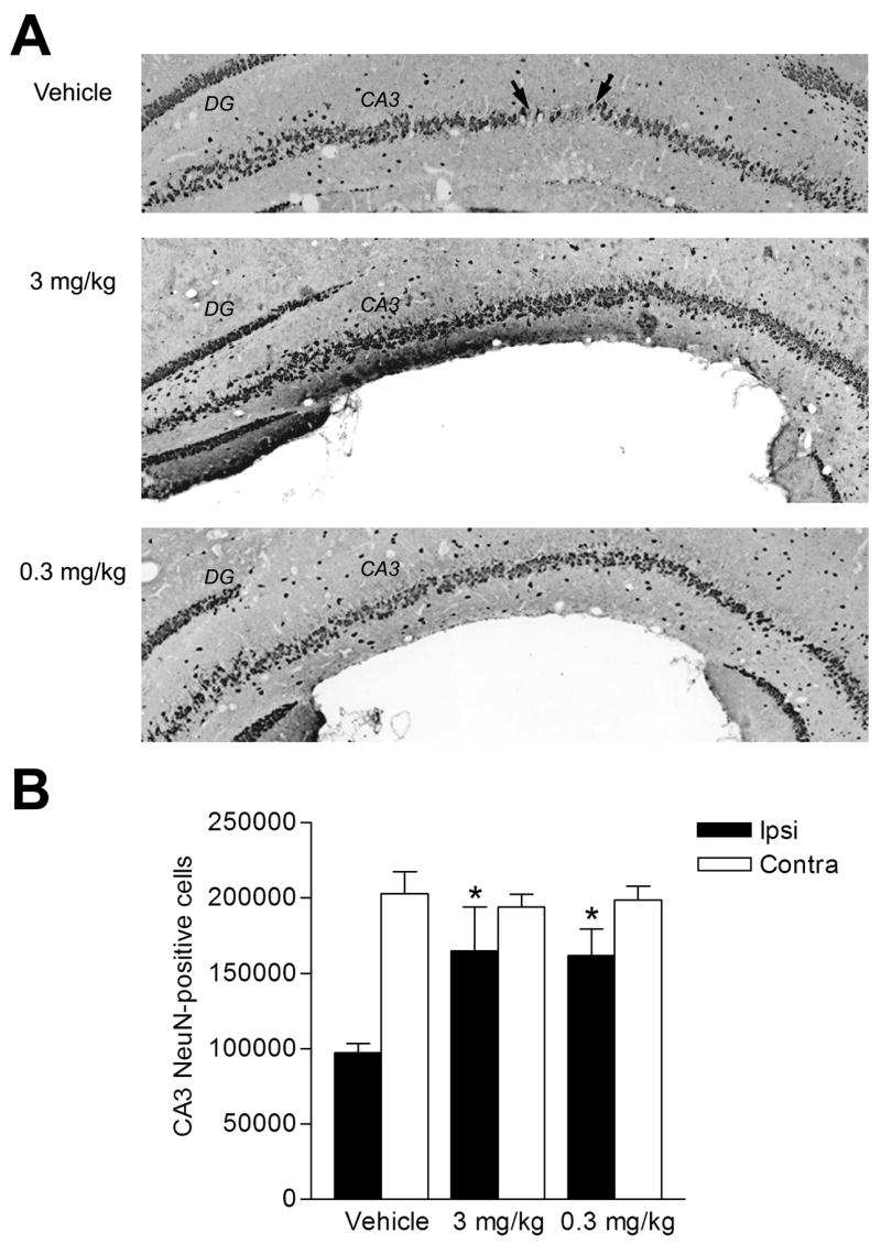
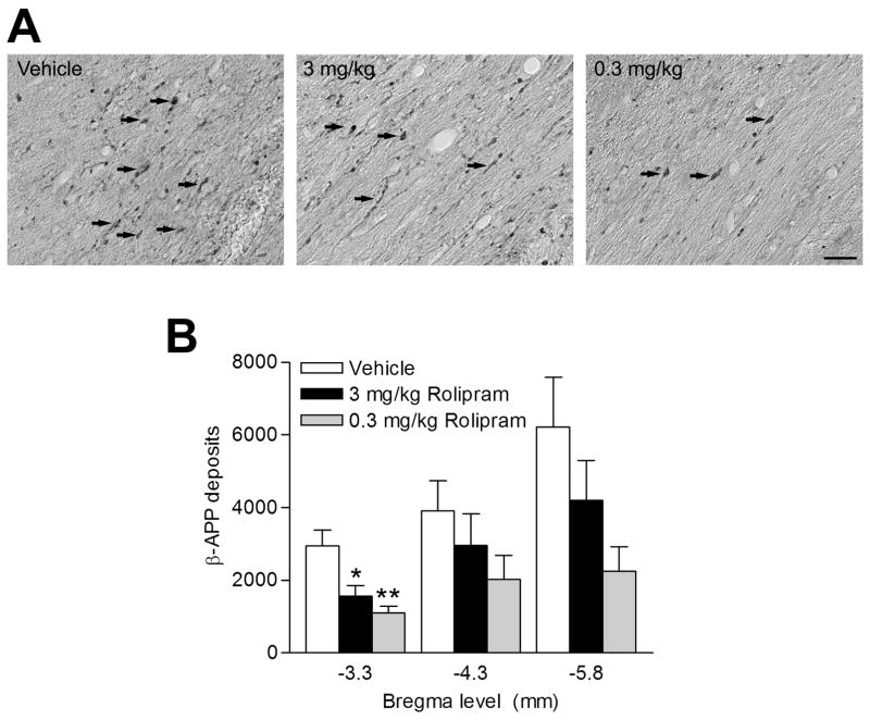
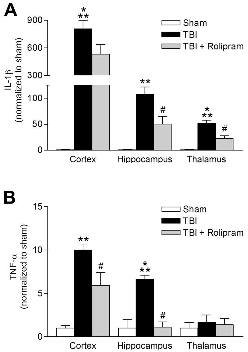
Traumatic brain injury (TBI) results in both focal and diffuse brain pathologies that are exacerbated by the inflammatory response and progress from hours to days after the initial injury. Using a clinically relevant model of TBI, the parasagittal fluid-percussion brain injury (FPI) model, we found injury-induced impairments in the cyclic AMP (cAMP) signaling pathway. Levels of cAMP were depressed in the ipsilateral parietal cortex and hippocampus, as well as activation of its downstream target, protein kinase A, from 15 min to 48 h after moderate FPI. To determine if preventing hydrolysis of cAMP by administration of a phosphodiesterase (PDE) IV inhibitor would improve outcome after TBI, we treated animals intraperitoneally with rolipram (0.3 or 3.0 mg/kg) 30 min prior to TBI, and then once per day for 3 days. Rolipram treatment restored cAMP to sham levels and significantly reduced cortical contusion volume and improved neuronal cell survival in the parietal cortex and CA3 region of the hippocampus. Traumatic axonal injury, characterized by beta-amyloid precursor protein deposits in the external capsule, was also significantly reduced in rolipram-treated animals. Furthermore, levels of the pro-inflammatory cytokines, interleukin-1beta (IL-1beta) and tumor necrosis factor-alpha (TNF-alpha), were significantly decreased with rolipram treatment. These results demonstrate that the cAMP-PKA signaling cascade is downregulated after TBI, and that treatment with a PDE IV inhibitor improves histopathological outcome and decreases inflammation after TBI.


Figures

Fig 1
Fig 1
cAMP levels decrease after TBI. The ipsilateral parietal cortex (A; n=4–10), hippocampus (B; n=4–11), and thalamus (C; n=5–10) were assayed by ELISA for cAMP levels after moderate parasagittal FPI. cAMP levels were significantly decreased in the cortex at 24 hr (n=4, *p<0.05) and 48 hr (n=6, **p<0.01) after TBI, and at 15 min (n=6, ***p<0.001), 1 hr (n=6, **p<0.01), and 4 hr (n=5, *p<0.05) after TBI in the hippocampus as compared to sham levels. Data represent mean ± SEM.
Fig 2
Fig 2
cAMP immunostaining after TBI. The ipsilateral parietal cortex of sham surgery animals (A) and TBI animals (B) were immunostained with cAMP (red) and NeuN (green). Images were from animals perfused 5 min after surgery. There was co-localization of cAMP with NeuN. The CA1 region of the hippocampus of sham surgery animals (C) and TBI animals 5 min after trauma (D) were immunostained for cAMP and NeuN. In TBI animals, cAMP levels were modestly reduced in NeuN-positive cells. Images are representative of 3 animals in each group. Scale bar, 50 μm.
Fig 3
Fig 3
PKA activation is downregulated after TBI. The ipsilateral parietal cortex (A; n=3–8) and hippocampus (B; n=3–6) were western blotted at the indicated times after TBI for activated, phosphorylated PKA (pPKA). PKA activation was decreased significantly in the cortex at 1 hr (n=5, *p<0.05), 24 hr (n=3, ***p<0.001), and 48 hr (n=5, *p<0.05) after TBI as compared to sham animals. In the hippocampus, phosphorylated PKA levels were significantly decreased at 4 hr (n=7, **p<0.01) after TBI as compared to sham animals. Data represent mean ± SEM.
Fig. 4
Fig. 4
Rolipram treatment increased cAMP levels and phosphorylation of CREB. Animals were treated with vehicle (5% DMSO in saline) or rolipram (0.3 mg/kg, i.p.) 30 min prior to moderate parasagittal FPI. At 24 hr after injury, the animals were treated once more with vehicle or rolipram (0.3 mg/kg, i.p.), then sacrificed 30 min later. (A) cAMP levels were measured by ELISA in the ipsilateral parietal cortex and hippocampus. cAMP levels were increased to sham levels in the parietal cortex although this was not statistically significant (n=3 for each group). In the hippocampus, cAMP levels were significantly increased in TBI animals that received rolipram as compared to TBI animals that received vehicle (n=3 for each group, *p<0.05). (B) Phosphorylated CREB and total CREB levels were assayed by western blotting in the ipsilateral parietal cortex and hippocampus 24 hr after injury. Total CREB levels significantly increased in the parietal cortex in rolipram-treated animals as compared to vehicle-treated animals (n=3 for each group, *p<0.05). Phosphorylated CREB levels significantly increased in the hippocampus in rolipram-treated animals as compared to vehicle-treated animals (n=3 for each group, *p<0.05). Data represent mean ± SEM.
Fig 5
Fig 5
Rolipram treatment decreased cortical contusions. Rats received vehicle (5% DMSO in saline) or rolipram i.p. 30 min prior to moderate parasagittal FPI. After TBI, the animals received vehicle or rolipram for 3 days and were then perfused for analysis at 30 min after their final injection. The brains were sectioned and stained with H&E, and the cortical contusion area was imaged. Representative sections at bregma level -5.8 mm are shown (A). Cortical contusion volume (B) and contusion areas at specific bregma levels (C) were quantified by stereology. The epicenter of the injury was at -3.8 mm bregma. The lower dose of rolipram, 0.3 mg/kg, significantly reduced total cortical contusion volume (n=6, *p<0.05) as compared to vehicle-treated animals and contusion area near the epicenter of the injury (bregma -3.3 mm, *p<0.05) as well as in the penumbra (bregma -6.8 mm, **p<0.01) as compared to vehicle-treated animals (n=9). Although the higher dose of 3.0 mg/kg rolipram reduced contusion volume as compared to vehicle-treated animals, this was not statistically significant (n=8). Data represent mean ± SEM.
Fig 6
Fig 6
Cortical neuron survival was improved with rolipram treatment. The parietal cortex overlying the contusion area between bregma levels -3.3 mm and -6.8 mm was immunostained with NeuN to identify surviving neurons. Animals were treated with vehicle, 0.3 mg/kg rolipram, or 3.0 mg/kg rolipram i.p. 30 min prior to injury, followed by once per day for 3 days. (A) Shown are representative images at bregma level -5.8 mm. (B) Total cortical neuron survival on the ipsilateral side (Ipsi) was significantly improved with both 0.3 mg/kg rolipram (n=5, **p<0.01) and 3.0 mg/kg rolipram (n=8, **p<0.01) as compared to vehicle-treated animals (n=9). There were no significant differences in total numbers of neurons on the contralateral side with rolipram treatment (Contra). (C) Quantification of neuronal survival at specific bregma levels illustrates that 3.0 mg/kg rolipram increased neuronal survival at bregma levels -4.3 mm and -5.8 mm (n=8, **p<0.01) when compared to vehicle-treated animals (n=9). Rolipram at 0.3 mg/kg improved cortical neuron survival at all bregma levels tested (n=6; -3.3 mm, *p<0.05; -4.3 mm, *p<0.05; -5.8 mm, **p<0.01). Data represent mean ± SEM. Scale bar, 50 μm.
Fig 7
Fig 7
Survival of CA3 hippocampal neurons with rolipram. (A) Animals received rolipram or vehicle 30 min prior to TBI, then once per day for 3 days. Sections were immunostained with NeuN. Bregma level -5.8 mm is shown. Arrows denote boundaries of an area of neuronal dropout. (B) Significant survival of CA3 neurons on the ipsilateral side (Ipsi) was seen with 0.3 mg/kg rolipram (n=4, *p<0.05) and 3.0 mg/kg rolipram (n=8, *p<0.05) as compared to vehicle-treated animals (n=9). There were no significant differences in neuronal survival on the contralateral side (Contra). Data represent mean ± SEM.
Fig 8
Fig 8
The external capsule was stained with β-APP to assess axonal pathology. Animals were treated with vehicle, 0.3 mg/kg rolipram, or 3.0 mg/kg rolipram i.p. 30 min prior to injury and once per day for 3 days. (A) Shown is bregma level -5.8 mm. Arrows demarcate β-APP deposits in the external capsule. (B) Both 0.3 mg/kg (n=6, **p<0.01) and 3.0 mg/kg rolipram (n=8, *p<0.05) modestly reduced β-APP deposits at bregma level -3.3 mm as compared to vehicle-treated animals (n=8). Data represent mean ± SEM. Scale bar, 25 μm.
Fig 9
Fig 9
Pro-inflammatory cytokine levels are attenuated with rolipram treatment. (A) IL-1β levels were assayed by ELISA at 3 hr after TBI. There was a significant increase in IL-1β levels in the ipsilateral parietal cortex (n=6, ***p<0.001), hippocampus (n=6, **p<0.01), and thalamus (n=8, ***p<0.001) as compared to sham animals (n=3–5). Rolipram treatment (0.3 mg/kg) 30 min prior to TBI and 30 min prior to sacrifice significantly reduced IL-1β levels in the hippocampus (n=7, #p<0.05) and thalamus (n=9, #p<0.001), but not in the parietal cortex (n=7), as compared to vehicle-treated TBI animals. (B) TNF-α levels significantly increased in the parietal cortex (n=6, **p<0.01) and hippocampus (n=6, ***p<0.001) at 3 hr after TBI as compared to sham animals. This increase in TNF-α was significantly reduced in rolipram-treated TBI animals (parietal cortex n=7, #p<0.05; hippocampus n=6, #p<0.001) as compared to vehicle-treated TBI animals. Data represent mean ± SEM.

References

    1. Armstead WM. Role of impaired cAMP and calcium-sensitive K+ channel function in altered cerebral hemodynamics following brain injury. Brain Res. 1997;768:177–184. - PubMed
    1. Atkins CM, Chen S, Alonso OF, Dietrich WD, Hu BR. Activation of calcium/calmodulin-dependent protein kinases after traumatic brain injury. J Cereb Blood Flow Metab. 2006;26:1507–1518. - PubMed
    1. Baer M, Dillner A, Schwartz RC, Sedon C, Nedospasov S, Johnson PF. Tumor necrosis factor-α transcription in macrophages is attenuated by an autocrine factor that preferentially induces NF-κB p50. Mol Cell Biol. 1998;18:5678–5689. - PMC - PubMed
    1. Barad M, Bourtchouladze R, Winder DG, Golan H, Kandel E. Rolipram, a type IV-specific phosphodiesterase inhibitor, facilitates the establishment of long-lasting long-term potentiation and improves memory. Proc Natl Acad Sci USA. 1998;95:15020–15025. - PMC - PubMed
    1. Bell MJ, Kochanek PM, Carcillo JA, Mi Z, Schiding JK, Wisniewski SR, Clark RS, Dixon CE, Marion DW, Jackson E. Interstitial adenosine, inosine, and hypoxanthine are increased after experimental traumatic brain injury in the rat. J Neurotrauma. 1998;15:163–170. - PubMed

Publication types

MeSH terms