. 2007 Nov;133(5):1627-36.
doi: 10.1053/j.gastro.2007.08.003. Epub 2007 Aug 2.

Viral and host factors induce macrophage activation and loss of toll-like receptor tolerance in chronic HCV infection

Affiliations

Viral and host factors induce macrophage activation and loss of toll-like receptor tolerance in chronic HCV infection

Angela Dolganiuc et al. Gastroenterology. 2007 Nov.

Abstract

Background & aims: Persistent inflammation contributes to progression of liver damage in chronic HCV (cHCV) infection. Repeated exposure to toll-like receptor (TLR) ligands results in tolerance, a protective mechanism aimed at limiting inflammation.

Methods: Monocytes/macrophages were repeatedly stimulated via proinflammatory cytokine-inducing TLRs and evaluated for activation markers.

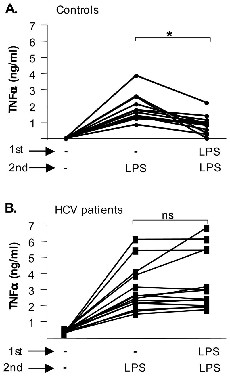
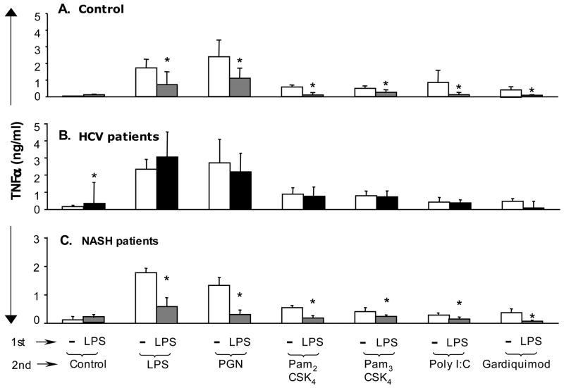
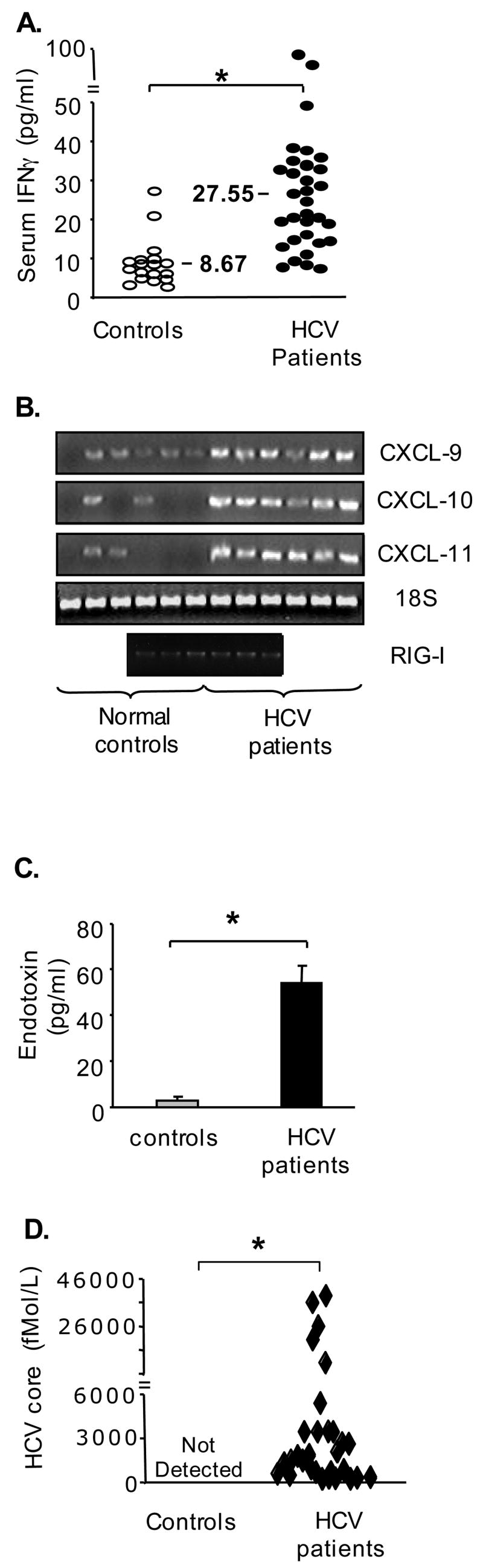
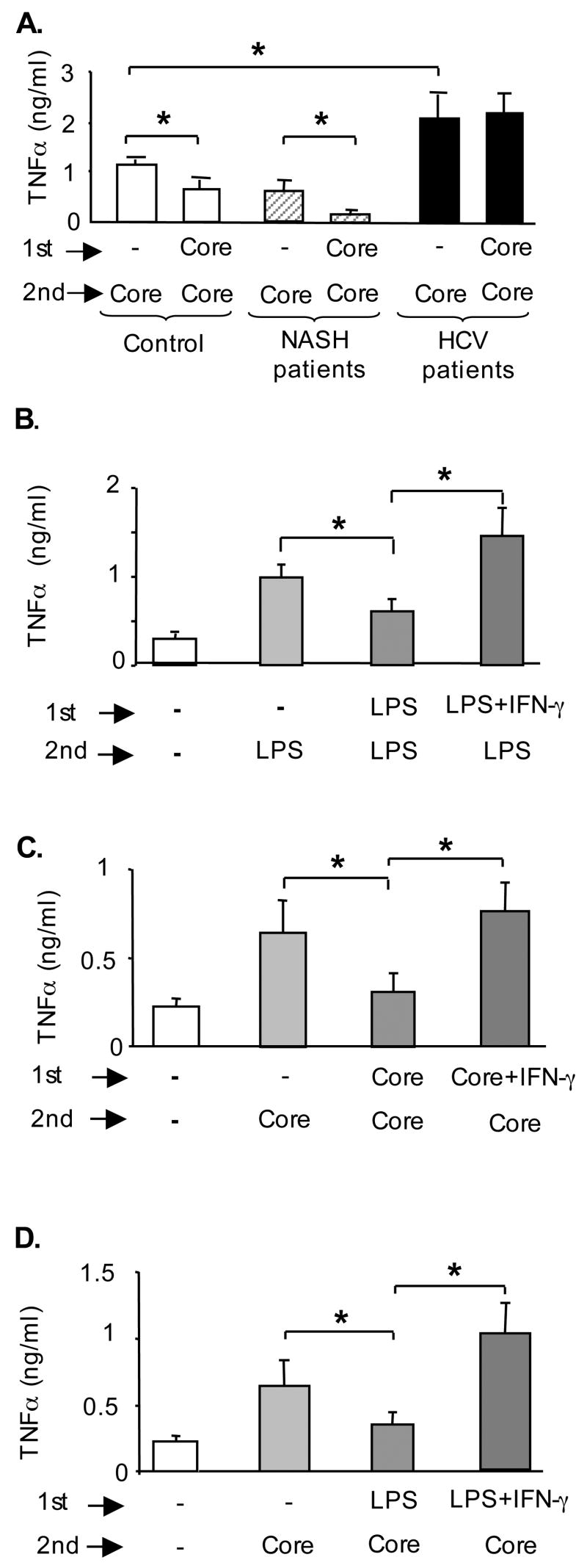
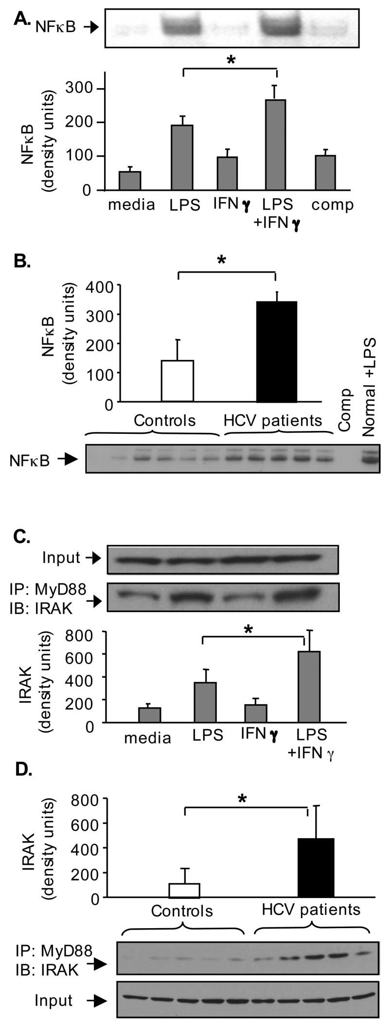
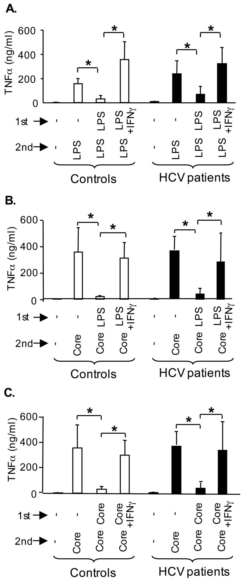
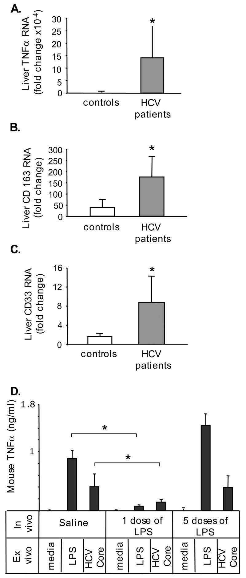
Results: Unlike monocytes of controls or patients with nonalcoholic steatohepatitis, the monocytes of cHCV patients were hyperresponsive and failed to show homo- or heterotolerance to TLR ligands, manifested by elevated tumor necrosis factor (TNF)-alpha production. Serum levels of interferon (IFN)-gamma, endotoxin (TLR4 ligand), and HCV core protein (TLR2 ligand) were elevated in cHCV patients suggesting potential mechanisms for in vivo monocyte preactivation. Treatment of normal monocytes with IFN-gamma resulted in loss of tolerance to lipopolysaccharide (LPS) or HCV core protein. Furthermore, we found increased levels of MyD88-IRAK1 complexes and nuclear factor (NF)-kappaB activity both in monocytes of cHCV patients and in normal monocytes that lost TLR tolerance after IFN-gamma + LPS pretreatment. In vitro differentiation of TLR non-tolerant cHCV monocytes into macrophages restored their capacity to exhibit TLR tolerance to LPS and HCV core protein, and this could be reversed by administration of IFN-gamma. cHCV patients exhibited increased TNF-alpha in the circulation and in the liver. In cHCV livers, we found Kupffer cell/macrophage activation indicated by increased CD163 and CD33 expression.

Conclusions: We identified that host-derived factors (IFN-gamma and endotoxin) and viral factors (HCV core protein) act in tandem to induce and maintain monocyte/macrophage activation, thus favoring persistent inflammation in patients with cHCV infection.


Figures

Figure 1
Figure 1. Monocytes of cHCV patients fail to develop homo-tolerance to TLR4
Monocytes of controls (n=16) (A) and HCV patients (n=15) (B) were kept in medium or stimulated with TLR4 ligand LPS (100ng/ml) for 24 hrs (1st stimulation) and re-challenged with LPS (100ng/ml) for an additional 24 hrs (2nd stimulation). The production of TNFα in culture supernatants was analyzed in ELISA. Average±SD is shown (*p<0.05).
Figure 2
Figure 2. Monocytes of cHCV patients, unlike controls and NASH patients, fail to develop hetero-tolerance to TNFα-inducing TLR ligands
Monocytes from controls (n=16) (A), HCV patients (n=15) (B) and NASH patients (n=6) (C) were cultured in medium or stimulated with LPS (100ng/ml) for 24 hrs (1st stimulation) and re-challenged with LPS (100ng/ml), PGN (5μg/ml), Pam2CSK4 (100ng/ml), Pam3CSK4 (100ng/ml), poly I:C (100ng/ml) or Gardiquimod (5μg/ml) for an additional 24 hrs (2nd stimulation). The production of TNFα in culture supernatants was analyzed in ELISA. Average±SD is shown (* p<0.05).
Figure 3
Figure 3. cHCV patients exhibit elevated levels of IFNγ, endotoxin, and HCV core protein in the peripheral circulation
(A) The levels of IFNγ in plasma of HCV-infected patients (n=30) and controls (n=18) were quantified using a specific ELISA. The asterisk (*) represents p<0.05. (B) The levels of RNA coding for CXCL9, CXCL10, CXCL11 and RIG-I in freshly isolated monocytes were quantified using PCR. Each band represents a pooled sample from 2 controls or 3 cHCV patients. (C) The levels of endotoxin in plasma of cHCV patients (n=30) and controls (n=18) were quantified using LAL assay; average±SE is shown, the asterisk (*) indicates p<0.05. (D) The levels of HCV core protein in the plasma of HCV-infected patients (n=30) and control (n=18) were quantified using HCV core ELISA; the asterisk (*) represents p<0.05.
Figure 4
Figure 4. Tolerance to LPS and to HCV core protein is disrupted by IFNγ treatment
(A) Monocytes from controls (n=16) and HCV patients (n=15) were kept in medium or stimulated for 24 hrs (1st stimulation) and re-challenged with HCV core protein for additional 24 hrs (2nd stimulation). The production of TNFα was analyzed in culture supernatants using ELISA and the * indicates p<0.05 (panels A–D). (B) Monocytes of controls (n=3) were stimulated with LPS (100pg/ml) for 24 hrs (1ststimulation) and re-challenged with LPS for 24 hrs (2nd stimulation). IFNγ (100pg/ml) was added 4 hrs before and during the 1st stimulation. (C) Monocytes of controls (n=3) were stimulated with HCV core (10000fMol/L) for24 hrs (1st stimulation) and re-challenged with HCV core (25000fMol/L), for 24 hrs (2nd stimulation). IFNγ (100pg/ml) was added for 4 hrs before and during the 1st stimulation. (D) Monocytes of controls (n=3) were stimulated with LPS (100pg/ml) for 24 hrs (1st stimulation) and re-challenged with HCV core (25000 fMol/L) for 24 hrs (2nd stimulation). IFNγ (100pg/ml) was added for 4 hrs before and during the 1st stimulation.
Figure 5
Figure 5. HCV-infected patients’ monocytes show elevated NFκB activity and increased frequency of MyD88/IRAK complexes, similar to IFNγ+LPS-stimulated normal monocytes
(A) Normal monocytes were pre-treated in vitro with IFNγ followed by stimulation with LPS for 1 hour, as indicated. Five μg of nuclear protein were analyzed for NFκB binding capacity in EMSA using a radioactive labeled NFκB-specific oligonucleotide. A cold NFκB oligonucleotide incubated with nuclear protein from LPS-stimulated sample was used to determine the specificity of NFκB band (Comp). A representative EMSA gel is shown on the top and densitometric analysis of n=4 is shown on the bottom. (B) The NFκB binding capacity of normal and cHCV monocytes was analyzed in EMSA, as described above. Each band represents 5μf of nuclear proteins from a pool of 2 controls or 3 HCV patients. Normal monocytes (corresponding to the pool #2) were stimulated with LPS (100ng/ml for 1hr in vitro) and used as positive control for this assay (Normal+LPS). A cold NFκB oligonucleotide, incubated with nuclear protein from LPS-stimulated sample was used to determine the specificity of NFκB band (Comp). The densitometric analysis of the EMSA gel is shown on the top as average±SD and a representative gel is shown at the bottom. (C) Normal monocytes were pre-treated in vitro with IFNγ followed by stimulation with LPS for 1 hour. Five hundred μg of protein were incubated with anti-MyD88 antibody at +4°C overnight; the loading of equal protein amounts included in the immunoprecipitation assay was conformed by immunoblotting against β-actin (input). The amount of IRAK1 immunopreciptated with anti-MyD88 antibodies was detected by western blot (bottom band) and quantified by densitometric analysis; shown is average±SD from n=3. (D) Equal amounts (500 μg) of total protein from freshly isolated monocytes of controls (n=10) and cHCV patients (n=15) were incubated with anti-MyD88 antibody at +4°C overnight; each band represents a pool of 2 controls or 3 HCV patients. The amount of IRAK1 immunopreciptated with anti-MyD88 antibodies was detected and quantified as above; shown is average±SD.
Figure 6
Figure 6. IFNγ is capable of breaking TLR tolerance in macrophages
(A) Macrophages of controls (open bars) and cHCV patients (black bars) were differentiated in vitro, then stimulated with LPS (10pg/ml) for 24 hrs (1st stimulation) and re-challenged with LPS (100pg/ml), for additional 24 hrs (2nd stimulation). Where indicated IFNγ (100pg/ml) was added during the differentiation and 1st stimulation. The production of TNFα in the culture supernatants was analyzed in ELISA from n=6 (A–C). (B) Macrophages were differentiated as above, then stimulated in vitro with LPS (10pg/ml) for 24 hrs (1st stimulation) and re-challenged with HCV core (25000 fMol/L) for additional 24 hrs (2nd stimulation). (C) Macrophages were differentiated as above, then stimulated with HCV core (10000 fMol/L) for 24 hrs (1st stimulation) and re-challenged with HCV core (25000 fMol/L), for additional 24 hrs (2nd stimulation).
Figure 7
Figure 7. The patients with cHCV infection exhibit elevated levels of TNFα, CD163 and CD33 in livers
The levels of RNA coding for TNFα(A), CD163 (B) and CD33 (C) were analyzed in livers of controls (n=3) and HCV patients (n=12) using real-time quantitative PCR. The asterisk (*) represents p<0.05. (D) The mice were injected i/p with saline, LPS (5mg/kg) one time (the “1 dose of LPS” group) or 5 times (the “5 dose of LPS” group received 1mg/kg LPS every 3 days, total of 5 doses) and the Kupffer cells were isolated. In vitro the cells were stimulated with LPS (100ng/ml) or HCV core protein (25000 fMol/L) and the TNFα in culture supernatants was quantified using specific ELISA. The asterisk (*) represents p<0.05 using t-test from n=4 in saline and n=3 in LPS-treated groups.

References

    1. Kim WR, Brown RS, Jr, Terrault NA, El-Serag H. Burden of liver disease in the United States: summary of a workshop. Hepatology. 2002;36:227–242. - PubMed
    1. Ward S, Lauer G, Isba R, Walker B, Klenerman P. Cellular immune responses against hepatitis C virus: the evidence base 2002. Clin Exp Immunol. 2002;128:195–203. - PMC - PubMed
    1. Neuman MG, Benhamou JP, Malkiewicz IM, Ibrahim A, Valla DC, Martinot-Peignoux M, Asselah T, Bourliere M, Katz GG, Shear NH, Marcellin P. Kinetics of serum cytokines reflect changes in the severity of chronic hepatitis C presenting minimal fibrosis. J Viral Hepat. 2002;9:134–140. - PubMed
    1. Takeda K, Kaisho T, Akira S. Toll-like receptors. Ann Rev Immunol. 2003;21:335–376. - PubMed
    1. Bartenschlager R, Frese M, Pietschmann T. Novel insights into hepatitis C virus replication and persistence. Adv Virus Res. 2004;63:71–180. - PubMed

Publication types

MeSH terms