. 2007 Dec;36(4):484-500.
doi: 10.1016/j.mcn.2007.08.008. Epub 2007 Aug 23.

Gephyrin clustering is required for the stability of GABAergic synapses

Affiliations

Gephyrin clustering is required for the stability of GABAergic synapses

Wendou Yu et al. Mol Cell Neurosci. 2007 Dec.

Abstract

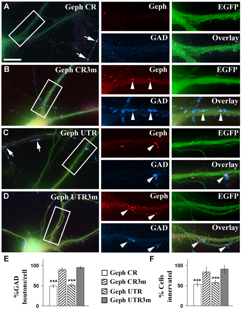
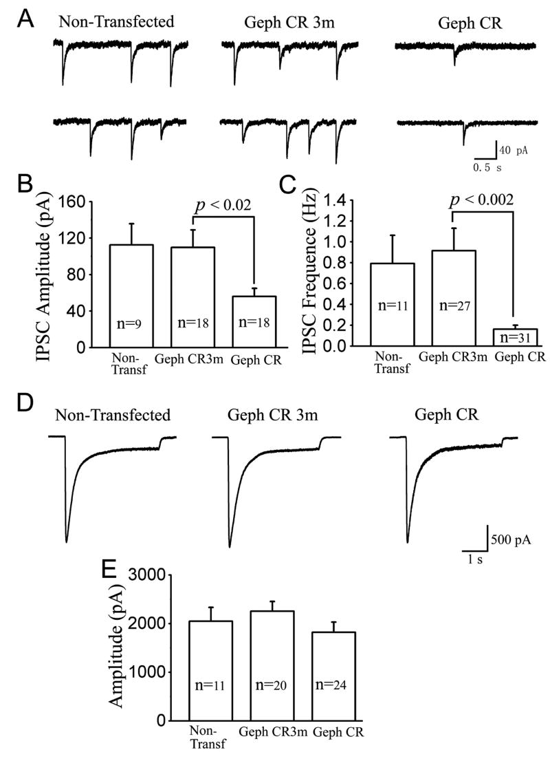
Although gephyrin is an important postsynaptic scaffolding protein at GABAergic synapses, the role of gephyrin for GABAergic synapse formation and/or maintenance is still under debate. We report here that knocking down gephyrin expression with small hairpin RNAs (shRNAs) in cultured hippocampal pyramidal cells decreased both the number of gephyrin and GABA(A) receptor clusters. Similar results were obtained by disrupting the clustering of endogenous gephyrin by overexpressing a gephyrin-EGFP fusion protein that formed aggregates with the endogenous gephyrin. Disrupting postsynaptic gephyrin clusters also had transsynaptic effects leading to a significant reduction of GABAergic presynaptic boutons contacting the transfected pyramidal cells. Consistent with the morphological decrease of GABAergic synapses, electrophysiological analysis revealed a significant reduction in both the amplitude and frequency of the spontaneous inhibitory postsynaptic currents (sIPSCs). However, no change in the whole-cell GABA currents was detected, suggesting a selective effect of gephyrin on GABA(A) receptor clustering at postsynaptic sites. It is concluded that gephyrin plays a critical role for the stability of GABAergic synapses.


Figures

Fig. 1
Fig. 1. Gephyrin knock down by shRNAs reduces the density of both gephyrin and γ2-GABAAR clusters
A, The gephyrin shRNAs used in this study. The three point mutations introduced in the control shRNAs are shown in red (see materials and methods); B-E, Cultured hippocampal neurons were co-transfected with pEGFP-N1 and Geph CR (B), or Geph CR3m (C), or Geph UTR (D), or Geph UTR3m (E) shRNAs. Triple-label immunofluorescence was done using mAb to gephyrin (red color) and rabbit anti-γ2 GABAAR antibodies (blue color). EGFP fluorescence of transfected neurons is shown in green color. The smaller panels at the right side of each figure show at higher magnification the corresponding boxed area. Arrows in B and D show dendrites of a non-transfected neuron which has much higher density of gephyrin clusters and γ2-GABAAR clusters than the dendrites of the sister neurons (green color) transfected with Geph CR or Geph UTR shRNAs respectively. Arrowheads show gephyrin clusters that colocalize with γ2-GABAAR clusters. Scale bar: 10 μm for large panels; 5 μm for the small panels. F, Immunoblots with anti-gephyrin and anti-α tubulin antibodies of hippocampal cultures transfected by nucleofection with Geph CR, Geph CR3m, Geph UTR, or Geph UTR3m shRNAs. G. Quantification of the immunoblots shown in F. Values for Geph CR and Geph UTR represent percentages of the normalized intensity of the gephyrin protein bands (normalized to the density of α-tubulin) when compared to that of the corresponding 100% control value (pyramidal cells transfected with Geph CR3m or Geph UTR3m shRNAs).
Fig. 2
Fig. 2. Gephyrin knock down reduces the clustering of various GABAAR subunits to different extent
A-F, Cultured hippocampal neurons were co-transfected with pEGFP-N1 and Geph CR (A, C, and E) or Geph CR3m (B, D, and F) shRNAs. Triple label immunofluorescence was done by using combinations of the mouse mAb to gephyrin and rabbit anti-α1, or rabbit anti-α2 GABAAR subunit antibodies or the combination of rabbit anti-γ2 and the mouse mAb to β2/3. Note that the neurons transfected (EGFP fluorescence) with Geph CR (C and E) show a high reduction in the cluster density of gephyrin and α2, β2/3, and γ2-GABAARs when compared to neurons transfected with the control Geph CR3m (D and F). However, the reduction of α1 subunit containing GABAAR clusters (A) is small when compared with Geph CR3m (B). G and H: Quantification of the effect of the gephyrin shRNAs on gephyrin and the GABAAR cluster density. Values (mean±SEM) represent the percentages compared to the corresponding internal control (non-transfected sister cells in the same cultures). Significant differences with the corresponding internal control values are indicated by asterisks (**, p<0.01; ***, p<0.001 in Student's t-test). Comparisons between groups using one-way ANOVA Tukey test when compared at p<0.05 showed that the mutated shRNAs (Geph CR3m or Geph UTR3m) had no effect on the density of gephyrin and GABAAR clusters. The cluster densities (number of clusters/100 μm2; mean ± SEM) in sister non-transfected cells in the Geph CR shRNA transfection experiments were 21.1±0.9 (632) for gephyrin, 20.9±0.9 (628) for γ2, 23.9±0.8 (717) for α1, 15.7±0.8 (472) for α2, and 17.7±0.8 for β2/3 (531) (The number of counted clusters is shown in parenthesis). Similar values for the non-transfected cells were obtained in the other transfection experiments. Arrowheads show colocalizing gephyrin and GABAAR clusters. Scale bar: 5 μm.
Fig. 3
Fig. 3. Overexpression of gephyrin-EGFP decreases endogenous gephyrin and GABAAR -γ2 clusters
Cultured hippocampal neurons were transfected with gephyrin-EGFP. A-D, Triple-label immunofluorescence, at 1 day (A), 3 days (B), and 5 days (C, D) after transfection, with a mouse mAb to gephyrin (red color) and rabbit anti-γ2 (blue color). Note that the pyramidal cells overexpressing gephyrin-EGFP form large filamentous aggregates of gephyrin-EGFP in the soma and proximal dendrites (crossed arrows). Arrows in C, D show that the dendrites of non-transfected neurons have normal density of gephyrin and γ2-GABAAR clusters, while transfected neurons expressing gephyrin-EGFP (green color) show reduced cluster density of gephyrin or γ2-GABAAR in cell dendrites. White arrowheads show colocalizing gephyrin clusters and γ2-GABAAR clusters. Black arrowheads show γ2-GABAARclusters without colocalizing gephyrin clusters. Gephyrin-EGFP fluorescence was not detected in gephyrin clusters of normal aspect, which are presumably formed by endogenous gephyrin (anti-Geph+/EGFP-). E-G, Time-course of the reduction of gephyrin and γ2-GABAAR clusters after transfection of pyramidal cells with gephyrin-EGFP. Overexpression of gephyrin-EGFP led to a reduction in the density of the endogenous gephyrin (Geph+/EGFP-) clusters to 51.5±3.2%, 31.8±3.2%, 14.6±1.9% (mean±SEM) at 1, 3, 5 days respectively after transfection when compared with control (100%) sister non-transfected neurons from the same culture (1 day 11.4±0.5; 3 day 14.1±0.5; 5 day 17.3±0.7 clusters/100 μm2, Fig 3F). Overexpression of gephyrin-EGFP also led to a delayed reduction (with respect to gephyrin clusters) in the density of the γ2-GABAAR clusters to 95.0±4.3%, 50.1±4.3%, and 22.4±2.5% at 1, 3, and 5 days respectively after transfection when compared with control (100%) sister non-transfected neurons from the same culture (1 day 12.6±0.5; 3 day 15.2±0.7; 5 day 17.7±0.6 clusters/100 μm2, Fig 3G). Significant differences with the corresponding control are indicated by asterisks (*** p<0.001 in Student's t test). Scale bar: 10 μm for the large panels; 5 μm for the small panels.
Fig. 4
Fig. 4. Overexpression of non-tagged gephyrin increases the density of both gephyrin and GABAAR -γ2 clusters
Cultured hippocampal neurons were co-transfected with EGFP and non-tagged gephyrin. A, Triple-label immunofluorescence was done at 1, 3 and 5 days after transfection, using a mAb to gephyrin and a rabbit anti-γ2 antibody. B and C, The density of gephyrin and γ2-GABAAR clusters is increased in the gephyrin transfected cells when compared with non-transfected sister cells at 1, 3 or 5 days after transfection respectively. (* p<0.05, in Student's t test). Scale bar: 5 μm.
Fig. 5
Fig. 5. The knock down of gephyrin in pyramidal cells decreases the GABAergic innervation that these cells receive
A-D, Cultured hippocampal neurons were co-transfected with pEGFP-N1 and Geph CR (A), or Geph CR3m (B), or Geph UTR (C), or Geph UTR3m (D) shRNAs. Triple-label immunofluorescence was done using a mouse mAb to gephyrin and a sheep anti-GAD antibody. Green color shows the EGFP fluorescence of transfected neurons. Arrows in A and C point to dendrites of non-transfected neurons, having a normal density of gephyrin clusters and GAD+ boutons. Arrowheads show presynaptic GAD+ boutons apposed to postsynaptic gephyrin clusters. Note that the pyramidal cells transfected with Geph CR (A) or Geph UTR (C) shRNAs show a lower density of gephyrin clusters and GAD+ boutons than the corresponding controls Geph CR3m (B) or Geph UTR3m (D) respectively. E, F, Quantification of the effect of the gephyrin shRNAs on the density of GAD+ boutons contacting pyramidal cells (E) and on the number of pyramidal cells showing synaptic GAD+ boutons (F) expressed as percentage of control (mean ± SEM). Control values were determined in non-transfected sister cells in the same cultures. The non-transfected pyramidal cells of the Geph CR shRNA transfection experiment had 57.5±4.5 GAD+ boutons/cell (mean ± SEM, n=30 cells). The percentage of innervated non-transfected pyramidal cells by GAD+ terminals was 89±6% (n=54 cells). There is significant decrease in the GAD+ terminals contacting the transfected pyramidal cells whose gephyrin has been knocked down, compared to non-transfected cells or cells transfected with the mutated shRNAs (***, p<0.001, Student's t test). However, there are no significant differences in the density of GAD+ boutons contacting the pyramidal cells transfected with gephyrin CR3m or gephyrin UTR3m shRNAs and the non-transfected cells (one-way ANOVA Tukey test when compared at p<0.05). Scale bar: 10 μm for large panels; 5 μm for small panels.
Fig. 6
Fig. 6. Transfection of pyramidal cells with gephyrin-EGFP leads to decreased GABAergic innervation while transfection with non-tagged gephyrin leads to little or no effect on GABAergic innervation
A-B, Triple label immunofluorescence of cultured hippocampal pyramidal cells were transfected with gephyrin-EGFP using a mouse mAb to gephyrin (red) and a sheep anti-GAD antibody (blue). Crossed arrows in A show aggregates of gephyrin-EGFP. There is a significant reduction of gephyrin clusters and GAD+ boutons contacting the pyramidal neurons overexpressing gephyrin-EGFP (A), when compared with the sister non-transfected cells from the same culture (B). Arrowheads show presynaptic GAD+ boutons apposed to postsynaptic gephyrin clusters. There is no apposition of gephyrin-EGFP aggregates with GAD+ boutons. C, D, Quantification shows that overexpression of gephyrin-EGFP reduces the density of GAD+ boutons contacting the transfected pyramidal cells and the number of these cells that are contacted by GAD+ terminals (*** p<0.001, in Student's t test). E, F, Triple-label immunofluorescence of pyramidal cells co-transfected with EGFP and non-tagged gephyrin. Arrowheads show gephyrin clusters apposed to GAD+ boutons. G, H, Quantification shows that pyramidal cells transfected with non-tagged gephyrin had little or no increase in either the density of GAD+ boutons (* p=0.041, Student's t test) or the percentage of pyramidal cells innervated by GAD+ terminals (p=0.063). Scale bar: 10 μm for large panels; 5 μm for small panels.
Fig. 7
Fig. 7. The knock down of gephyrin by gephyrin shRNA decreases inhibitory synaptic currents
A, Representative recordings of spontaneous synaptic responses from non-transfected (left panel), Geph CR3m-transfected (middle panel), and Geph CR-transfected neurons (right panel). Cells with typical sIPSCs are presented. B, Bar graphs showing that the average sIPSC amplitude was similar between non-transfected (112.5 ± 23.2 pA) and Geph CR3m groups (109.8 ± 19.0 pA; p>0.9). In contrast, the sIPSC amplitude in the Geph CR group was greatly decreased (56.0 ± 9.0 pA; p<0.02) comparing to the Geph CR3m group. C, Bar graphs showing that the average sIPSC frequency was similar between non-transfected (0.79 ± 0.27 Hz) and the Geph CR3m group (0.92 ± 0.22 Hz; p>0.7), but the sIPSC frequency in the Geph CR group was significantly reduced (0.16 ± 0.04 Hz; p<0.002). D, Typical recordings showing whole-cell currents induced by rapid application of GABA (20 μM) from non-transfected (left panel), Geph CR3m-transfected (middle panel), and Geph CR-transfected neurons (right panel). E, Summarized data showing no difference in the GABA current amplitude among non-transfected (2045 ± 288 pA), Geph CR3m transfected (2252 ± 202 pA; p>0.6), and Geph CR transfected neurons (1823 ± 207 pA; p>0.1).
Fig. 8
Fig. 8. The knock down of gephyrin with gephyrin shRNA doesn't affect the density of glutamatergic synapses
A-D, Cultured hippocampal neurons were co-transfected with EGFP and Geph CR (A, C) or Geph CR3m (B, D) shRNAs. Triple-label immunofluorescence was done using a combination of a mAb to gephyrin (blue color) and a rabbit anti-GluR1 (A and B, red color), or a guinea pig anti-vGlut1 (C and D, red color). Transfected neurons show green fluorescence. Pyramidal cells transfected with Geph CR shRNAs show a high reduction of gephyrin clusters (A, blue, black arrowheads), compared to neurons transfected with Geph CR3m (B, blue, black arrowheads). However, the same neurons showed no obvious difference in the density of GluR1 clusters (A, red, white arrowheads) or vGlut1+ boutons (C, red, white arrowheads), compared to the neurons transfected with Geph CR3m (B and D, respectively). E-F, Quantification of the density of GluR1, GluR2/3, NR1, PSD-95 clusters and vGlut1+ boutons in pyramidal cells transfected with gephyrin shRNA. Values (mean±SEM) represent the percentages compared to the corresponding internal control (non-transfected sister cells in the same culture). There was no significant difference (p>0.05, Student's t test) in the cluster density of various glutamatergic markers between transfected and non-transfected cells. Comparisons between groups using one-way ANOVA Tukey test showed that the Geph CR, Geph UTR, and the mutated shRNAs (Geph CR3m and Geph UTR3m) had no effect on the density of GluR1, GluR2/3, NR1, PSD-95 clusters or vGlut1 boutons (when compared at p<0.05). The cluster densities (number of clusters/100 μm2; mean ± SEM, 100%) in the controls of the Geph CR shRNA transfection experiments were 16.0±0.5 (481) for GluR1, 16.5±0.7 (508) for GluR2/3, 16.2±0.1 (501) for NR1, 15.7±0.7 (471) for PSD-95, 13.4±0.5 (402) for vGlut1 (the number of clusters or vGlut1+ boutons is shown in parentheses). Similar control values were obtained in the other transfection experiments. Scale bar: 10 μm for large panels; 5 μm for small panels.

Similar articles

Cited by

References

    1. Alvarez VA, Ridenour DA, Sabatini BL. Retraction of synapses and dendritic spines induced by off-target effects of RNA interference. J Neurosci. 2006;26:7820–7825. - PMC - PubMed
    1. Boucard AA, Chubykin AA, Comoletti D, Taylor P, Sudhof TC. A splice code for trans-synaptic cell adhesion mediated by binding of neuroligin 1 to alpha- and beta-neurexins. Neuron. 2005;48:229–236. - PubMed
    1. Bruckner K, Pablo Labrador J, Scheiffele P, Herb A, Seeburg PH, Klein R. EphrinB ligands recruit GRIP family PDZ adaptor proteins into raft membrane microdomains. Neuron. 1999;22:511–524. - PubMed
    1. Chandra D, Korpi ER, Miralles CP, De Blas AL, Homanics GE. GABAA receptor gamma 2 subunit knockdown mice have enhanced anxiety-like behavior but unaltered hypnotic response to benzodiazepines. BMC Neurosci. 2005;6:30. - PMC - PubMed
    1. Charrier C, Ehrensperger MV, Dahan M, Levi S, Triller A. Cytoskeleton regulation of glycine receptor number at synapses and diffusion in the plasma membrane. J Neurosci. 2006;26:8502–8511. - PMC - PubMed

Publication types

MeSH terms