Caveolin-1 is critical for the maturation of tumor blood vessels through the regulation of both endothelial tube formation and mural cell recruitment
- PMID: 17916598
- PMCID: PMC2043522
- DOI: 10.2353/ajpath.2007.060968
Caveolin-1 is critical for the maturation of tumor blood vessels through the regulation of both endothelial tube formation and mural cell recruitment
Abstract
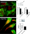
In the normal microvasculature, caveolin-1, the structural protein of caveolae, modulates transcytosis and paracellular permeability. Here, we used caveolin-1-deficient mice (Cav(-/-)) to track the potential active roles of caveolin-1 down-modulation in the regulation of vascular permeability and morphogenesis in tumors. In B16 melanoma-bearing Cav(-/-) mice, we found that fibrinogen accumulated in early-stage tumors to a larger extent than in wild-type animals. These results were confirmed by the observations of a net elevation of the interstitial fluid pressure and a relative deficit in albumin extravasation in Cav(-/-) tumors (versus healthy tissues). Immunostaining analyses of Cav(-/-) tumor sections further revealed a higher density of CD31-positive vascular structures and a dramatic deficit in alpha-smooth muscle actin-stained mural cells. The increase in blood plasma volume in Cav(-/-) tumors was confirmed by dynamic contrast enhanced-magnetic resonance imaging and found to be associated with a more rapid tumor growth. Finally, an in vitro wound test and the aorta ring assay revealed that silencing caveolin expression could directly impair the migration and the outgrowth of smooth muscle cells/pericytes, particularly in response to platelet-derived growth factor. In conclusion, a decrease in caveolin abundance, by promoting angiogenesis and preventing its termination by mural cell recruitment, appears as an important control point for the formation of new tumor blood vessels. Caveolin-1 therefore has the potential to be a marker of tumor vasculature maturity that may help adjusting anticancer therapies.
Figures


Similar articles
-
Vascular permeability and pathological angiogenesis in caveolin-1-null mice.Am J Pathol. 2009 Oct;175(4):1768-76. doi: 10.2353/ajpath.2009.090171. Epub 2009 Sep 3. Am J Pathol. 2009. PMID: 19729487 Free PMC article.
-
Caveolin-1-deficient mice have increased tumor microvascular permeability, angiogenesis, and growth.Cancer Res. 2007 Mar 15;67(6):2849-56. doi: 10.1158/0008-5472.CAN-06-4082. Cancer Res. 2007. PMID: 17363608
-
Endothelial caveolin-1 regulates pathologic angiogenesis in a mouse model of colitis.Gastroenterology. 2009 Feb;136(2):575-84.e2. doi: 10.1053/j.gastro.2008.10.085. Epub 2008 Nov 7. Gastroenterology. 2009. PMID: 19111727 Free PMC article.
-
Caveolin, caveolae, and endothelial cell function.Arterioscler Thromb Vasc Biol. 2003 Jul 1;23(7):1161-8. doi: 10.1161/01.ATV.0000070546.16946.3A. Epub 2003 Apr 10. Arterioscler Thromb Vasc Biol. 2003. PMID: 12689915 Review.
-
Role of pericytes in angiogenesis: focus on cancer angiogenesis and anti-angiogenic therapy.Neoplasma. 2016;63(2):173-82. doi: 10.4149/201_150704N369. Neoplasma. 2016. PMID: 26774138 Review.
Cited by
-
Vascular permeability and pathological angiogenesis in caveolin-1-null mice.Am J Pathol. 2009 Oct;175(4):1768-76. doi: 10.2353/ajpath.2009.090171. Epub 2009 Sep 3. Am J Pathol. 2009. PMID: 19729487 Free PMC article.
-
Vascular permeability, vascular hyperpermeability and angiogenesis.Angiogenesis. 2008;11(2):109-19. doi: 10.1007/s10456-008-9099-z. Epub 2008 Feb 22. Angiogenesis. 2008. PMID: 18293091 Free PMC article. Review.
-
Endothelial-specific overexpression of caveolin-1 accelerates atherosclerosis in apolipoprotein E-deficient mice.Am J Pathol. 2010 Aug;177(2):998-1003. doi: 10.2353/ajpath.2010.091287. Epub 2010 Jun 25. Am J Pathol. 2010. PMID: 20581061 Free PMC article.
-
Cav-1 Ablation in Pancreatic Stellate Cells Promotes Pancreatic Cancer Growth through Nrf2-Induced shh Signaling.Oxid Med Cell Longev. 2020 Apr 20;2020:1868764. doi: 10.1155/2020/1868764. eCollection 2020. Oxid Med Cell Longev. 2020. PMID: 32377291 Free PMC article.
-
Brain pericytes: emerging concepts and functional roles in brain homeostasis.Cell Mol Neurobiol. 2011 Mar;31(2):175-93. doi: 10.1007/s10571-010-9605-x. Cell Mol Neurobiol. 2011. PMID: 21061157 Free PMC article.
References
-
- Less JR, Skalak TC, Sevick EM, Jain RK. Microvascular architecture in a mammary carcinoma: branching patterns and vessel dimensions. Cancer Res. 1991;51:265–273. - PubMed
-
- McDonald DM, Baluk P. Significance of blood vessel leakiness in cancer. Cancer Res. 2002;62:5381–5385. - PubMed
-
- Jain RK. Molecular regulation of vessel maturation. Nat Med. 2003;9:685–693. - PubMed
Publication types
MeSH terms
Substances
LinkOut - more resources
Full Text Sources
Other Literature Sources
Molecular Biology Databases

