

Neural drive to human genioglossus in obstructive sleep apnoea
- PMID: 17916615
- PMCID: PMC2375464
- DOI: 10.1113/jphysiol.2007.139584
Neural drive to human genioglossus in obstructive sleep apnoea
Abstract
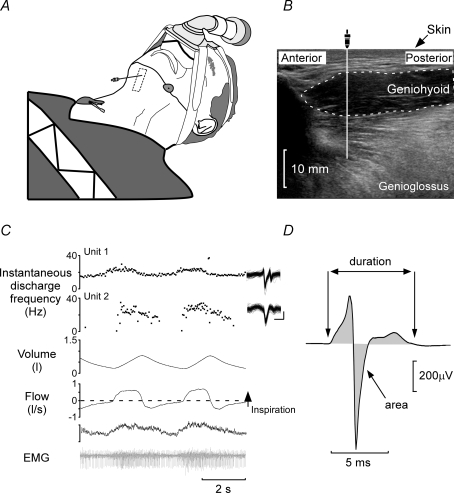
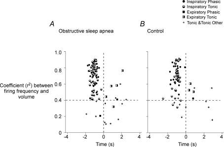
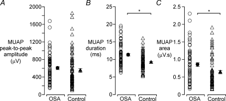
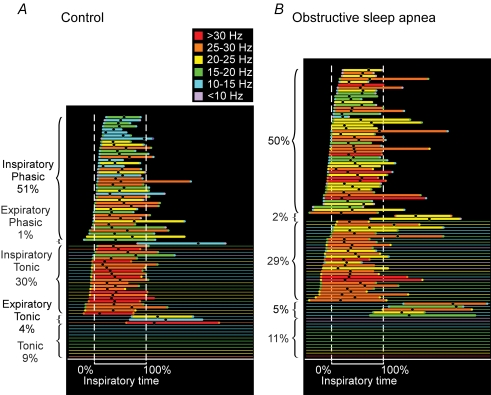
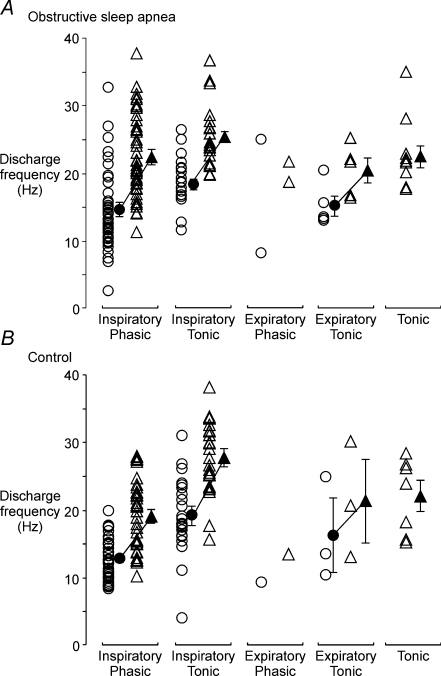
One postulated mechanism for obstructive sleep apnoea (OSA) is insufficient drive to the upper-airway musculature during sleep, with increased (compensatory) drive during wakefulness. This generates more electromyographic activity in upper airway muscles including genioglossus. To understand drives to upper airway muscles, we recorded single motor unit activity from genioglossus in male groups of control (n = 7, 7 +/- 2 events h(-1)) and severe OSA (n = 9, 54 +/- 4 events h(-1)) subjects. One hundred and seventy-eight genioglossus units were recorded using monopolar electrodes. Subjects were awake, supine and breathing through a nasal mask. The distribution of the six types of motor unit activity in genioglossus (Inspiratory Phasic, Inspiratory Tonic, Expiratory Phasic, Expiratory Tonic, Tonic and Tonic Other) was identical in both groups. Single unit action potentials in OSA were larger in area (by 34%, P < 0.05) and longer in duration (by 23%, P < 0.05). Inspiratory units were recruited earlier in OSA than control subjects. In control subjects, Inspiratory Tonic units peaked earlier than Inspiratory Phasic units, while in OSA subjects, Inspiratory Tonic and Phasic units peaked simultaneously. Onset frequencies did not differ between groups, but the peak discharge frequency for Inspiratory Phasic units was higher in OSA (22 +/- 1 Hz) than control subjects (19 +/- 1 Hz, P = 0.003), but conversely, the peak discharge frequency of Inspiratory Tonic units was higher in control subjects (28 +/- 1 Hz versus 25 +/- 1 Hz, P < 0.05). Increased motor unit action potential area indicates that neurogenic changes have occurred in OSA. In addition, the differences in the timing and firing frequency of the inspiratory classes of genioglossus motor units indicate that the output of the hypoglossal nucleus may have changed.
Figures
Similar articles
-
Compartmental inspiratory genioglossus electromyographic activity in supine, awake individuals with and without obstructive sleep apnoea.J Physiol. 2025 May;603(9):2877-2893. doi: 10.1113/JP287943. Epub 2025 Apr 15. J Physiol. 2025. PMID: 40231764 Free PMC article.
-
Tonic and phasic respiratory drives to human genioglossus motoneurons during breathing.J Neurophysiol. 2006 Apr;95(4):2213-21. doi: 10.1152/jn.00940.2005. Epub 2005 Nov 23. J Neurophysiol. 2006. PMID: 16306175
-
Regional associations between inspiratory tongue dilatory movement and genioglossus activity during wakefulness in people with obstructive sleep apnoea.J Physiol. 2023 Dec;601(24):5795-5811. doi: 10.1113/JP285187. Epub 2023 Nov 20. J Physiol. 2023. PMID: 37983193 Free PMC article.
-
Discharge properties of upper airway motor units during wakefulness and sleep.Prog Brain Res. 2014;212:59-75. doi: 10.1016/B978-0-444-63488-7.00004-5. Prog Brain Res. 2014. PMID: 25194193 Review.
-
The output from human inspiratory motoneurone pools.J Physiol. 2008 Mar 1;586(5):1257-64. doi: 10.1113/jphysiol.2007.145789. Epub 2007 Nov 1. J Physiol. 2008. PMID: 17974589 Free PMC article. Review.
Cited by
-
Sleep-wake control of the upper airway by noradrenergic neurons, with and without intermittent hypoxia.Prog Brain Res. 2014;209:255-74. doi: 10.1016/B978-0-444-63274-6.00013-8. Prog Brain Res. 2014. PMID: 24746052 Free PMC article.
-
Hypoglossal nerve conduction findings in obstructive sleep apnea.Muscle Nerve. 2010 Aug;42(2):257-61. doi: 10.1002/mus.21690. Muscle Nerve. 2010. PMID: 20544939 Free PMC article.
-
Recruitment and rate-coding strategies of the human genioglossus muscle.J Appl Physiol (1985). 2010 Dec;109(6):1939-49. doi: 10.1152/japplphysiol.00812.2010. Epub 2010 Oct 14. J Appl Physiol (1985). 2010. PMID: 20947713 Free PMC article.
-
Sleeping tongue: current perspectives of genioglossus control in healthy individuals and patients with obstructive sleep apnea.Nat Sci Sleep. 2018 Jun 15;10:169-179. doi: 10.2147/NSS.S143296. eCollection 2018. Nat Sci Sleep. 2018. PMID: 29942169 Free PMC article. Review.
-
Compartmental inspiratory genioglossus electromyographic activity in supine, awake individuals with and without obstructive sleep apnoea.J Physiol. 2025 May;603(9):2877-2893. doi: 10.1113/JP287943. Epub 2025 Apr 15. J Physiol. 2025. PMID: 40231764 Free PMC article.
References
-
- American Academy of Sleep Medical Task Force. Sleep-related breathing disorders in adults: Recommendations for syndrome definition and measurement techniques in clinical research. Sleep. 1999;22:667–689. - PubMed
-
- Bailey EF, Fregosi RF. Coordination of intrinsic and extrinsic tongue muscles during spontaneous breathing in the rat. J Appl Physiol. 2004;96:440–449. - PubMed
-
- Bailey EF, Rice AD, Fuglevand AJ. Firing patterns of human genioglossus motor units during voluntary tongue movement. J Neurophysiol. 2007;97:933–936. - PubMed
Publication types
MeSH terms
Grants and funding
LinkOut - more resources
Full Text Sources