. 2007 Nov;17(11):1562-71.
doi: 10.1101/gr.6772807. Epub 2007 Oct 4.

Breed relationships facilitate fine-mapping studies: a 7.8-kb deletion cosegregates with Collie eye anomaly across multiple dog breeds

Affiliations

Breed relationships facilitate fine-mapping studies: a 7.8-kb deletion cosegregates with Collie eye anomaly across multiple dog breeds

Heidi G Parker et al. Genome Res. 2007 Nov.

Abstract

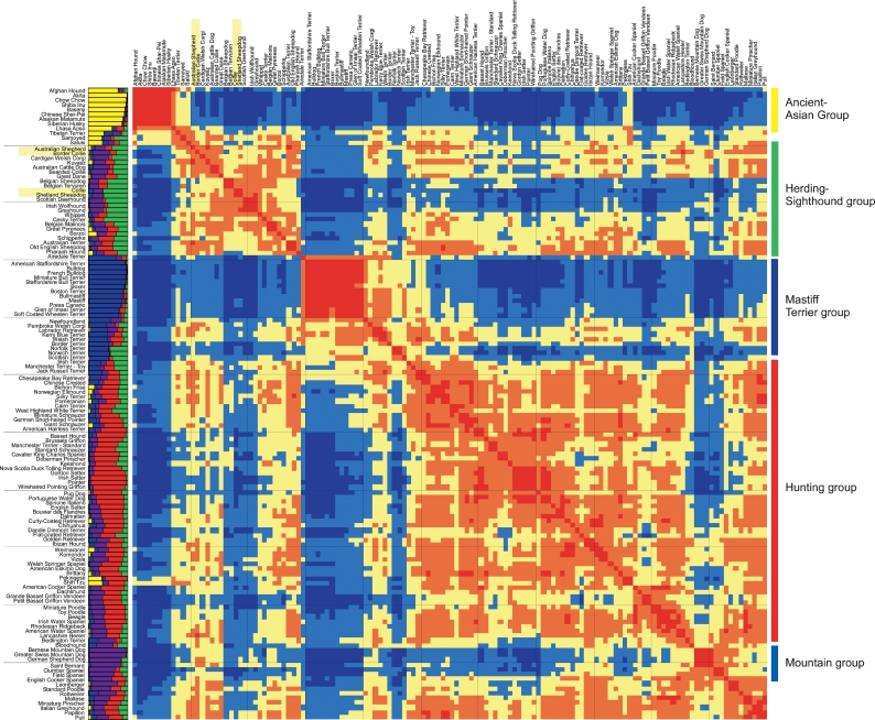
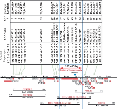
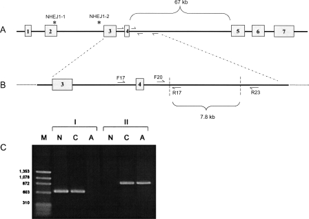
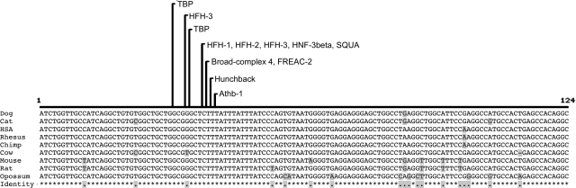
The features of modern dog breeds that increase the ease of mapping common diseases, such as reduced heterogeneity and extensive linkage disequilibrium, may also increase the difficulty associated with fine mapping and identifying causative mutations. One way to address this problem is by combining data from multiple breeds segregating the same trait after initial linkage has been determined. The multibreed approach increases the number of potentially informative recombination events and reduces the size of the critical haplotype by taking advantage of shortened linkage disequilibrium distances found across breeds. In order to identify breeds that likely share a trait inherited from the same ancestral source, we have used cluster analysis to divide 132 breeds of dog into five primary breed groups. We then use the multibreed approach to fine-map Collie eye anomaly (cea), a complex disorder of ocular development that was initially mapped to a 3.9-cM region on canine chromosome 37. Combined genotypes from affected individuals from four breeds of a single breed group significantly narrowed the candidate gene region to a 103-kb interval spanning only four genes. Sequence analysis revealed that all affected dogs share a homozygous deletion of 7.8 kb in the NHEJ1 gene. This intronic deletion spans a highly conserved binding domain to which several developmentally important proteins bind. This work both establishes that the primary cea mutation arose as a single disease allele in a common ancestor of herding breeds as well as highlights the value of comparative population analysis for refining regions of linkage.


Figures

Figure 1.
Figure 1.
Population structure of 132 domestic dog breeds. On the left of the heatmap graph is a structure graph with each bar representing a breed comprised of four to five individuals. The bars are divided into K colors, where K is the number of populations assumed. The length of the colored segment shows the breed’s estimated proportion of membership in that cluster averaged over 20 runs with K = 5. The heatmap graph shows consistency of clustering between breeds across the same 20 runs of structure. (Red blocks) Breeds that cluster together in >80% of runs; (orange) breeds that cluster together in 60%–80% of all runs; (yellow) breeds that cluster in 40%–60% of runs; (light blue) breeds that cluster in 20%–40% of runs; and (dark blue) breeds that cluster in <20% of all runs. Breeds are ordered identically along both the X- and Y-axes of the color map. Order was determined by a dendrogram based on the distances displayed (dendrogram not shown). The names of the breeds are shown to the left of the structure graph and above the heatmap. The four breeds used to map cea are highlighted in yellow. Group names are listed to the right with colored lines indicating the location of the group on the heatmap.
Figure 2.
Figure 2.
Alignment of SNP haplotypes and a canine BAC contig to the CanFam2 genomic sequence for the cea interval on canine chromosome 37. Eight haplotypes, spanning >376 kb on CFA37, represent cea-transmitting chromosomes segregating in four different breeds and define a linkage disequilibrium interval common to all known cea-affected breeds. Haplotype 1 was observed in all cea-affected Rough and Smooth Collies and Australian Shepherds tested, and in some but not all Border Collies. Haplotypes 2 through 7 were identified in specific Border Collies, and haplotype 8 was only seen in European Shetland Sheepdogs. All haplotypes are identical, and all affected dogs are homozygous for SNPs rs8797935 through rs23961919 (the region bounded by vertical blue lines). (Blue box above the chromosome, the black line in the center of the figure) The position of the 7799-bp deletion in intron 4 of NHEJ1. (Red boxes below the chromosome line) Genes in the region. BACs are tiled below the genes and are identified by their number. The gene-specific probes used to pick the BACs and align them to the chromosome are listed in red above the BAC.
Figure 3.
Figure 3.
A two-step PCR protocol for genotyping the cea-associated deletion. (A) The NHEJ1 gene with the positions of the two linked SNPs designated by * and primer locations within intron four. (B) Expanded representation of the cea-associated deletion region (outlined by diagonal dashed lines), with flanking and internal primers. (C) Electrophoretogram demonstrating PCR results using primers shown in B on DNA from Normal (N), Carrier (C), and Affected (A) dogs. (Lane 1, M) A marker lane with sizes as indicated. Set I of PCR products (lanes 2–4) presents amplification results using primers NHEJ1-F17 and NHEJ1-R17. Set II (lanes 5–7) presents results using primers NHEJ1-F20 and NHEJ1-R23.
Figure 4.
Figure 4.
Comparative alignment of the 124-bp highly conserved region within the canine cea-associated deletion for nine diverse mammalian sequences. The most strongly conserved region includes a cluster of recognition domains for several DNA-binding proteins, conserved in all nine species. These sites are listed above the alignment with lines pointing to the starting position. Locations and sequence information for these conserved binding domains are listed in Supplemental Table 6. The sequence locations for alignment are dog, chr37:28,702,470–28,702,593 rc; human, chr2:219,715,427–219,715,550 rc; chimp, chr2b:225,076,398–225,076,521 rc; rhesus, chr12:82,998,197–82,998,320 rc; mouse, chr1:74,973,801–74,973,924 rc; rat, chr9:74,386,991–74,387,114 rc; cat, scaffold_100612:247,076–247,199; cow, chr2:65,088,541–65,088,663 rc; opossum, chr7:175,293,269–175,293,392.

References

    1. Alur R., Brooks B., Brooks B. Clinical and genetic analysis of coloboma: A review. Asian J. Exp. Sci. 2004;20:1–15.
    1. American College of Veterinary Ophthalmologists . Ocular disorders proven or suspected to be inherited in purebred dogs. Genetics Committee Report; Meridian, ID: 2007.
    1. Berryere T.G., Kerns J.A., Barsh G.S., Schmutz S.M., Kerns J.A., Barsh G.S., Schmutz S.M., Barsh G.S., Schmutz S.M., Schmutz S.M. Association of an Agouti allele with fawn or sable coat color in domestic dogs. Mamm. Genome. 2005;16:262–272. - PubMed
    1. Bimboim H.C., Doly J., Doly J. A rapid alkaline procedure for screening recombinant plasmid DNA. Nucleic Acids Res. 1979;7:1513–1523. doi: 10.1093/nar/7.6.1513. - DOI - PMC - PubMed
    1. Chandler K. Canine epilepsy: What can we learn from human seizure disorders? Vet. J. 2006;172:207–217. - PubMed

Publication types