. 2008 Feb;22(2):403-14.
doi: 10.1210/me.2007-0272. Epub 2007 Oct 4.

Prostaglandin F2alpha stimulates the expression and secretion of transforming growth factor B1 via induction of the early growth response 1 gene (EGR1) in the bovine corpus luteum

Affiliations

Prostaglandin F2alpha stimulates the expression and secretion of transforming growth factor B1 via induction of the early growth response 1 gene (EGR1) in the bovine corpus luteum

Xiaoying Hou et al. Mol Endocrinol. 2008 Feb.

Abstract

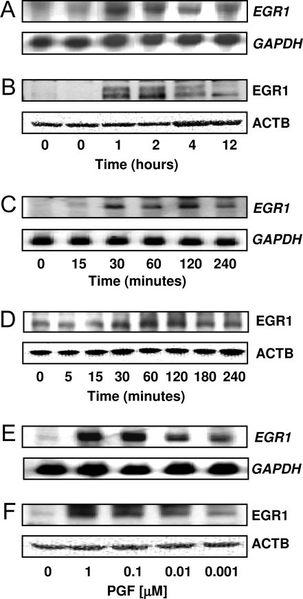
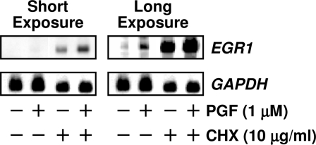
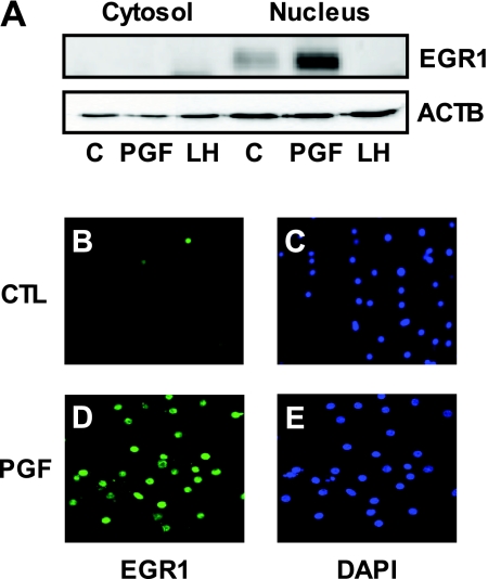
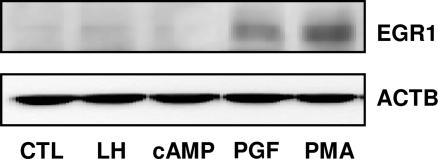
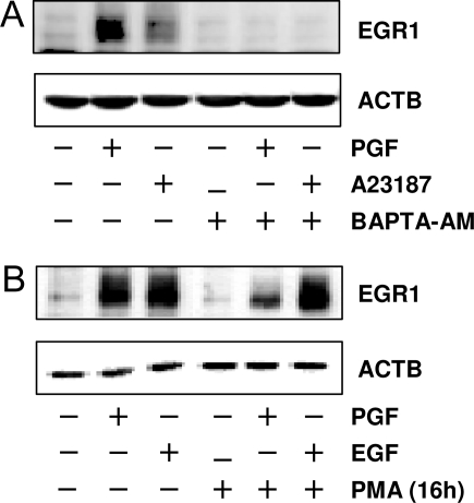
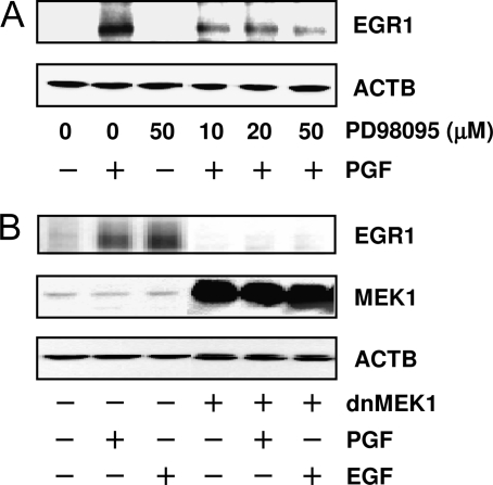
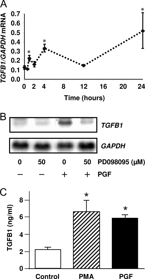
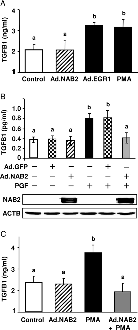
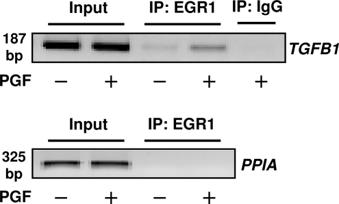
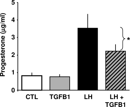
In most mammals, prostaglandin F2alpha (PGF2alpha) is believed to be a trigger that induces the regression of the corpus luteum (CL), whereby progesterone synthesis is inhibited, the luteal structure involutes, and the reproductive cycle resumes. Studies have shown that the early growth response 1 (EGR1) protein can induce the expression of proapoptotic proteins, suggesting that EGR1 may play a role in luteal regression. Our hypothesis is that EGR1 mediates the actions of PGF2alpha by inducing the expression of TGF beta1 (TGFB1), a key tissue remodeling protein. The levels of EGR1 mRNA and protein were up-regulated in the bovine CL during PGF2alpha-induced luteolysis in vivo and in PGF2alpha-treated luteal cells in vitro. Using chemical and genetic approaches, the RAF/MAPK kinase (MEK) 1/ERK pathway was identified as a proximal signaling event required for the induction of EGR1 in PGF2alpha-treated cells. Treatment with PGF2alpha increased the expression of TGFB1 mRNA and protein as well as the binding of EGR1 protein to TGFB1 promoter in bovine luteal cells. The effect of PGF2alpha on TGFB1 expression was mimicked by a protein kinase C (PKC)/RAF/MEK1/ERK activator or adenoviral-mediated expression of EGR1. The stimulatory effect of PGF2alpha on TGFB1 mRNA and TGFB1 protein secretion was inhibited by blockade of MEK1/ERK signaling and by adenoviral-mediated expression of NAB2, an EGR1 binding protein that inhibits EGR1 transcriptional activity. Treatment of luteal cells with TGFB1 reduced progesterone secretion, implicating TGFB1 in luteal regression. These studies demonstrate that PGF2alpha stimulates the expression of EGR1 and TGFB1 in the CL. We suggest that EGR1 plays a role in the expression of genes whose cognate proteins coordinate luteal regression.


Figures

Figure 1
Figure 1
PGF2α Induces EGR1 Expression in Vivo and in Vitro A and B, Angus crossbred cows (two to four animals per time point) were injected im with 25 mg Lutalyse or saline during the mid-luteal phase and CL were removed at 0, 1, 2, 4, and 12 h after injection. The total RNA and protein were extracted as described in Materials and Methods. A, Northern blot analysis of EGR1 mRNA. The same blot was also probed for GAPDH mRNA as a loading control. B, Western blot analysis using an EGR1-specific antibody (1:1000). The same blot was also probed with β-actin (ATCB) (1:5000) as a loading control. C and D, Primary cultures of bovine luteal cells were treated with PGF2α (1 μm) for 5, 15, 30, 60, 120, 180, and 240 min. Cells were extracted and subjected to Northern and Western analysis described above. E and F, Luteal cells were treated for 1 h with increasing concentrations (1 nm–1 μm) of PGF2α). The EGR1 mRNA (C and E) and EGR1 protein (D and F) are shown.
Figure 2
Figure 2
PGF2α-Induced EGR1 mRNA Does Not Require de Novo Protein Synthesis Luteal cells were pretreated with control media or CHX (10 μg/ml) for 4 h before adding PGF2α (1 μm) for another 60 min. Total RNA was extracted and subjected to Northern blot analysis described in Fig. 1. The Northern blot was subjected to a short and long exposure to demonstrate various levels of EGR1 mRNA.
Figure 3
Figure 3
Nuclear Localization of EGR1 Protein A, Bovine luteal cells were treated with or without PGF2α (1 μm) or LH (100 ng/ml) for 1 h and then cytoplasmic (Cytosol) and nuclear (Nucleus) fractions were prepared using the Nuclear Extract Kit according to the manufacturer’s instructions. Proteins (40 μg) were separated on 10% SDS-PAGE and transferred to PVDF membranes. EGR1 protein levels were examined by Western blot using the EGR1 antibody (1:1000) and β-actin (ACTB) (1:5000) served as a loading control. B–E, Bovine luteal cells were cultured in four-well Lab-Tek glass chamber slides as described in Materials and Methods. After 24 h in serum-free media, cells were treated with or without PGF2α (1 μm) for 1 h. Photomicrographs showed EGR1 immunofluorescence (green) present in the nucleus of luteal cells after PGF2αtreatment. 4′, 6-Diamidino-2-phenylindole (DAPI) (blue) was used to stain nucleus. Immunofluorescence localization was repeated on cell preparations from three separate ovaries and representative photographs are presented. CTL, Control.
Figure 4
Figure 4
PGF2α and PMA, But Not LH or cAMP, Elevate EGR1 Protein in Luteal Cells Bovine luteal cells were treated with LH (100 ng/ml), 8-Br-cAMP (2 mm), PGF2α (1 μm) or PMA (20 nm), for 90 min. EGR1 protein levels were examined by Western blot using an antibody specific for EGR1 (1:1000) as described in Fig. 1. The same blot was also probed with β-actin (ACTB) (1:5000) as a loading control. CTL, Control.
Figure 5
Figure 5
PGF2α Induces EGR1 Expression through Calcium and PKC-Dependent Pathway A, Luteal cells were pretreated with BAPTA-AM (25 μm) for 1 h before treatment for 1 h with PGF2α (1 μm) or calcium ionophore A23187 (1 μm). B, Luteal cells were pretreated with PMA (20 nm) for 16 h to deplete phorbol ester-responsive isoforms of PKC. Cells were then treated with either PGF2α (1 μm) or EGF (10 ng/ml) for another 60 min. EGR1 protein levels were examined by Western blot as described in Fig. 1. β-Actin (ACTB) (1:5000) served as a loading control.
Figure 6
Figure 6
PGF2α-Induced EGR1 Protein Elevation Was MEK1/ERK Dependent A, Luteal cells were pretreated with the MEK1 inhibitor PD98095 (50, 25, and 10 μm) for 1 h before treatment for 1 h with PGF2α (1 μm). EGR1 protein was detected by Western blot. Results were representative of three experiments. β-Actin (ACTB) (1:5000) served as a loading control. B, Luteal cells were infected with Ad expressing dominant-negative mutant MEK1 (Ad.dnMEK1) for 24 h as described in Materials and Methods. Cells were then stimulated with either control media, PGF2α (1 μm) or EGF (10 ng/ml) for 1 h. EGR1 protein was detected by Western blot as described in Fig. 1. The membranes were also blotted with antibodies against MEK1 (1:000) to demonstrate the expression of the ectopic protein and for β-actin (ACTB) (1:5000) to verify equal protein loading.
Figure 7
Figure 7
PGF2α Induces TGFB1 Expression in Vivo and in Vitro A, Angus crossbred cows were injected im with 25 mg Lutalyse or saline during the mid-luteal phase and the CL were removed at 0.5, 1, 2, 4, 12, and 24 h after injection. Levels of TGFB1 mRNA in bovine CL were quantified by real-time RT-PCR using total CL RNA as described in Materials and Methods. The results are presented in terms of TGFB1 mRNA normalized to levels of GAPDH mRNA. Each time point represents the mean ± sem (n = 3–6 CL each collected from different cows). *, P < 0.05, vs. untreated control. B, Bovine luteal cells were pretreated with PD98095 (50 μm) for 1 h before treatment of PGF2α (1 μm) for 24 h. TGFB1 mRNA was detected by Northern blot. The same blot was probed for GAPDH mRNA as a loading control. Similar results were observed in replicate experiments. C, Luteal cells were treated with control media, PGF2α (1 μm) or PMA (20 nm) for 24 h. The conditioned media were collected for TGFB1 assay utilizing a TGFB1 ELISA as described in Materials and Methods. Each experiment was performed in duplicate, and three separated experiments were performed. *, P < 0.05, vs. control.
Figure 8
Figure 8
EGR1 Mediates PGF2α-Induced TGFB1 Expression A, Bovine luteal cells were infected with or without Ad (Ad.NAB2 or Ad.EGR1) for 24 h in the presence of serum and then serum-starved for 24 h. On the day of the experiment, the cells were preequilibrated in fresh media for 3 h, and luteal cells were treated without or with PMA (20 nm) for 24 h. TGFB1 in the conditioned media was determined by ELISA. Each experiment was performed in duplicate, and five separate experiments were performed. B, Luteal cells were infected with or without Ad (Ad.GFP or Ad.NAB2) and treated with or without PGF2α (1 μm) for 24 h as described above. TGFB1 in the conditioned media was determined by ELISA. Each experiment was performed in duplicate, and results are the average of four experiments. ACTB, β-Actin. C, Luteal cells were infected with or without Ad.NAB2 and treated with or without PMA (20 nm) for 24 h as described above. TGFB1 in the conditioned media were determined by ELISA. Each experiment was performed in duplicate, and results are the average of five experiments. A–C, Bars with similar letters are not statistically different; bars with different letters (a vs. b) are significantly different, P < 0.05.
Figure 9
Figure 9
EGR1 Binds to TGFB1 Promoter in Response to PGF2α in Bovine Luteal Cells Luteal cells were treated with or without PGF2α (1 μm) for 60 min and DNA and protein cross-linked with formaldehyde as described in Materials and Methods. Extracts were treated with EGR1 antibodies or IgG and ChIP assays performed using the primer pairs specific for the proximal promoter region of TGFB1 or cyclophilin A (peptidylprolyl isomerase A, PIAA) as described in Materials and Methods. Similar results were observed a replicate experiment using a separate cell preparation. IP, Immunoprecipitation.
Figure 10
Figure 10
TGFB1 Inhibits LH-Induced Progesterone Synthesis in Bovine Luteal Cells Bovine luteal cells were treated with or without LH (100 ng/ml) in the presence or absence of TGFB1 (1 ng/ml) for 6 h. The conditioned medium was collected and progesterone levels were determined by RIA. Results are presented as mean ± sem of four experiments each conducted in triplicate. *, P < 0.05 vs. LH. CTL, Control.

References

    1. Davis JS, Rueda BR 2002 The corpus luteum: an ovarian structure with maternal instincts and suicidal tendencies. Front Biosci 7:d1949–d1978 - PubMed
    1. Stocco C, Telleria C, Gibori G 2007 The molecular control of corpus luteum formation, function, and regression. Endocr Rev 28:117–149 - PubMed
    1. Sakamoto K, Ezashi T, Miwa K, Okuda-Ashitaka E, Houtani T, Sugimoto T, Ito S, Hayaishi O 1994 Molecular cloning and expression of a cDNA of the bovine prostaglandin F2 α receptor. J Biol Chem 269:3881–3886 - PubMed
    1. Davis JS 1987 Stimulation of intracellular free Ca2+ by luteinizing hormone in isolated bovine luteal cells. Adv Exp Med Biol 219:671–675 - PubMed
    1. Chen DB, Westfall SD, Fong HW, Roberson MS, Davis JS 1998 Prostaglandin F2α stimulates the Raf/MEK1/mitogen-activated protein kinase signaling cascade in bovine luteal cells. Endocrinology 139:3876–3885 - PubMed

Publication types

MeSH terms

Substances