. 2008 Jan;22(1):196-205.
doi: 10.1210/me.2007-0241. Epub 2007 Oct 4.

Fibroblast growth factor receptor-1 signaling in pancreatic islet beta-cells is modulated by the extracellular matrix

Affiliations

Fibroblast growth factor receptor-1 signaling in pancreatic islet beta-cells is modulated by the extracellular matrix

Dawn M Kilkenny et al. Mol Endocrinol. 2008 Jan.

Abstract

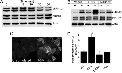
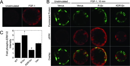
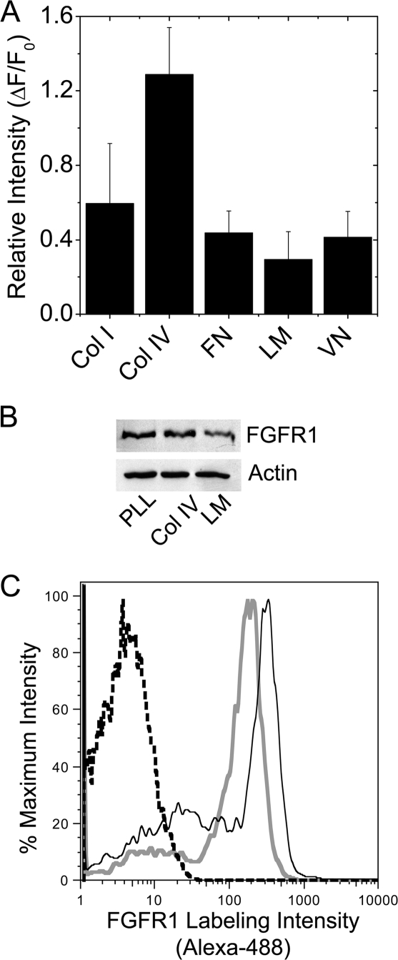
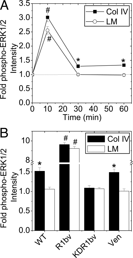
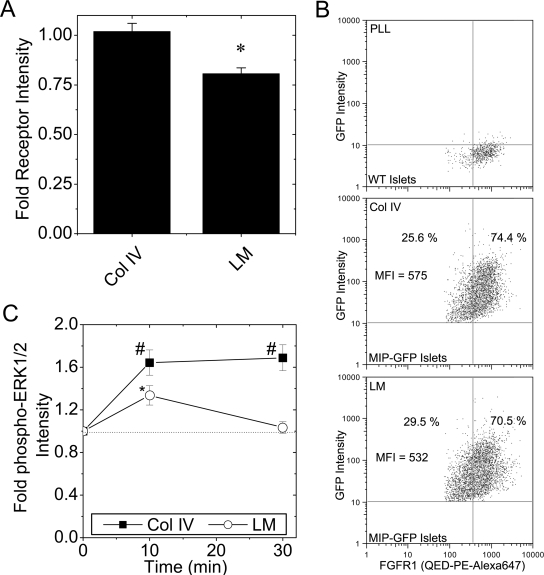
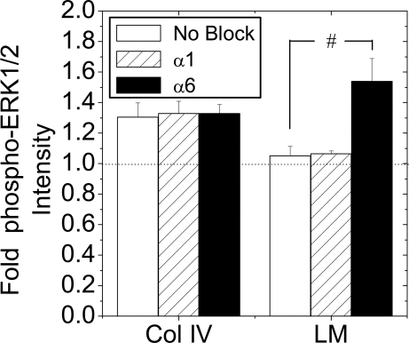
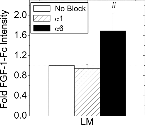
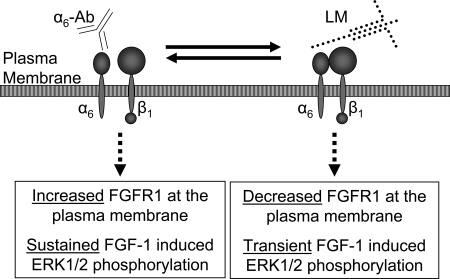
Maintenance of pancreatic beta-cell mass depends on extracellular stimuli that promote survival and proliferation. In the islet, these stimuli come from the beta-cell microenvironment and include extracellular matrix deposited by associated vascular endothelial cells. Fibroblast growth factor receptor-1 (FGFR1) has recently been implicated as a signaling pathway that is important for normal beta-cell function. We would like to understand how extracellular matrix and FGFR1 signaling interact to promote beta-cell survival and proliferation. To examine beta-cell-specific receptor responses, we created lentiviral vectors with rat insulin promoter-driven expression of Venus fluorescent protein-tagged full-length (R1betav) and kinase-deficient (KDR1betav) FGFR1. Significant FGF-1-dependent activation of ERK1/2 was observed in betaTC3 cells, dispersed beta-cells, and beta-cells in intact islets. This response was enhanced by R1betav expression and reduced by KDR1betav expression. Plating-dispersed beta-cells on collagen type IV resulted in enhanced expression of endogenous FGFR1 that was associated with sustained activation of ERK1/2. Conversely, plating cells on laminin reduced expression of FGFR1, and this reduction was associated with transient activation of ERK1/2. Addition of neutralizing antibodies to inhibit beta-cell attachment to laminin via alpha(6)-integrin increased high-affinity FGF-1-binding at the plasma membrane and resulted in sustained ERK1/2 activity similar to cells plated on collagen type IV. These data show that the FGF-stimulated beta-cell response is negatively affected by alpha(6)-integrin binding to laminin and suggest regulation associated with vascular endothelial cell remodeling.


Figures

Figure 1
Figure 1
FGF-1-Induced ERK1/2 Phosphorylation in βTC3 Cells Is Enhanced by R1βv Expression and Reduced by KDR1βv Expression A, Western immunoblotting of βTC3 whole-cell lysates (10 μg/lane) stimulated with FGF-1 (10 ng/ml; times as indicated) using antibodies recognizing phospho-ERK1/2 (top), ERK1/2 (middle), and β-actin (loading control, bottom). B, Western immunoblotting of whole-cell lysates (10 μg/lane) from βTC3 cells expressing Venus, R1βv, or KDR1βv in the absence (−) or presence (+) of FGF-1 (10 ng/ml for 10 min) using antibodies to detect phosphoERK1/2, ERK1/2, and β-actin. Representative blots are shown. C, Immunofluorescent detection of phospho-ERK1/2 in WT βTC3 cells in the absence (left) or presence (right) of FGF-1. D, Fold change in phospho-ERK1/2 immunofluorescence for cells stimulated with FGF-1 (as shown in panel C) compared with nonstimulated control cells. Data were collected from five fields of view for each sample and are plotted as the mean fold increase in phospho-ERK1/2 intensity compared with control cells ± sem from four independent experiments. *, P < 0.05; #, P < 0.02 (Student’s two-tailed t test). Ven, Venus.
Figure 2
Figure 2
FGFR1 Enhances ERK1/2 Activation in β-Cells of Intact Islets A, Phospho-ERK1/2 immunofluorescence of unstimulated or FGF-1 stimulated (10 ng/ml for 10 min) WT islets. B, Islets expressing β-cell specific Venus, R1βv, or KDR1β stimulated with FGF-1 (10 ng/ml for 10 min) and immunostained for phospho-ERK1/2. Representative images of Venus protein expression (top), phospho-ERK1/2 immunofluorescence (middle), and the overlay (bottom) are shown for islets expressing Venus, R1βv, and KDR1βv (as indicated). C, Fold change in phospho-ERK1/2 immunofluorescence in construct expressing β-cells. Data are plotted as mean fold change phospho-ERK1/2 intensity (FGF-1 treatment compared with nonstimulated islets) ± sem for WT, R1βv-, KDR1βv-, and Venus-expressing islets. The experiment was performed in triplicate, and intensity measurements were acquired from five to seven islets per treatment. *, P < 0.05; #, P < 0.02 (Student’s two-tailed t test). Ven, Venus.
Figure 3
Figure 3
βTC3 cell FGFR1 Expression Is Reduced by Attachment to Laminin A, FGFR1-associated immunofluorescence in WT βTC3 cells plated overnight on collagen type I (Col I), collagen type IV (Col IV), fibronectin (FN), laminin (LM), and vitronectin (VN) (n = 4). B, Western immunoblotting of whole-cell lysates (5 μg/lane) from βTC3 cells plated overnight on poly-l-lysine (PLL), Col IV, and LM using an antibody to detect the N terminus of FGFR1 (3472, Cell Signaling Technology). β-Actin is shown as a loading control (representative blot is shown; n = 3). C, Enhanced cell surface labeling was detected in βTC3 cells plated on Col IV (solid black line) vs. LM (solid gray line) by flow cytometry using an antibody to detect the N terminus of FGFR1 (M2F12, QED Biosciences). Unstained control cell intensity is indicated by the black dashed line (representative plot shown; n = 5)
Figure 4
Figure 4
FGF-1 Stimulated βTC3 Cell ERK1/2 Phosphorylation Is Altered by ECM Attachment A, FGF-1-induced phospho-ERK1/2 immunofluorescence of WT cells grown on collagen type IV or laminin. Data are plotted as fold change in phospho-ERK1/2 immunofluorescence intensity compared with nonstimulated control (time 0) ± sem for three to four independent experiments (minimum of 70 cells analyzed per time point). The dotted line shown at 1-fold change phospho-ERK1/2 intensity is shown to assist data interpretation. B, Similar experiment as panel A. including R1βv-, KDR1βv-, and Venus-expressing cells after 30 min of FGF-1 stimulation (10 ng/ml). Data are plotted as fold change in intensity compared with unstimulated control cells ± sem from three independent experiments. *, P < 0.05; #, P < 0.02 (Student’s two-tailed t test). Col, Collagen; LM, laminin;Ven, Venus.
Figure 5
Figure 5
Dispersed β-Cell FGFR1 Expression and ERK1/2 Phosphorylation Is Reduced by Attachment to Laminin A, FGFR1-associated immunofluorescence (flg) in dispersed islet β-cells cultured overnight on collagen type IV or laminin. Data are plotted as fold change in intensity compared with cells plated on control poly-l-lysine ± sem from three mice on separate days. B, FGFR expression was detected by flow cytometry in insulin (GFP)-positive β-cells dispersed on Col IV and LM. WT islets labeled for FGFR1 using the N terminus-specific antibody complexed with Zenon PE-Alexa647 were used to set the quadrants (top). MIP-GFP islets were dispersed on collagen type IV (Col IV, middle) and laminin (LM, bottom). FGFR1-mean fluorescence intensity (MFI) of the GFP-positive cells is shown as well as the percent GFP-positive cells that were FGFR1 negative and positive. A representative plot is shown; n = 2. C, Changes in ERK1/2 phosphorylation in dispersed islet β-cells plated on collagen type IV and laminin. Cells were stimulated with FGF-1 (10 ng/ml, times as indicated), and phospho-ERK1/2-associated fluorescence intensity was measured in β-cells costaining positively for insulin. Data are plotted as fold change in phospho-ERK1/2 immunofluorescence intensity compared with nonstimulated control (time 0) ± sem from four mice on separate days. The dotted line shown at 1-fold change phospho-ERK1/2 intensity is shown to assist data interpretation. *, P < 0.05; #, P < 0.02 (Student’s two-tailed t test). PLL, Poly-l-lysine.
Figure 6
Figure 6
FGF-1 Induced ERK1/2 Activation in the Presence of Neutralizing Integrin Antibodies Islets were dispersed to single β-cells and cultured overnight on collagen type IV (Col IV) or laminin (LM) in the presence or absence (No block) of integrin-neutralizing antibodies (α1 or α6; 10 μg/ml). Cells were serum starved in 0.2% fetal bovine serum for 6 h before treatment with 10 ng/ml FGF-1 for 30 min. Data are plotted as fold change in phospho-ERK1/2 immunofluorescence intensity compared with nonstimulated control ± sem from four mice on separate days. *, P < 0.05 (Student’s two-tailed t test).
Figure 7
Figure 7
FGF-1-Fc Fusion Protein Receptor Binding in the Presence of Integrin-Neutralizing Antibodies Islets were dispersed to single β-cells and cultured overnight on laminin (LM) in the presence or absence (No block) of integrin neutralizing antibodies (α1 or α6; 10 μg/ml). Cells were incubated with FGF-1-Fc (100 ng/ml, 1 h on ice) and fusion protein was removed from low-affinity binding sites by washing quickly with heparin (250 μg/ml). FGF-1-Fc and insulin were detected by immunofluorescence. Data are plotted as the mean FGF-1-Fc immunofluorescence intensity relative to no block control ± sem from four mice on separate days. The dotted line shown at 1-fold change phospho-ERK1/2 intensity is shown to assist data interpretation.*, P < 0.05 (Student’s two-tailed t test).
Figure 8
Figure 8
The Effect of β-Cell Attachment to Laminin through α6-Integrin A schematic representing a β-cell plasma membrane with α6β1-integrin either neutralized with antibody (α6-Ab) or bound to laminin (LM). Our data demonstrate that inhibiting integrin attachment with neutralizing antibody results in an increase in high-affinity FGF-1 binding (Increased FGFR1) and sustained FGF-1-stimulated ERK1/2 response. In contrast, α6β1 binding to laminin decreases high-affinity receptor at the plasma membrane (Decreased FGFR1) and results in a transient FGF-1-stimulated ERK1/2 response.

Similar articles

Cited by

References

    1. Ornitz DM, Itoh N 2001 Fibroblast growth factors. Genome Biol 2:REVIEWS3005 - PMC - PubMed
    1. Gospodarowicz D, Cheng J 1986 Heparin protects basic and acidic FGF from inactivation. J Cell Physiol 128:475–484 - PubMed
    1. Johnson DE, Williams LT 1993 Structural and functional diversity in the FGF receptor multigene family. Adv Cancer Res 60:1–41 - PubMed
    1. Cool SM, Sayer RE, van Heumen WR, Pickles JO, Nurcombe V 2002 Temporal and spatial expression of fibroblast growth factor receptor 4 isoforms in murine tissues. Histochem J 34:291–297 - PubMed
    1. Arany E, Hill DJ 2000 Ontogeny of fibroblast growth factors in the early development of the rat endocrine pancreas. Pediatr Res 48:389–403 - PubMed

Publication types

MeSH terms