

Ordered phosphorylation governs oscillation of a three-protein circadian clock
- PMID: 17916691
- PMCID: PMC2427396
- DOI: 10.1126/science.1148596
Ordered phosphorylation governs oscillation of a three-protein circadian clock
Abstract
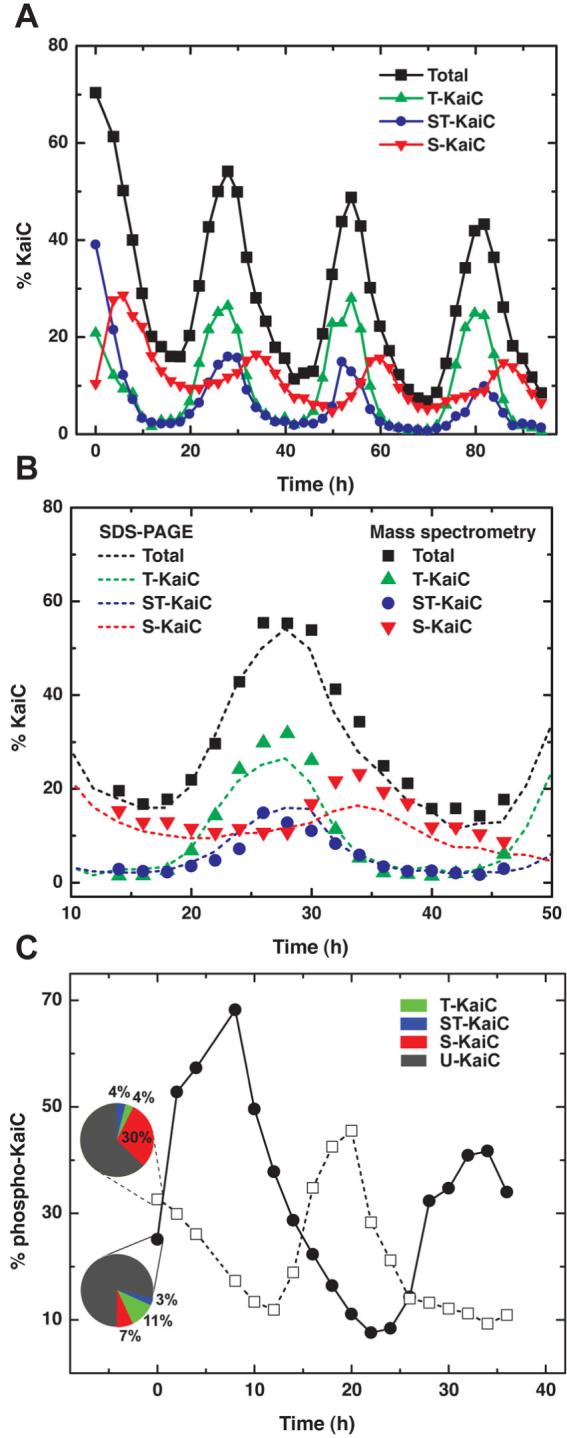
The simple circadian oscillator found in cyanobacteria can be reconstituted in vitro using three proteins-KaiA, KaiB, and KaiC. The total phosphorylation level of KaiC oscillates with a circadian period, but the mechanism underlying its sustained oscillation remains unclear. We have shown that four forms of KaiC differing in their phosphorylation state appear in an ordered pattern arising from the intrinsic autokinase and autophosphatase rates of KaiC and their modulation by KaiA. Kinetic and biochemical data indicate that one of these phosphoforms inhibits the activity of KaiA through interaction with KaiB, providing the crucial feedback that sustains oscillation. A mathematical model constrained by experimental data quantitatively reproduces the circadian period and the distinctive dynamics of the four phosphoforms.
Figures


Comment in
-
Systems biology. A clock with a flip switch.Science. 2007 Nov 2;318(5851):757-8. doi: 10.1126/science.1150740. Science. 2007. PMID: 17975056 No abstract available.
Similar articles
-
A mathematical model for the Kai-protein-based chemical oscillator and clock gene expression rhythms in cyanobacteria.J Biol Rhythms. 2007 Feb;22(1):69-80. doi: 10.1177/0748730406295749. J Biol Rhythms. 2007. PMID: 17229926
-
The day/night switch in KaiC, a central oscillator component of the circadian clock of cyanobacteria.Proc Natl Acad Sci U S A. 2008 Sep 2;105(35):12825-30. doi: 10.1073/pnas.0800526105. Epub 2008 Aug 26. Proc Natl Acad Sci U S A. 2008. PMID: 18728181 Free PMC article.
-
A cyanobacterial circadian clock based on the Kai oscillator.Cold Spring Harb Symp Quant Biol. 2007;72:47-55. doi: 10.1101/sqb.2007.72.029. Cold Spring Harb Symp Quant Biol. 2007. PMID: 18419262 Review.
-
Autonomous synchronization of the circadian KaiC phosphorylation rhythm.Nat Struct Mol Biol. 2007 Nov;14(11):1084-8. doi: 10.1038/nsmb1312. Epub 2007 Oct 28. Nat Struct Mol Biol. 2007. PMID: 17965725
-
Structural insights into a circadian oscillator.Science. 2008 Oct 31;322(5902):697-701. doi: 10.1126/science.1150451. Science. 2008. PMID: 18974343 Free PMC article. Review.
Cited by
-
Diurnal Regulation of Cellular Processes in the Cyanobacterium Synechocystis sp. Strain PCC 6803: Insights from Transcriptomic, Fluxomic, and Physiological Analyses.mBio. 2016 May 3;7(3):e00464-16. doi: 10.1128/mBio.00464-16. mBio. 2016. PMID: 27143387 Free PMC article.
-
Costs of Clock-Environment Misalignment in Individual Cyanobacterial Cells.Biophys J. 2016 Aug 23;111(4):883-891. doi: 10.1016/j.bpj.2016.07.008. Biophys J. 2016. PMID: 27558731 Free PMC article.
-
Perturbation-based analysis and modeling of combinatorial regulation in the yeast sulfur assimilation pathway.Mol Biol Cell. 2012 Aug;23(15):2993-3007. doi: 10.1091/mbc.E12-03-0232. Epub 2012 Jun 13. Mol Biol Cell. 2012. PMID: 22696683 Free PMC article.
-
Systematic analysis of negative and positive feedback loops for robustness and temperature compensation in circadian rhythms.NPJ Syst Biol Appl. 2023 Feb 11;9(1):5. doi: 10.1038/s41540-023-00268-7. NPJ Syst Biol Appl. 2023. PMID: 36774353 Free PMC article.
-
Active output state of the Synechococcus Kai circadian oscillator.Proc Natl Acad Sci U S A. 2013 Oct 1;110(40):E3849-57. doi: 10.1073/pnas.1315170110. Epub 2013 Sep 16. Proc Natl Acad Sci U S A. 2013. PMID: 24043774 Free PMC article.
References
Publication types
MeSH terms
Substances
Grants and funding
LinkOut - more resources
Full Text Sources
Other Literature Sources
