

Glia promote local synaptogenesis through UNC-6 (netrin) signaling in C. elegans
- PMID: 17916735
- PMCID: PMC2741089
- DOI: 10.1126/science.1143762
Glia promote local synaptogenesis through UNC-6 (netrin) signaling in C. elegans
Abstract
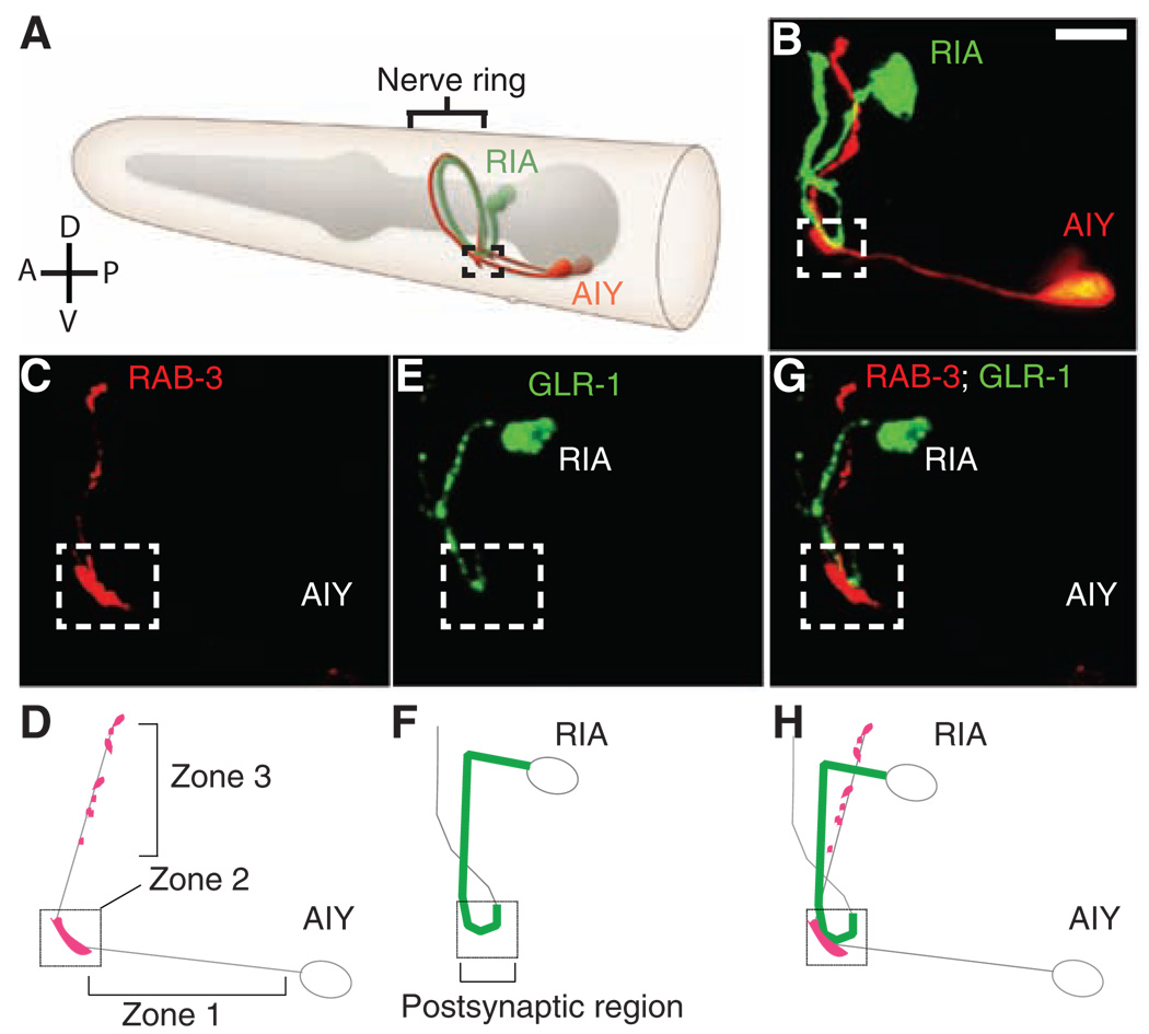
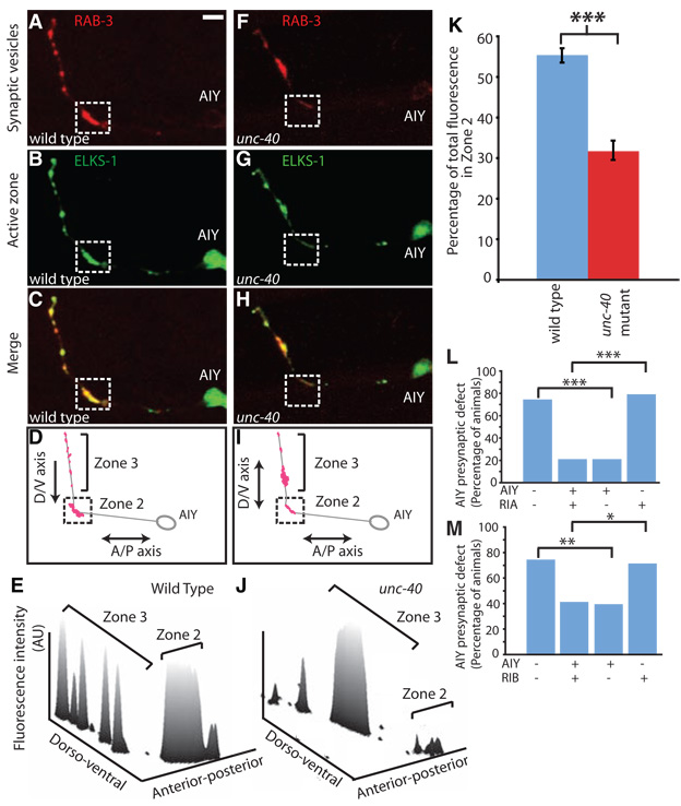
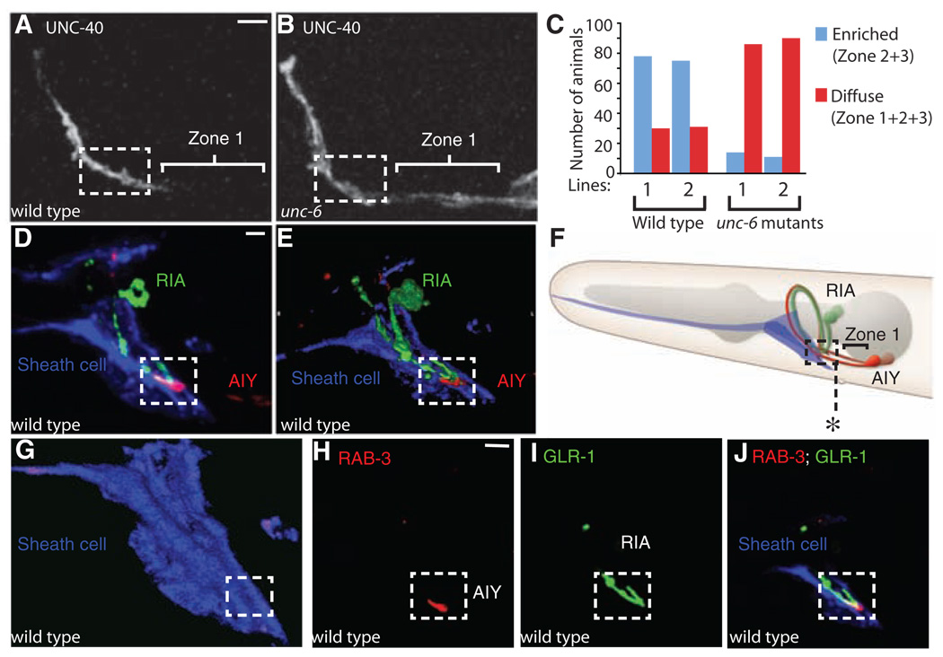
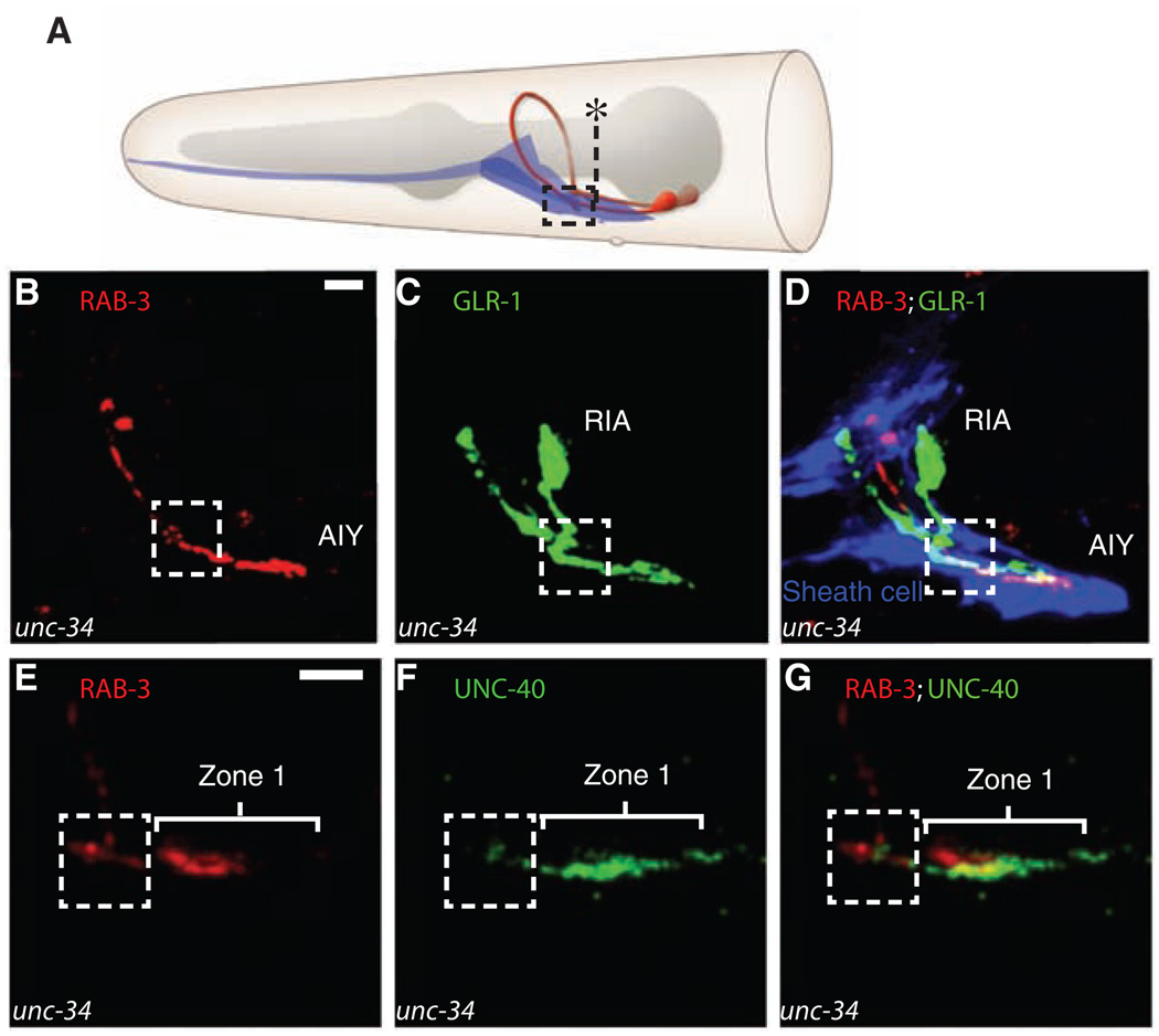
Neural circuits are assembled through the coordinated innervation of pre- and postsynaptic partners. We show that connectivity between two interneurons, AIY and RIA, in Caenorhabditis elegans is orchestrated by a pair of glial cells that express UNC-6 (netrin). In the postsynaptic neuron RIA, the netrin receptor UNC-40 (DCC, deleted in colorectal cancer) plays a conventional guidance role, directing outgrowth of the RIA process ventrally toward the glia. In the presynaptic neuron AIY, UNC-40 (DCC) plays an unexpected and previously uncharacterized role: It cell-autonomously promotes assembly of presynaptic terminals in the immediate vicinity of the glial cell endfeet. These results indicate that netrin can be used both for guidance and local synaptogenesis and suggest that glial cells can function as guideposts during the assembly of neural circuits in vivo.
Figures
Similar articles
-
A conserved juxtacrine signal regulates synaptic partner recognition in Caenorhabditis elegans.Neural Dev. 2011 Jun 10;6:28. doi: 10.1186/1749-8104-6-28. Neural Dev. 2011. PMID: 21663630 Free PMC article.
-
The dual role of the ligand UNC-6/Netrin in both axon guidance and synaptogenesis in C. elegans.Cell Adh Migr. 2009 Jul-Sep;3(3):268-71. doi: 10.4161/cam.3.3.8398. Epub 2009 Jul 12. Cell Adh Migr. 2009. PMID: 19377288 Free PMC article.
-
UNC-6/Netrin induces neuronal asymmetry and defines the site of axon formation.Nat Neurosci. 2006 Apr;9(4):511-8. doi: 10.1038/nn1666. Epub 2006 Mar 5. Nat Neurosci. 2006. PMID: 16520734 Free PMC article.
-
Localization mechanisms of the axon guidance molecule UNC-6/Netrin and its receptors, UNC-5 and UNC-40, in Caenorhabditis elegans.Dev Growth Differ. 2012 Apr;54(3):390-7. doi: 10.1111/j.1440-169X.2012.01349.x. Dev Growth Differ. 2012. PMID: 22524608 Review.
-
Netrins and netrin receptors.Cell Mol Life Sci. 1999 Oct 1;56(1-2):62-8. doi: 10.1007/s000180050006. Cell Mol Life Sci. 1999. PMID: 11213262 Free PMC article. Review.
Cited by
-
Neuronal target identification requires AHA-1-mediated fine-tuning of Wnt signaling in C. elegans.PLoS Genet. 2013 Jun;9(6):e1003618. doi: 10.1371/journal.pgen.1003618. Epub 2013 Jun 27. PLoS Genet. 2013. PMID: 23825972 Free PMC article.
-
Mitochondrial hydrogen peroxide positively regulates neuropeptide secretion during diet-induced activation of the oxidative stress response.Nat Commun. 2021 Apr 16;12(1):2304. doi: 10.1038/s41467-021-22561-x. Nat Commun. 2021. PMID: 33863916 Free PMC article.
-
DCC Confers Susceptibility to Depression-like Behaviors in Humans and Mice and Is Regulated by miR-218.Biol Psychiatry. 2017 Feb 15;81(4):306-315. doi: 10.1016/j.biopsych.2016.08.017. Epub 2016 Aug 18. Biol Psychiatry. 2017. PMID: 27773352 Free PMC article.
-
Role of netrin-1 in the organization and function of the mesocorticolimbic dopamine system.J Psychiatry Neurosci. 2011 Sep;36(5):296-310. doi: 10.1503/jpn.100171. J Psychiatry Neurosci. 2011. PMID: 21481303 Free PMC article. Review.
-
Daniel Colon-Ramos: observing and making connections. Interview by Caitlin Sedwick.J Cell Biol. 2013 Oct 28;203(2):168-9. doi: 10.1083/jcb.2032pi. J Cell Biol. 2013. PMID: 24165932 Free PMC article.
References
-
- Salie R, Niederkofler V, Arber S. Neuron. 2005;45:189. - PubMed
-
- Waites CL, Craig AM, Garner CC. Annu. Rev. Neurosci. 2005;28:251. - PubMed
-
- White JG, Southgate E, Thomson JN, Brenner S. Philos. Trans. R. Soc. London Ser. B. 1986;314:1. - PubMed
-
- White JG, Southgate E, Thomson JN, Brenner S. Cold Spring Harbor Symp. Quant. Biol. 1983;48:633. - PubMed
Publication types
MeSH terms
Substances
Grants and funding
LinkOut - more resources
Full Text Sources
Other Literature Sources
