. 2008 Jan;26(1):99-107.
doi: 10.1634/stemcells.2007-0563. Epub 2007 Oct 4.

Toll-like receptors on human mesenchymal stem cells drive their migration and immunomodulating responses

Affiliations

Toll-like receptors on human mesenchymal stem cells drive their migration and immunomodulating responses

Suzanne L Tomchuck et al. Stem Cells. 2008 Jan.

Abstract

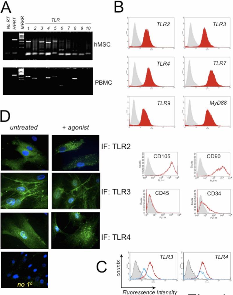
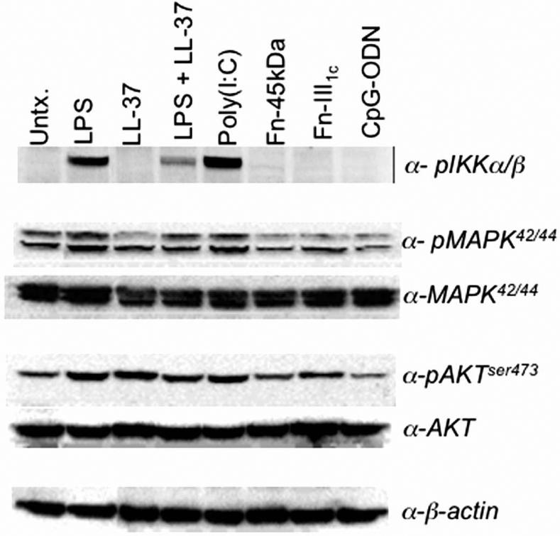
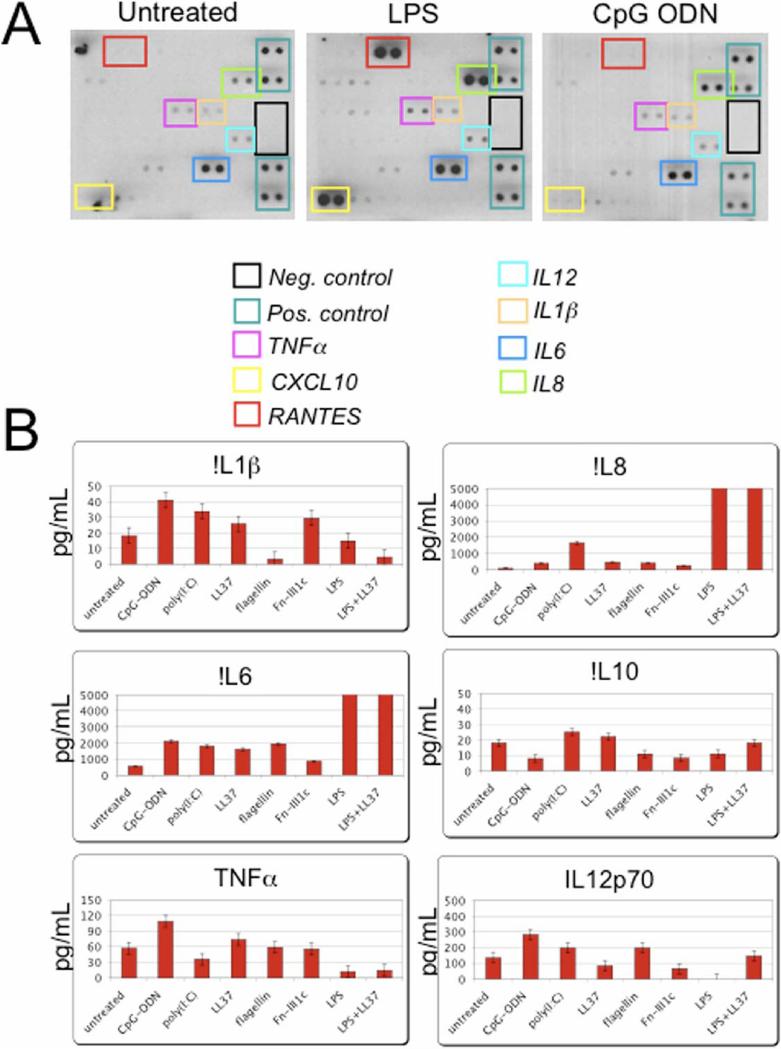
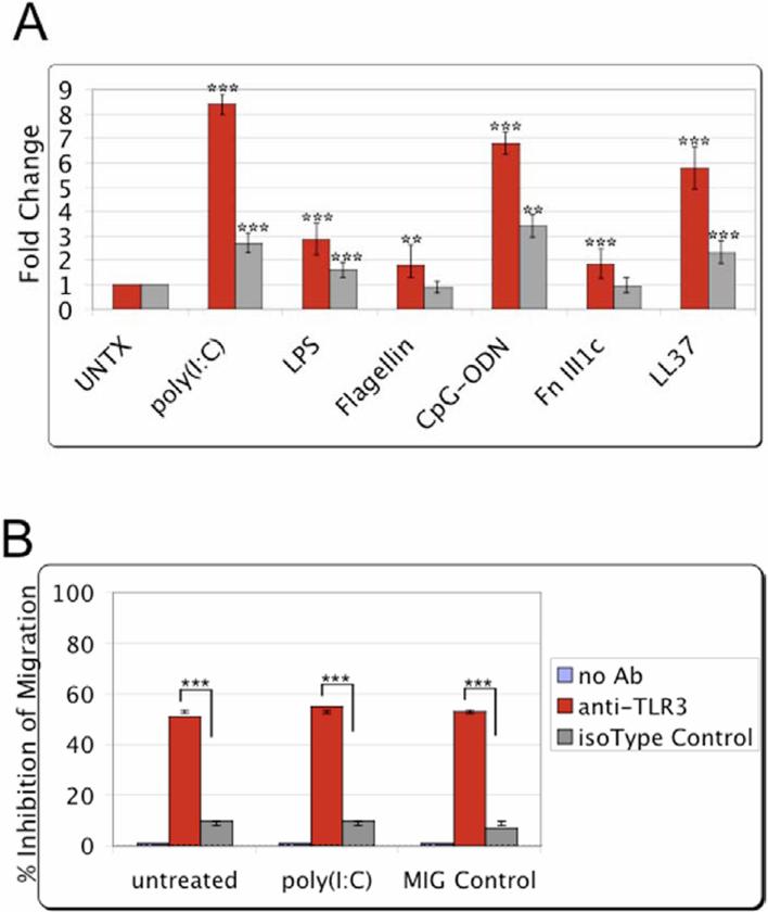
Adult human bone marrow-derived mesenchymal stem cells (hMSCs) are under study as therapeutic delivery agents that assist in the repair of damaged tissues. To achieve the desired clinical outcomes for this strategy requires a better understanding of the mechanisms that drive the recruitment, migration, and engraftment of hMSCs to the targeted tissues. It is known that hMSCs are recruited to sites of stress or inflammation to fulfill their repair function. It is recognized that toll-like receptors (TLRs) mediate stress responses of other bone marrow-derived cells. This study explored the role of TLRs in mediating stress responses of hMSCs. Accordingly, the presence of TLRs in hMSCs was initially established by reverse transcription-polymerase chain reaction assays. Flow cytometry and fluorescence immunocytochemical analyses confirmed these findings. The stimulation of hMSCs with TLR agonists led to the activation of downstream signaling pathways, including nuclear factor kappaB, AKT, and MAPK. Consequently, activation of these pathways triggered the induction and secretion of cytokines, chemokines, and related TLR gene products as established from cDNA array, immunoassay, and cytokine antibody array analyses. Interestingly, the unique patterns of affected genes, cytokines, and chemokines measured identify these receptors as critical players in the clinically established immunomodulation observed for hMSCs. Lastly, hMSC migration was promoted by TLR ligand exposure as demonstrated by transwell migration assays. Conversely, disruption of TLRs by neutralizing TLR antibodies compromised hMSC migration. This study defines a novel TLR-driven stress and immune modulating response for hMSCs that is critical to consider in the design of stem cell-based therapies.


Figures

Figure 1
Figure 1. Human Mesenchymal Stem Cells (hMSCs) Express Toll-like Receptors (TLRs) and downstream signaling molecule, MyD88
A. RNA was isolated from hMSCs and PBMC and analyzed by RT-PCR for expression of TLR 1−10. HPRT was used as loading control. hMSC expressed TLR 1, 2, 3, 4, 5, 6, and 9 whereas PBMC expressed TLR 1, 2, 4, 5, and 8 (n>3). B. Human MSC donor pools were routinely examined by flow cytometry for expression of discriminating MSC cell surface markers as described in Materials and Methods. Shown in bottom panels are representative findings for hMSCs: positive expression of CD90 and CD105 and mostly negative expression of CD45 and CD34 (n>12). HuMSCs were examined also by flow cytometry for expression of TLR2, 3, 4, 7, 9 and MyD88 as indicated by red filled-in curves on the top panels (n>7). The grey filled-in line represents hMSCs incubated with corresponding isotype antibody controls. C. Representative flow cytometry analysis of hMSCs for TLR3 and TLR4 after ligand stimulation for 30 min (Blue line) or without stimulation (constitutive expression, Red line). D. Antibody staining of TLRs was performed following fixation and membrane permeabilization of the ligand-treated hMSCs seeded on chamber slides. Samples were pre-treated for 1 h with ligands: 1 μM poly(I:C) (TLR3) or 10 ng/mL LPS (TLR2 & 4) prior to harvest and immunofluorescence (IF). As a control, the primary antibody was omitted from staining procedure (no 1o, n>3).
Figure 2
Figure 2
hMSC stimulation by discrete TLR-ligands affects established TLR-downstream signaling components. Downstream signaling potential by the stimulated TLRs within the hMSCs was assessed by Western blot analysis. LPS, CpG-ODN, and poly(I:C) represented exogenous ligands. Fibronectin-derived fragments (Fn-45kDa, -III1c) and the human secreted antimicrobial peptide LL-37 served as the endogenous danger signals (n>3).
Figure 3
Figure 3
The hMSCs secrete various cytokines and chemokines following stimulation with different TLR ligands. Primary hMSC cultures were treated with various TLR-ligands: 1μM poly(I:C); 10ng/mL LPS; 5μM flagellin; 1μM CpG-ODN; 1μg/mL fibronectin III1c; 5μg/mL LL-37; or LPS and LL-37 combined; as noted, for 24 h (A.) or 48 h (B.), prior to harvesting and concentrating the spent culture medium as indicated in Materials and Methods. A. Cytokine antibody array assay (n>3) and B. Fluorescence bead immunoassay (n>4) analysis revealed that TLR-stimulation induced unique cytokine and chemokine secretion profiles dependent on ligand used to treat hMSCs. Error bars indicate +/− standard error of the mean (SEM). Values exceeding the bar graph scale are enumerated above the corresponding bars.
Figure 4
Figure 4
TLR stimulation promotes the migration of the treated hMSCs. MSC migration towards TLR-specific ligands was examined by transwell migration assay. After overnight incubation, migration towards the various TLR-ligands was visualized and recorded by fluorescence microscopy. Migration was quantified from the obtained micrographs by counting the number of fluorescently labeled cells remaining after removal of non-migrating cells. A. Bar graph of the obtained results normalized to untreated (without TLR-ligand or chemotactic factor) control samples for two donors (red and gray). Error bars indicate SEM. Statistical significance analyzed for samples compared with the untreated control samples (n=6). ***p>0.001, **p>0.01 B. hMSC migration was examined following pre-incubation of the cells for 1 h with a human TLR3 specific monoclonal antibody or an isotype IgG control in the transwell migration assay as indicated. The equilibrated chemoattractant wells were loaded with growth medium in combination with poly(I:C), the TLR3 ligand, or a routinely used chemoattractant as a migration control (MIG control). After overnight incubation, migration was quantified as in A. Error bars indicate SEM. Statistical significance analyzed for samples compared with the isotype IgG treated control samples (n=3). ***p>0.001, **p>0.01.

References

    1. Wright SD. Toll, a new piece in the puzzle of innate immunity. J Exp Med. 1999 Feb 15;189(4):605–609. - PMC - PubMed
    1. West AP, Koblansky AA, Ghosh S. Recognition and signaling by toll-like receptors. Annu Rev Cell Dev Biol. 2006;22:409–437. - PubMed
    1. Sabroe I, Read RC, Whyte MK, Dockrell DH, Vogel SN, Dower SK. Toll-like receptors in health and disease: complex questions remain. J Immunol. 2003 Aug 15;171(4):1630–1635. - PubMed
    1. Miggin SM, O'Neill LA. New insights into the regulation of TLR signaling. J Leukoc Biol. 2006 Aug;80(2):220–226. - PubMed
    1. Anders HJ, Banas B, Schlondorff D. Signaling danger: toll-like receptors and their potential roles in kidney disease. J Am Soc Nephrol. 2004 Apr;15(4):854–867. - PubMed

Publication types