

CD40-CD40 ligand interactions in human microglia induce CXCL8 (interleukin-8) secretion by a mechanism dependent on activation of ERK1/2 and nuclear translocation of nuclear factor-kappaB (NFkappaB) and activator protein-1 (AP-1)
- PMID: 17918746
- PMCID: PMC2639790
- DOI: 10.1002/jnr.21525
CD40-CD40 ligand interactions in human microglia induce CXCL8 (interleukin-8) secretion by a mechanism dependent on activation of ERK1/2 and nuclear translocation of nuclear factor-kappaB (NFkappaB) and activator protein-1 (AP-1)
Abstract
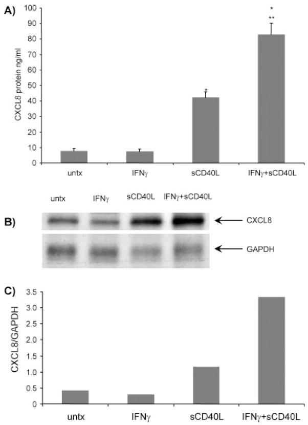
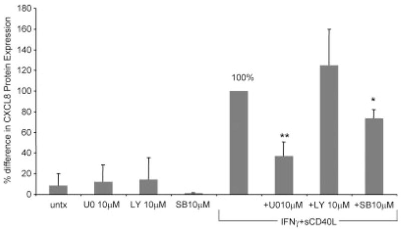
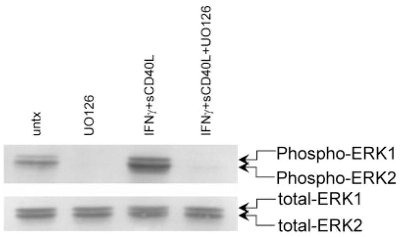
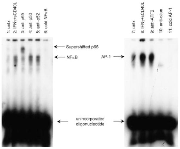
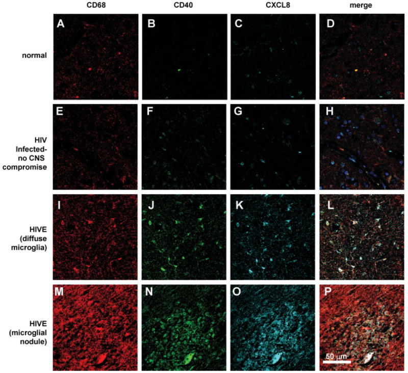
CXCL8 is a CXC chemokine that recruits leukocytes to sites of inflammation. Expression of CXCL8 in the CNS has been demonstrated in neuroinflammatory diseases, including human immunodeficiency virus (HIV-1) encephalitis, but the mechanism of secretion of this chemokine is not fully understood. CD40 is a 50-kDa protein on the surface of microglia, and we have previously shown that it is increased in expression in HIV-1-infected brain tissue as well as by interferon-gamma (IFNgamma) in tissue culture. We examined the expression and regulation of CXCL8 in cultured human fetal microglia after ligation of CD40 with soluble trimeric CD40 ligand (sCD40L) as well as the expression of CXCL8 on microglia in HIV encephalitic brain tissue sections. Treatment of cultured microglia with IFNgamma + sCD40L resulted in significant induction of CXCL8. This expression was mediated by activation of the ERK1/2 MAPK pathway, as demonstrated by ELISA and Western blot using a specific inhibitor (U0126). Gel shift analyses demonstrated that NFkappaB and AP-1, but not C/EBPbeta, mediate microglial CXCL8 production. We also found increased colocalization of CXCL8 with CD68/CD40-positive cells in HIV encephalitic brain tissue compared with HIV-infected nonencephalitic and normal tissue. Thus, CD40-CD40L interactions facilitate chemokine expression, leading to the influx of inflammatory cells into the CNS. These events can lead to the pathology that is associated with neuroinflammatory diseases.
(c) 2007 Wiley-Liss, Inc.
Figures
Similar articles
-
CD40-CD40L interactions induce chemokine expression by human microglia: implications for human immunodeficiency virus encephalitis and multiple sclerosis.Am J Pathol. 2002 Feb;160(2):559-67. doi: 10.1016/S0002-9440(10)64875-4. Am J Pathol. 2002. PMID: 11839576 Free PMC article.
-
Ligation of CD40 stimulates the induction of nitric-oxide synthase in microglial cells.J Biol Chem. 2001 Nov 30;276(48):44527-33. doi: 10.1074/jbc.M106771200. Epub 2001 Sep 10. J Biol Chem. 2001. PMID: 11551948 Free PMC article.
-
The expression of intercellular adhesion molecule-1 induced by CD40-CD40L ligand signaling in orbital fibroblasts in patients with Graves' ophthalmopathy.Invest Ophthalmol Vis Sci. 2010 Sep;51(9):4652-60. doi: 10.1167/iovs.09-3789. Epub 2010 Jan 27. Invest Ophthalmol Vis Sci. 2010. PMID: 20107176
-
CXCL8 as a Potential Therapeutic Target for HIV-Associated Neurocognitive Disorders.Curr Drug Targets. 2016;17(1):111-21. doi: 10.2174/1389450116666150626124544. Curr Drug Targets. 2016. PMID: 26112047 Review.
-
Molecular regulation of CD40 gene expression in macrophages and microglia.Brain Behav Immun. 2004 Jan;18(1):7-12. doi: 10.1016/j.bbi.2003.09.001. Brain Behav Immun. 2004. PMID: 14651941 Review.
Cited by
-
CD40 Ligand-CD40 Interaction Is an Intermediary between Inflammation and Angiogenesis in Proliferative Diabetic Retinopathy.Int J Mol Sci. 2023 Oct 25;24(21):15582. doi: 10.3390/ijms242115582. Int J Mol Sci. 2023. PMID: 37958563 Free PMC article.
-
Microglia activation by SIV-infected macrophages: alterations in morphology and cytokine secretion.J Neurovirol. 2012 Jun;18(3):213-21. doi: 10.1007/s13365-012-0100-7. Epub 2012 Apr 26. J Neurovirol. 2012. PMID: 22535448 Free PMC article.
-
Arterial Baroreflex Dysfunction Promotes Neuroinflammation by Activating the Platelet CD40L/Nuclear Factor Kappa B Signaling Pathway in Microglia and Astrocytes.Neurochem Res. 2023 Jun;48(6):1691-1706. doi: 10.1007/s11064-022-03852-1. Epub 2023 Jan 2. Neurochem Res. 2023. PMID: 36592325 Free PMC article.
-
SOLUBLE CD40 LIGAND IN DEMENTIA.Drugs Future. 2009 Apr 1;34(4):333-340. doi: 10.1358/dof.2009.034.04.1358595. Drugs Future. 2009. PMID: 19777117 Free PMC article.
-
Neuroinflammation and EIF2 Signaling Persist despite Antiretroviral Treatment in an hiPSC Tri-culture Model of HIV Infection.Stem Cell Reports. 2020 Apr 14;14(4):703-716. doi: 10.1016/j.stemcr.2020.02.010. Epub 2020 Mar 26. Stem Cell Reports. 2020. PMID: 32220329 Free PMC article.
References
-
- Albright AV, Soldan SS, Gonzalez-Scarano F. Pathogenesis of human immunodeficiency virus-induced neurological disease. J Neurovirol. 2003;9:222–227. - PubMed
-
- Aloisi F, De Simone R, Columba-Cabezas S, Penna G, Adorini L. Functional maturation of adult mouse resting microglia into an APC is promoted by granulocyte-macrophage colony-stimulating factor and interaction with Th1 cells. J Immunol. 2000;164:1705–1712. - PubMed
-
- Armitage RJ, Fanslow WC, Strockbine L, Sato TA, Clifford KN, Mac-duff BM, Anderson DM, Gimpel SD, Davis-Smith T, Maliszewski CR, et al. Molecular and biological characterization of a murine ligand for CD40. Nature. 1992a;357:80–82. - PubMed
-
- Armitage RJ, Sato TA, Macduff BM, Clifford KN, Alpert AR, Smith CA, Fanslow WC. Identification of a source of biologically active CD40 ligand. Eur J Immunol. 1992b;22:2071–2076. - PubMed
Publication types
MeSH terms
Substances
Grants and funding
LinkOut - more resources
Full Text Sources
Research Materials
Miscellaneous
