. 2007 Nov 15;311(2):396-407.
doi: 10.1016/j.ydbio.2007.08.046. Epub 2007 Aug 31.

TCF4 deficiency expands ventral diencephalon signaling and increases induction of pituitary progenitors

Affiliations

TCF4 deficiency expands ventral diencephalon signaling and increases induction of pituitary progenitors

Michelle L Brinkmeier et al. Dev Biol. .

Abstract

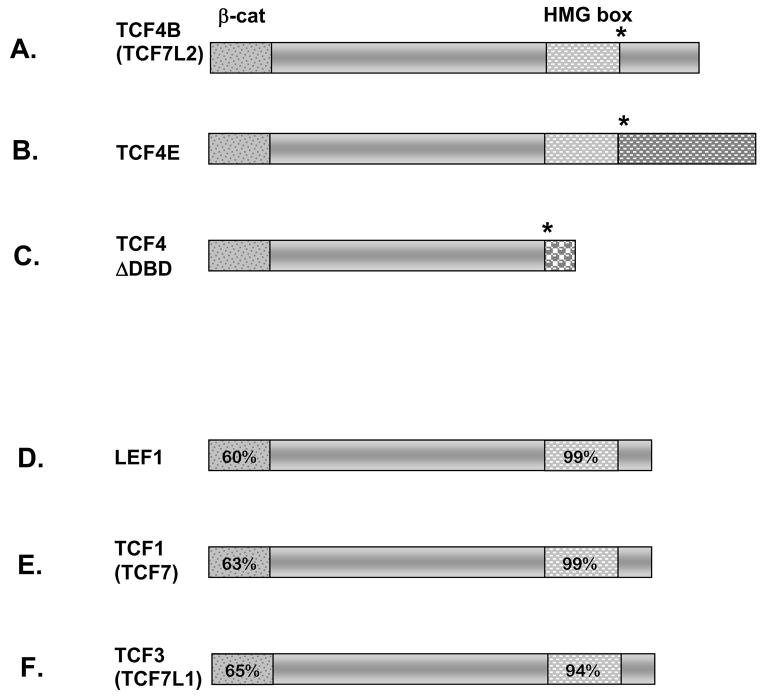
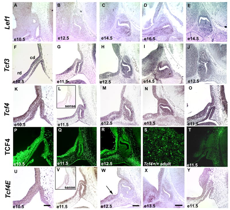
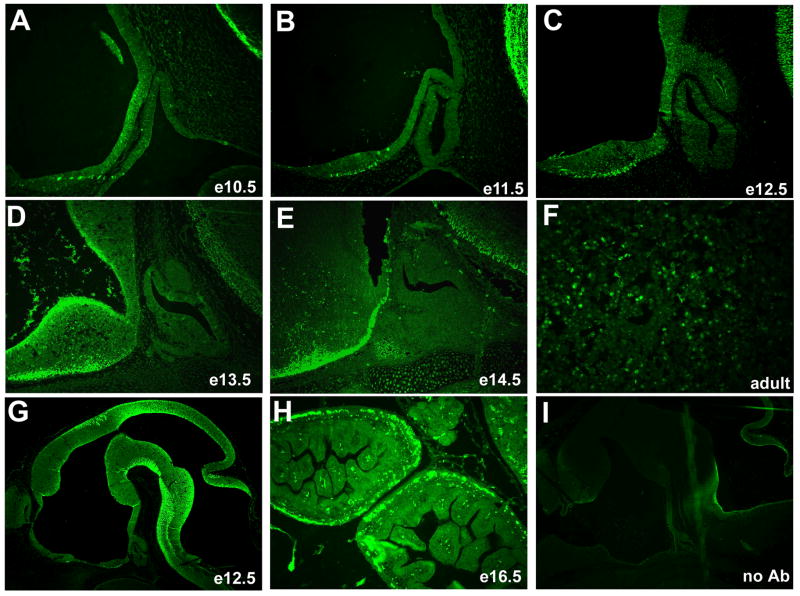
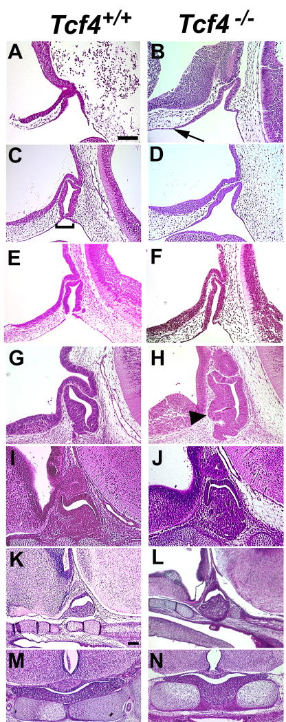
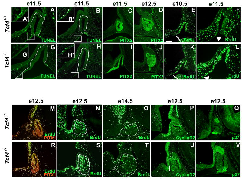
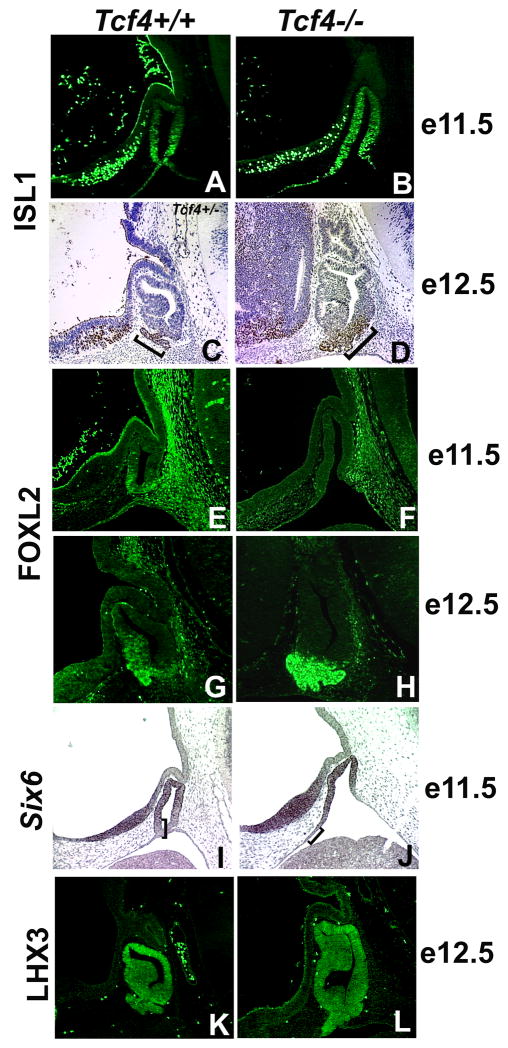
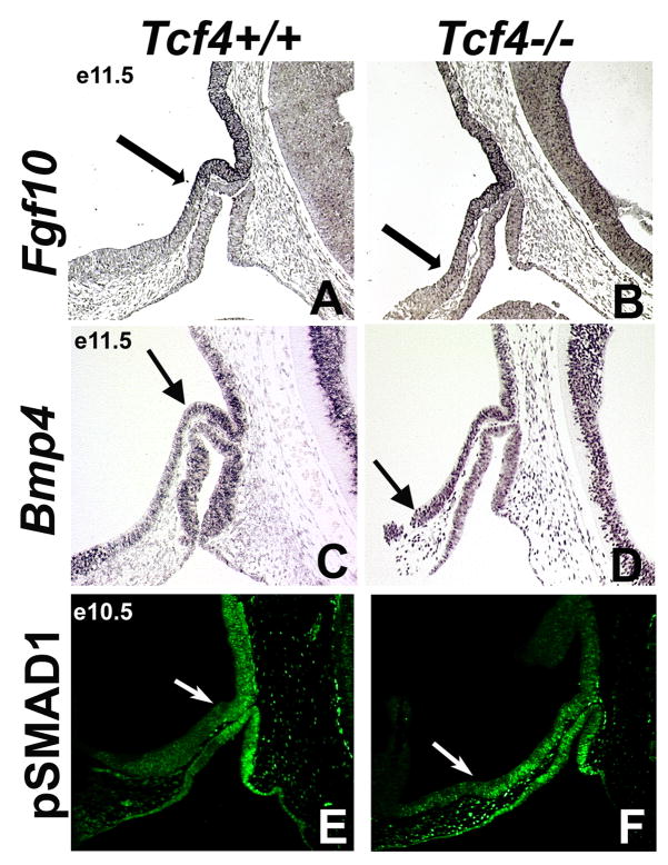
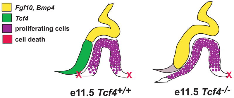
The anterior and intermediate lobes of the pituitary gland are formed from Rathke's pouch. FGF, BMP and WNT signals emanating from the ventral diencephalon influence pouch growth and development. In order to examine the role of canonical WNT signaling during pituitary development we examined the pituitary expression of the TCF/LEF family of transcription factors, which mediate WNT signaling through the binding of beta-catenin. We report here the expression of several members of this family during pituitary development and the functional role of one member, TCF4 (TCF7L2), in the induction of the pituitary primordium. TCF4 is expressed in the ventral diencephalon early in pituitary development, rostral to a domain of BMP and FGF expression. Tcf4 deficient mice express Fgf10 and Bmp4; however, the Bmp and Fgf expression domains are expanded rostrally. As a result, additional pituitary progenitor cells are recruited into Rathke's pouch in Tcf4 mutants. Mutants also exhibit an expansion of the Six6 expression domain within Rathke's pouch, which may increase the number of proliferating pouch cells, resulting in a greatly enlarged anterior pituitary gland. This suggests that TCF4 negatively regulates pituitary growth through two mechanisms. The first mechanism is to restrict the domains of BMP and FGF signaling in the ventral diencephalon, and the second mechanism is the restriction of Six6 within Rathke's pouch. Thus, TCF4 is necessary both intrinsically and extrinsically to Rathke's pouch to ensure the proper growth of the pituitary gland.


Figures

Figure 1
Figure 1
TCF4 isoforms and conservation within the TCF/LEF family. Amino acid comparison of TCF/LEF family members in the mouse reveals a high degree of homology within the β-catenin binding domain and HMG box (patterned boxes in panels A–F). Percentages depicted in the conserved domains were determined by comparing the coding sequence to TCF4E (A–C). Alternative splicing results in three Tcf4 mRNAs and protein isoforms. TCF4, officially named TCF7L2, can be spliced (*) downstream of the HMG box to generate the TCF4B and TCF4E proteins, panels A and B. Panel C shows an alternative splice site (*) before the HMG box generates a protein that is missing the DNA binding domain, TCF4ΔDBD. The unique amino acid domains in the carboxy termini of TCF4B, TCF4E, and TCF4ΔDBD isoforms are depicted with different patterns.
Figure 2
Figure 2
The temporal and spatial expression pattern of Lef1, Tcf3, and Tcf4 in the developing pituitary gland. In situ hybridization was used to analyze Lef1, Tcf3, Tcf4, and Tcf4E expression in the developing anterior pituitary gland and the adjacent dorsal, caudal domain (cd) and ventral, rostral domain (rd) of the ventral diencephalon. Immunohistochemistry utilizing an antibody that recognizes all TCF4 isoforms was used to confirm developmental expression. The scale bar in panel U represents 50μm and should be applied to e10.5 (A, F, K, P, and, U), and e11.5 (B, G, L, O, Q, T, V, and Y) pituitary sections. The scale bar for the e12.5 (B, H, J, M, R, and V), e13.5 (N and X) and e14.5 (C and I) pituitary sections represents 100μm and is shown in panel W. The arrow in e12.5 sections labeled with Tcf4E marks the boundary between the caudal and rostral domains of the ventral diencephalon. The insets in panels L and V represent sense probes from Tcf4 and Tcf4E, respectively. Panel S is a high magnification (400X) image of an adult pituitary gland.
Figure 3
Figure 3
Activated, nuclear localized β-catenin predominates in the ventral diencephalon of developing mice. Immunohistochemistry reveals little or no staining in the developing pituitary gland from e10.5 through e13.5, with some staining of the rostral tip area at e14.5 (A–E, 200X). Activated β-catenin can be easily detected in the adult pituitary gland (F, 400X). Activated β-catenin can also be deteced in other known regions of WNT signaling such as e12.5 brain (G, 50X) and e16.5 intestine (H, 100X). Sections incubated without primary antibody do not show any nuclear staining (I, e13.5 brain, 50X).
Figure 4
Figure 4
Pituitary dysmorphology is evident by e10.5 in Tcf4−/− mice. Hematoxylin and eosin staining were used to determine the onset of pituitary dysmorphology in Tcf4−/− mice compared to Tcf4+/+ controls. Rathke’s pouch is thickened and expanded rostrally beginning at e10.5 (panel A, B, arrow). Thinning of the oral ectoderm and the separation of Rathke’s pouch (panel C, bracket) is evident in Tcf4+/+ mice at e11.5 (panel C, E) but is typically delayed in Tcf4−/− mice (panel D). One example of the separation of the pouch from the oral ectoderm has been identified in a Tcf4−/− mouse at e11.5 (panel F). The thickened and expanded Rathke’s pouch is separated from the oral ectoderm by e12.5 in Tcf4−/− mice and the additional cells are incorporated into the anterior pituitary (panel H, arrowhead). Sagittal sections from fetuses at e10.5 through e16.5 (panels A–L) are oriented with rostral on the left and e18.5 (panels M, N) are coronal sections. Panels from e10.5 through e14.5 are the same magnification; e16.5 and e18.5 are at a lower magnification. The scale bars in panels A and K represent 100μm.
Figure 5
Figure 5
Rathke’s pouch dysmorphology in Tcf4−/− mice is caused by an inappropriate separation from the oral ectoderm and an increased zone of proliferating cells. TUNEL staining showed apoptotic cells at the rostral and caudal edges where Rathke’s pouch separates from the oral ectoderm in e11.5 Tcf4+/+ mice (A, B). TUNEL staining is absent on the rostral side of Rathke’s pouch in the majority of Tcf4−/− mice (panel G and inset G′). Separation of Rathke’s pouch from the oral ectoderm and TUNEL positive cells can be identified on rare occasions Tcf4−/− mouse (panel H, inset H′). The rostral edge of apoptosis is boxed in panel A, B, G, H and shown in the higher magnification insets A′, B′, G′, H′. The cells forming Rathke’s pouch are outlined in white in the TUNEL panels (A, B, G, H). The expression of PITX2 at e11.5 and e12.5 was used to mark Rathke’s pouch cells (C, D, I, J). Proliferation of cells in Rathke’s pouch between e10.5 and e14.5 was analyzed in Tcf4+/+ and Tcf4−/− mice (E, F, K, L–O. R–T). BrdU immunohistochemistry reveals a zone of increased proliferation on the rostral side of Rathke’s pouch in Tcf4−/− mice (K, L, R. S). The arrows in e10.5 denote the rostral ventral boundary of BrdU detection (E, K), which is expanded in Tcf4−/− mice. The arrowhead in e11.5 panels F and L denote the expanded boundary between proliferating and nonproliferating cells in Rathke’s pouch of Tcf4−/− mice compared to Tcf4+/+ mice. The additional proliferating cells in Tcf4−/− mice are incorporated into the rostral ventral side of Rathke’s pouch by e12.5 in both parasagittal (R) and midsagittal (S) sections. Proliferating cells are absent in comparable regions of Rathke’s pouch in Tcf4+/+ mice (M, N). Parasagittal sections were co-immunostained with PITX1 in red to clearly identify Rathke’s pouch cells (M, R). There were no differences in proliferation at e14.5 between Tcf4+/+ and Tcf4−/− mice (O, T). Rathke’s pouch is outlined in e12.5 and e14.5 midsagittal sections (N, O, S, T). CyclinD2 immunoreactivity, marking the G1 phase of the cell cycle, was used to confirm the additional region of proliferation at e12.5 (P vs. U, bracket). p27 immunoreactivity, marking post mitotic cells, is present in the portion of Rathke’s pouch that ceases proliferation in both Tcf4+/+ and Tcf4−/− mice at 12.5 (Q, V). Panels C, D, E, I, J, K, and M–V were taken at the same magnification, and the scale bar in panel E represent 100μm. Panels A, B, F, G, H, and L were taken at the same magnification, and the scale bar in panel F represents 50μm.
Figure 6
Figure 6
Differentiation of ventral Rathke’s pouch cells is delayed in Tcf4−/− mice. Immunohistochemistry for ISL1 at e11.5 (A, B) and e12.5 (C, D) as well as FOXL2 at e11.5 (E, F) and e12.5 (G, H) were used to determine the onset of cell differentiation in the ventral aspect of Rathke’s pouch. In situ hybridization for Six6 at e11.5 (I, J) and immunohistochemistry for LHX3 at e12.5 (K, L) were used to analyze the progress of cell differentiation in the dorsal aspect of Rathke’s pouch. The brackets in panels I and J mark the region void of Six6 expression.
Figure 7
Figure 7
The boundaries of BMP and FGF signaling are expanded rostrally in Tcf4−/− mice. In situ hybridization for Fgf10 at e11.5 (A, B), Bmp4 at e11.5 (C, D), and immunohistochemistry for the phosophorylated form of SMAD1, pSMAD1, a protein activated by BMP, at e10.5 (E, F) were examined in Tcf4+/+ and Tcf4−/− mice. The boundaries Fgf10, Bmp4, and pSMAD1 expression in the ventral diencephalon are expanded rostrally in Tcf4−/− mice (A–F, arrows).
Figure 8
Figure 8
The rostral expansion of the FGF and BMP boundary in Tcf4−/− mice increases the amount of oral ectoderm specified to invaginate and become Rathke’s pouch. As Rathke’s pouch invaginates at e11.5, opposing boundaries of FGF/BMP and TCF4 expression are established within the ventral diencephalon. This pattern of expression in the ventral diencephalon likely influences the adjacent cells within Rathke’s pouch to proliferate. The ventral boundary on each side of Rathke’s pouch establishes the pinch off point from the oral ectoderm and determines the subsequent size of the pituitary gland. In Tcf4−/− mice, the absence of TCF4 signal in the ventral diencephalon results in ventral expansion of the FGF/BMP expression boundary. This establishes a larger region of proliferating cells in Rathke’s pouch, potentially leading to recruitment of additional oral ectoderm, and ultimately a larger pituitary gland.

References

    1. Ai D, Wang J, Amen M, Lu M, Amendt B, Martin J. Nuclear Factor-1 and TCF/LEF recognition elements regulate Pitx2 transcription in pituitary development. Mol Cell Biol. 2007 Epub ahead of print. - PMC - PubMed
    1. Baek SH, Kioussi C, Briata P, Wang D, Nguyen HD, Ohgi KA, Glass CK, Wynshaw-Boris A, Rose DW, Rosenfeld MG. Regulated subset of G1 growth-control genes in response to derepression by the Wnt pathway. Proc Natl Acad Sci U S A. 2003;100:3245–50. - PMC - PubMed
    1. Basch M, Bronner-Fraser M. Neural crest inducing signals. Adv Exp Med Biol. 2006;589:34–31. - PubMed
    1. Bellusci S, Grindley J, Emoto H, Itoh N, Hogan BL. Fibroblast growth factor 10 (FGF10) and branching morphogenesis in the embryonic mouse lung. Development. 1997;124:4867–78. - PubMed
    1. Bellusci S, Henderson R, Winnier G, Oikawa T, Hogan BL. Evidence from normal expression and targeted misexpression that bone morphogenetic protein (Bmp-4) plays a role in mouse embryonic lung morphogenesis. Development. 1996;122:1693–702. - PubMed

Publication types

MeSH terms

Substances