. 2007 Oct 23;17(20):1759-64.
doi: 10.1016/j.cub.2007.08.065. Epub 2007 Oct 4.

Drosophila Spd-2 recruits PCM to the sperm centriole, but is dispensable for centriole duplication

Affiliations

Drosophila Spd-2 recruits PCM to the sperm centriole, but is dispensable for centriole duplication

Carly I Dix et al. Curr Biol. .

Abstract

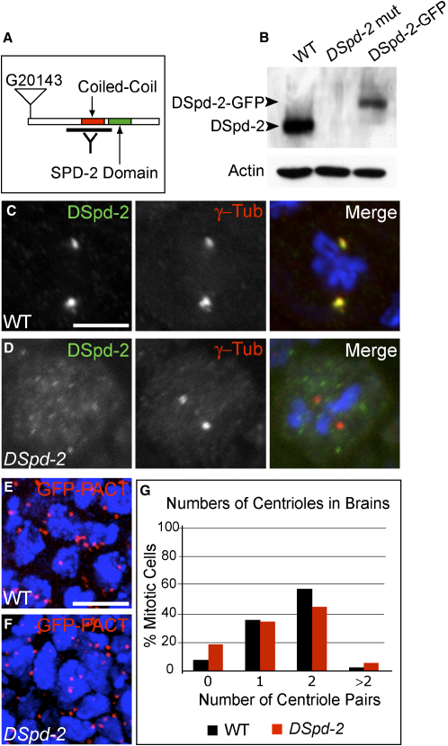
In C. elegans, genome-wide screens have identified just five essential centriole-duplication factors: SPD-2, ZYG-1, SAS-5, SAS-6, and SAS-4 [1-8]. These proteins are widely believed to comprise a conserved core duplication module [3, 9-14]. In worm embryos, SPD-2 is the most upstream component of this module, and it is also essential for pericentriolar material (PCM) recruitment to the centrioles [1, 4, 15, 16]. Here, we show that Drosophila Spd-2 (DSpd-2) is a component of both the centrioles and the PCM and has a role in recruiting PCM to the centrioles. DSpd-2 appears not, however, to be essential for centriole duplication in somatic cells. Moreover, PCM recruitment in DSpd-2 mutant somatic cells is only partially compromised, and mitosis appears unperturbed. In contrast, DSpd-2 is essential for proper PCM recruitment to the fertilizing sperm centriole, and hence for microtubule nucleation and pronuclear fusion. DSpd-2 therefore appears to have a particularly important role in recruiting PCM to the sperm centriole. We speculate that the SPD-2 family of proteins might only be absolutely essential for the recruitment of centriole duplication factors and PCM to the centriole(s) that enter the egg with the fertilizing sperm.


Figures

Figure 1
Figure 1
DSpd-2 Localizes to the Centrioles throughout the Cell Cycle and to the PCM in Mitosis (A) WT mitotic third-instar larval neuroblast stained for DSpd-2 (green), γ-tubulin (red), and DNA (blue). DSpd-2 localizes to a broad spot at the mitotic centrosome and largely colocalizes with the PCM marker γ-tubulin. (B) The behavior of DSpd-2-GFP in a syncytial embryo. DSpd-2-GFP is present at the centrosome throughout the embryonic cell cycle (see Movie S1). The embryo shown here is in telophase, and DSpd-2-GFP can be seen on both centrosomes after duplication. (C) Whole-mount third-instar larval brain expressing DSpd-2-GFP (red), stained for Phospho-Histone-H3 (green) to mark mitotic cells and DNA (blue). DSpd-2-GFP localizes to small dots (centrioles) in interphase cells (arrow) and to the PCM in mitotic cells (arrowhead). (D) A larval primary spermatocyte expressing DSpd-2-GFP (red). DNA is also shown (blue). DSpd-2-GFP localizes along the entire length of the two orthogonally arranged pairs of centriole barrels. Scale bars represent 10 μm in (A), (B), and (D) and 5 μm in (C).
Figure 2
Figure 2
DSpd-2 Is Not Essential for Centriole Duplication in Cells of the Drosophila Brain (A) A schematic representation of the DSpd-2 protein and the position of the G20143 P element insertion. DSpd-2 is 1146 amino acids (aa) in length and was identified in the basis of the SPD-2 Domain (green) of ∼200 aa . DSpd-2 also has an adjacent conserved coiled-coil region (red). The region against which the DSpd-2 antibody was raised is indicated (black line). (B) Western blot of protein extracts from third-instar larval brains, probed with DSpd-2 antibodies. Actin antibodies were used as a loading control. A band of the predicted size for DSpd-2 (125 kDa) is detected in WT but not DSpd-2 mutant brains, indicating antibody specificity. A larger band (of ∼155 kDa) is observed in DSpd-2 mutant brains expressing DSpd-2-GFP. (C and D) WT and DSpd-2 third-instar larval neuroblasts stained for DSpd-2 (green), γ-tubulin (red), and DNA (blue). DSpd-2 protein is detectable at the centrosome of WT (C) but not mutant (D) neuroblasts. (E and F) Maximum-intensity projections of stacks taken through WT (E) and DSpd-2 (F) whole-mount third-instar larval brains, expressing the centriole marker GFP-PACT (red). DNA is in blue. Large numbers of centrioles are observed in both WT and mutant brains. (G) A graph showing the percentage of mitotic (Phospho-Histone-H3 positive) cells with 0, 1, 2, or > 2 D-PLP-positive dots (centriole pairs). For WT cells, n = 546; for DSpd-2 cells, n = 484. Cells were counted from at least six brains for both the WT and mutant. A dramatic reduction in centriole number is not observed in DSpd-2 mutant brains compared to the WT. Scale bars represent 10 μm in (C) and (D) and 5 μm in (E) and (F).
Figure 3
Figure 3
Centrosome Maturation Is Inefficient in the Absence of DSpd-2 (A and B) WT (A) and DSpd-2 mutant (B) mitotic third-instar larval brains stained for Phospho-Histone-H3 (green), Cnn (red), and DNA (blue). WT cells strongly recruit Cnn to mitotic centrosomes, whereas Cnn recruitment in the DSpd-2 mutant is clearly reduced. (C) A graph showing the percentage of mitotic (Phospho-Histone-H3 positive) cells with strong, medium, weak, or no Cnn staining at the centrosome. Cells were counted in at least six brains for each sample. For the WT, n = 526; for DSpd-2, n = 359. (D) A graph showing the centrosomal intensity of γ-tubulin (black and red) and Cnn (blue and green) staining of individual centrosomes in WT and mutant fixed mitotic third-instar larval neuroblasts. (E and F) Stills from movies of WT (E) and mutant (F) neuroblasts expressing GFP-α-tubulin (taken from Movies S2 and S3, respectively). Both WT and mutant cells drive the assembly of a robust mitotic spindle from their centrosomes (WT n = 47, DSpd-2 n = 108; times relative to nuclear envelope breakdown [NEBD] are shown in top left). Scale bars represent 10 μm in (A) and (B) and 5 μm in (E) and (F).
Figure 4
Figure 4
DSpd-2 Is Essential for PCM Recruitment and MT Nucleation from the Sperm Centriole WT or DSpd-2 embryos expressing Asl-GFP (centriole marker) were stained to reveal the distribution of GFP, MTs, DNA, and Cnn. (A) The first mitotic spindle in a WT embryo. Shown are MTs (red), centrioles (green), and DNA (blue). A single centriole is visible at each spindle pole (arrowheads). (B) A DSpd-2 mutant embryo at the equivalent stage (based on polar body morphology; not shown) as the WT embryo shown in (A); MTs (red), Centrioles (green), and DNA (blue) are shown. The male and female pronuclei (marked) have failed in pronuclear fusion. Two centrioles (arrowheads) are associated with the male pronucleus (see inset). (C and D) WT and DSpd-2 sperm asters at anaphase or telophase of meiosis II. Shown are MTs (red), centrioles (green), and DNA (blue). WT embryos (C) have a large sperm aster associated with the centrioles, which by this time have clearly duplicated (arrowheads). Cnn protein is strongly recruited to these centrioles and the surrounding aster (see inset, far-right panel). In DSpd-2 embryos (D), centriole duplication has still occurred (arrowheads), but the sperm aster is very small, and Cnn is absent from the centrioles (see inset, far-right panel). Scale bars represent 10 μm in (A), (C), and (D) and 20 μm in (B). The inset in (B) is a 7× magnification of the image. The insets in (C) and (D) are 3× magnifications of the image.

References

    1. Pelletier L., Ozlu N., Hannak E., Cowan C., Habermann B., Ruer M., Muller-Reichert T., Hyman A.A. The Caenorhabditis elegans centrosomal protein SPD-2 is required for both pericentriolar material recruitment and centriole duplication. Curr. Biol. 2004;14:863–873. - PubMed
    1. Leidel S., Gonczy P. SAS-4 is essential for centrosome duplication in C elegans and is recruited to daughter centrioles once per cell cycle. Dev. Cell. 2003;4:431–439. - PubMed
    1. Leidel S., Delattre M., Cerutti L., Baumer K., Gonczy P. SAS-6 defines a protein family required for centrosome duplication in C. elegans and in human cells. Nat. Cell Biol. 2005;7:115–125. - PubMed
    1. Kemp C.A., Kopish K.R., Zipperlen P., Ahringer J., O'Connell K.F. Centrosome maturation and duplication in C. elegans require the coiled-coil protein SPD-2. Dev. Cell. 2004;6:511–523. - PubMed
    1. Dammermann A., Muller-Reichert T., Pelletier L., Habermann B., Desai A., Oegema K. Centriole assembly requires both centriolar and pericentriolar material proteins. Dev. Cell. 2004;7:815–829. - PubMed

Publication types

MeSH terms

Substances

LinkOut - more resources