. 2007 Oct;27(4):647-59.
doi: 10.1016/j.immuni.2007.07.023. Epub 2007 Oct 4.

Interleukin-22 but not interleukin-17 provides protection to hepatocytes during acute liver inflammation

Affiliations

Interleukin-22 but not interleukin-17 provides protection to hepatocytes during acute liver inflammation

Lauren A Zenewicz et al. Immunity. 2007 Oct.

Abstract

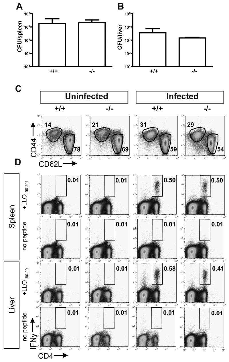
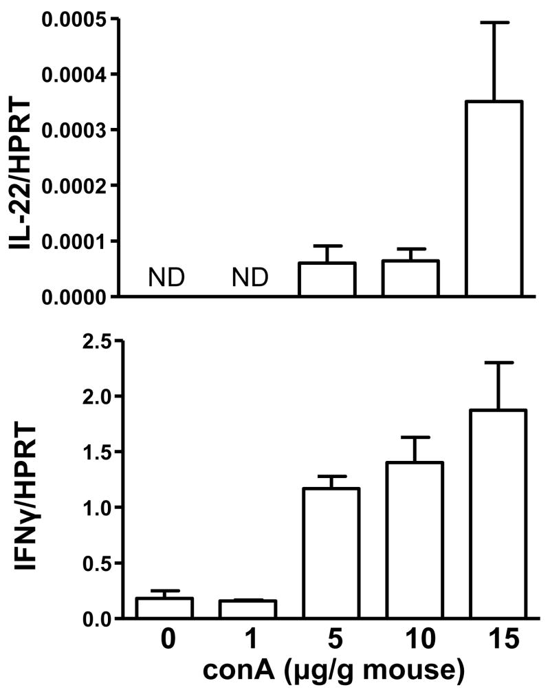
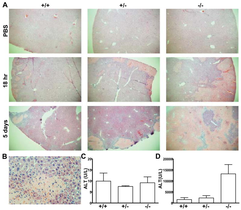
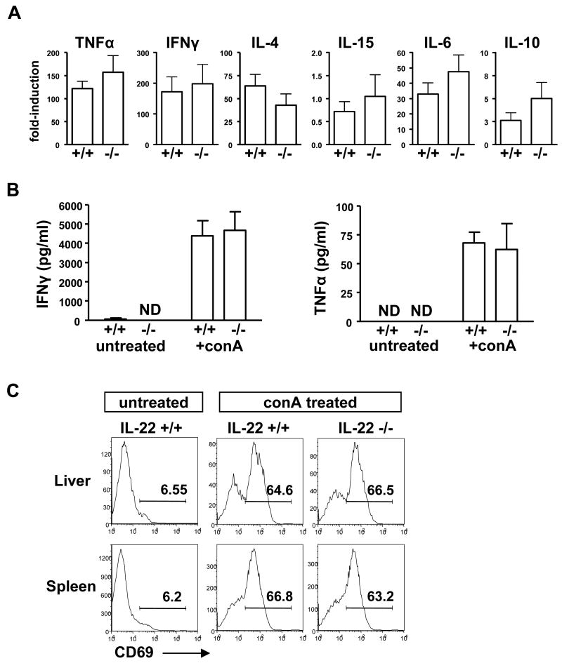
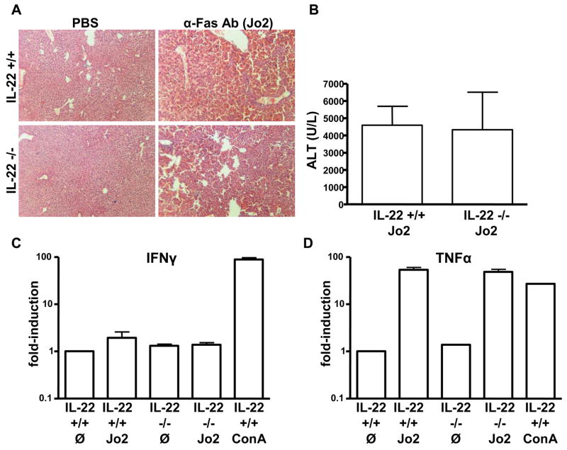
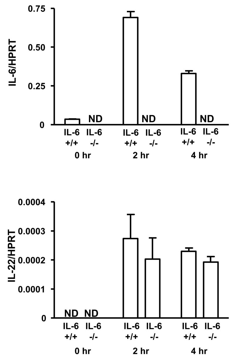
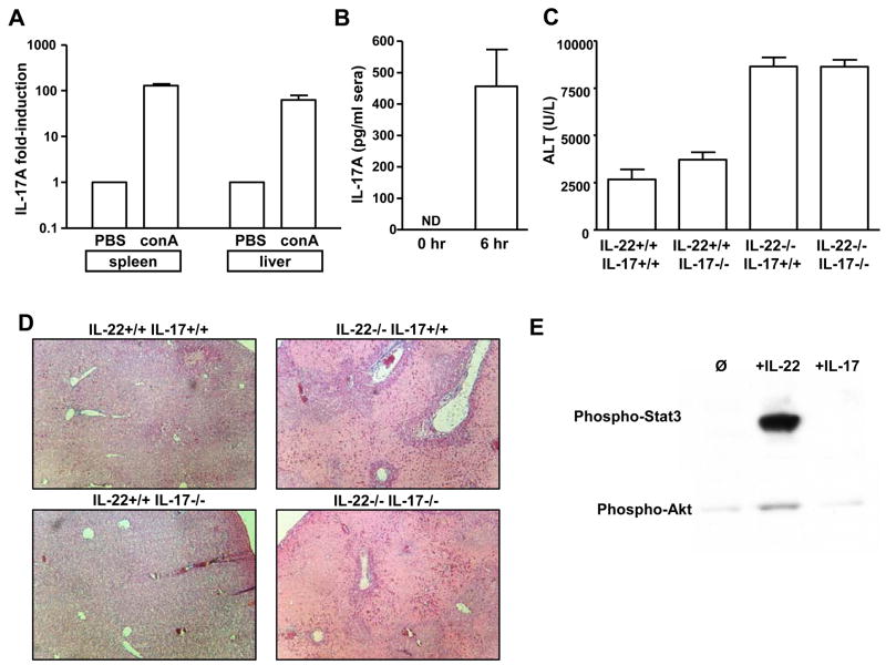
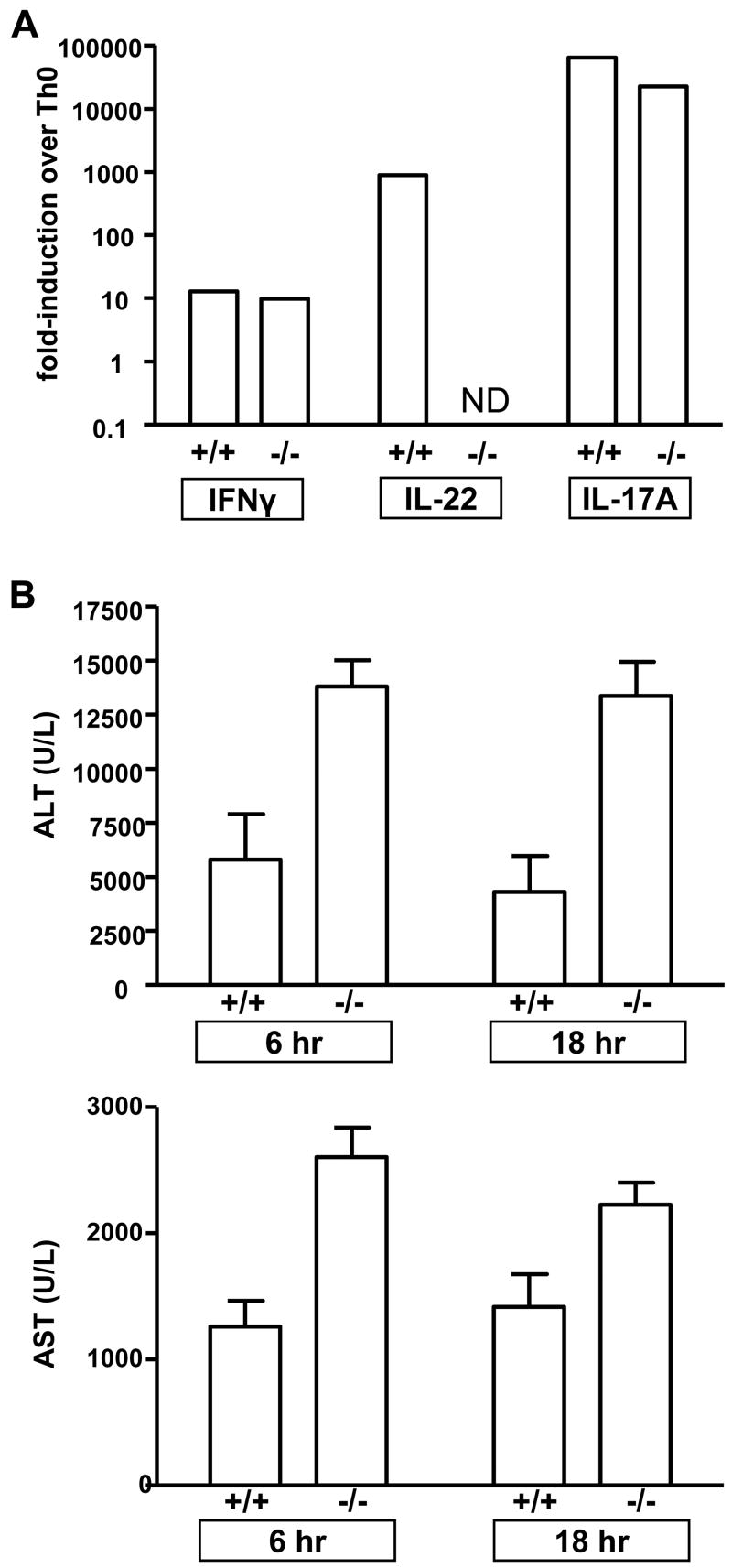
The cytokine interleukin-22 (IL-22) is primarily expressed by T helper 17 (Th17) CD4(+) T cells and is highly upregulated during chronic inflammatory diseases. IL-22 receptor expression is absent on immune cells, but is instead restricted to the tissues, providing signaling directionality from the immune system to the tissues. However, the role of IL-22 in inflammatory responses has been confounded by data suggesting both pro- and anti-inflammatory functions. Herein, we provide evidence that during inflammation, IL-22 played a protective role in preventing tissue injury. Hepatocytes from mice deficient in IL-22 were highly sensitive to the detrimental immune response associated with hepatitis. Additionally, IL-22-expressing Th17 cells provided protection during hepatitis in IL-22-deficient mice. On the other hand, interleukin-17 (IL-17), which is coexpressed with IL-22 and can induce similar cellular responses, had no observable role in liver inflammation. Our data suggest that IL-22 serves as a protective molecule to counteract the destructive nature of the immune response to limit tissue damage.


Conflict of interest statement

Conflict of Interest

LAZ and RAF declare no conflict of interest. GDY, DMV, AJM and MK were employees of Regeneron Pharmaceuticals at the time this work was performed.

Figures

Figure 1
Figure 1. IL-22 deficient mice have no defects in innate or adaptive immunity to Listeria monocytogenes infection
IL-22 wild-type (+/+) or deficient (−/−) mice were injected with 1×10⁴ CFU rLM-ova. Bacterial loads in the (A) spleens and (B) livers of infected mice 3 days post-infection. Bars represent mean±SD of four mice per group. Seven days post-infection, (C) surface expression of CD44 and CD62L was quantitated by FACS on CD4+ T cells from uninfected mice and rLM-ova-infected mice. Shown is a FACS plot from one representative mouse, number represents the percentage of CD4 T cells that are CD44high CD62Llow or CD44low CD62Lhigh. (D) To analyze the antigen-specific response, T cells from the spleen or liver were stimulated with LLO190–201 peptide or without peptide in the presence of GogiStop. IFNγ production was measured by intracellular cytokine staining. Percent shown is the percent of cells that are CD4+ IFNγ+. Plots are representative of four mice/group. Results are representative of two independent experiments.
Figure 2
Figure 2. Hepatocytes are highly responsive to IL-22
(A) IL-22R, IL-10Rα, IL-10Rβ expression in different cell subsets by real-time RT-PCR. Bars represent the gene/HPRT expression as determined by the relative quantification method (ΔΔCT). ND=not detected. (B) FL38B cells or (C) bone-marrow derived macrophages were left untreated (Ø) or stimulated for 20 min with 50 ng/ml recombinant murine IL-10 or IL-22. Phosphorylation or total levels of Stat3 and β-actin in the cell lysates were determined by western blotting with the respective antibodies. (D) FL38B cells were either serum starved for 5 hrs (−FBS) or remained in media with serum (+FBS) and then stimulated with 50 ng/ml recombinant IL-22 for 0, 10 or 30 min. Phosphorylation or total levels of Akt and β-actin in the cell lysates was determined by western blotting with the respective antibodies. Experiment was performed four times with similar results.
Figure 3
Figure 3. IL-22 mRNA is upregulated in the liver during conA-mediated hepatitis
C57BL/6 mice were intravenously injected with the indicated dose of conA and 2 hrs later mRNA was harvested from the livers. mRNA was reverse transcribed, and IL-22 and IFNγ cDNA were quantitated by real-time PCR. Bars represent the mean±SD expression of the cytokine/HPRT using the ΔΔCT method. Experiment was performed two times with similar results. ND=not detected.
Figure 4
Figure 4. IL-22 deficient mice are highly susceptible to conA-induced acute liver inflammation
IL-22 wild-type (+/+), heterozygous (+/−), and deficient mice (−/−) were intravenously injected with 10 μg/g conA or injected with PBS. (A) 18 hrs or 5 days post-injection mice were euthanized, and livers were harvested, fixed, sectioned and stained with H&E. Shown are representative sections from one of three mice per group. (B) Higher magnification of a lesion from an IL-22−/− mouse, 18 hrs post-conA. (C and D) ALT levels in the sera of mice injected 18 hrs previously with (C) PBS or (D) conA. Bars represent mean±SD of three mice/group. Experiment was performed three times with similar results.
Figure 5
Figure 5. IL-22 deficient and wild-type mice have equal immune responses during conA-mediated hepatitis
IL-22 wild-type (+/+) and deficient (−/−) mice were intravenously injected with 10 μg/ml conA or left untreated and 6 hrs later the resulting immune response was analyzed. (A) Cytokine mRNA levels in the liver were semi-quantitated by real-time RT-PCR using the ΔΔCT method. Bars represent mean±SD of four mice. (B) IFNγ and TNFα levels in the sera of mice were determined by ELISA. Bars represent mean±SD of six-eight mice per group. (C) Activation of CD4+ T cells from the livers and spleens was analyzed by surface expression of CD69 by FACS. Number represents the percentage of CD4 cells that are CD69high. Shown are representative FACS plots of one mouse of three mice/group.
Figure 6
Figure 6. Hepatocyte damage in IL-22 deficient mice is dependent on inflammation and not inherent susceptibility of hepatocytes to apoptosis
IL-22 deficient (IL-22 −/−) or control wild-type mice (IL-22 +/+) were injected intraperitoneally with 1 μg/g mouse of anti-Fas Ab (clone Jo-2) or PBS and were euthanized 6 hrs later. (A) Representative liver sections stained with H&E. (B) ALT levels in the sera of Jo2 injected mice, mean±SD of four mice/group. Fold-induction of (C) IFNγ and (D) TNFα mRNA levels in the livers of Jo2 injected (Jo2) or conA injected mice (ConA) compared to untreated mice (Ø) as quantitated by real-time RT-PCR using the ΔΔCT method. Experiment was performed three times with similar results.
Figure 7
Figure 7. IL-6 is not required for IL-22 induction during conA-mediated hepatitis
IL-6 wild-type (IL-6 +/+) or deficient mice (IL-6 −/−) were intravenously injected with 10 μg/g conA. 0, 2 or 4 hrs post-injection, liver lymphocytes were purified, RNA was harvested, reverse transcribed and IL-6 and IL-22 cDNA were quantitated by real-time PCR. Bars indicate mean±SD of the cytokine/HPRT ratio using the ΔΔCT method in 3 mice per group. ND=not detected. Experiment was performed twice with similar results.
Figure 8
Figure 8. IL-22 and IL-17 have non-homologous roles in conA-mediated hepatitis
(A) C57BL/6 mice were injected with 10 μg/ml conA or PBS alone. 2 hrs later IL-17 mRNA fold-induction in the spleen compared to PBS mice was quantitated by real-time RT-PCR using the ΔΔCT method. Bars indicate mean±SD. (B) IL-17A protein levels in the sera of C57BL/6 mice 6 hrs after conA injection. Bar indicates mean±SD. ND=not detected. (C and D) Wild-type (IL-22 +/+ IL-17 +/+), IL-22 deficient (IL-22 −/− IL-17 +/+), IL-17 deficient (IL-22 +/+ IL-17 −/−) or IL-22 IL-17 double deficient (IL-22 −/− IL-17 −/−) mice were intranvenously injected with 10 μg/g conA and 18 hrs later liver injury was examined. (B) H&E liver sections from the indicated mice. Shown are representative sections from one mouse of three per group. (C) ALT levels (mean±SD) in the sera of conA injected mice. (E) FL38B cells were stimulated for 20 min with 50 ng/ml of recombinant murine IL-22 or IL-17 or left unstimulated (Ø). Phosphorylation of Stat3 and Akt in the cell lysates was examined by western blotting with phospho-specific Abs as described in the Experimental Procedures.
Figure 9
Figure 9. Th17 differentiated IL-22 expressing cells protect hepatocytes during hepatitis
IL-22 wild-type (+/+) or deficient (−/−) naïve (CD44low, CD62Llow, CD25-) CD4 T cells were differentiated in vitro with plate-bound α-CD3/α-CD28 Ab for 5 days under Th0 or Th17 conditions. (A) Cells were stimulated with PMA and ionomycin or remained unstimulated. 5 hr post-stimulation, mRNA was harvested, reverse transcribed, and cDNA was quantitated by real-time RT-PCR. Fold-induction over unstimulated cells was calculated by the ΔΔCT method. ND=not detected. (B) 5×10⁶ IL-22 +/+ or IL-22−/− Th17 cells were injected into IL-22−/− mice, and then 6-12 hrs later, mice were injected with 10 μg/g conA. 6 and 18 hrs post-conA injection, ALT and AST levels in the sera were quantitated, mean±SD for 6 mice/group. A similar experiment yielded similar results.

Comment in

Similar articles

Cited by

References

    1. Aggarwal S, Xie MH, Maruoka M, Foster J, Gurney AL. Acinar cells of the pancreas are a target of interleukin-22. J Interferon Cytokine Res. 2001;21:1047–1053. - PubMed
    1. Andoh A, Zhang Z, Inatomi O, Fujino S, Deguchi Y, Araki Y, Tsujikawa T, Kitoh K, Kim-Mitsuyama S, Takayanagi A, et al. Interleukin-22, a member of the IL-10 subfamily, induces inflammatory responses in colonic subepithelial myofibroblasts. Gastroenterology. 2005;129:969–984. - PubMed
    1. Bettelli E, Carrier Y, Gao W, Korn T, Strom TB, Oukka M, Weiner HL, Kuchroo VK. Reciprocal developmental pathways for the generation of pathogenic effector TH17 and regulatory T cells. Nature. 2006;441:235–238. - PubMed
    1. Blum HE. Hepatocellular carcinoma: therapy and prevention. World J Gastroenterol. 2005;11:7391–7400. - PMC - PubMed
    1. Boniface K, Bernard FX, Garcia M, Gurney AL, Lecron JC, Morel F. IL-22 inhibits epidermal differentiation and induces proinflammatory gene expression and migration of human keratinocytes. J Immunol. 2005;174:3695–3702. - PubMed

Publication types

MeSH terms