. 2008 Jan 5;370(1):130-41.
doi: 10.1016/j.virol.2007.08.022. Epub 2007 Oct 24.

Selective induction of cell-mediated immunity and protection of rhesus macaques from chronic SHIV(KU2) infection by prophylactic vaccination with a conserved HIV-1 envelope peptide-cocktail

Affiliations

Selective induction of cell-mediated immunity and protection of rhesus macaques from chronic SHIV(KU2) infection by prophylactic vaccination with a conserved HIV-1 envelope peptide-cocktail

Pramod N Nehete et al. Virology. .

Abstract

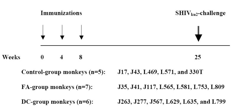
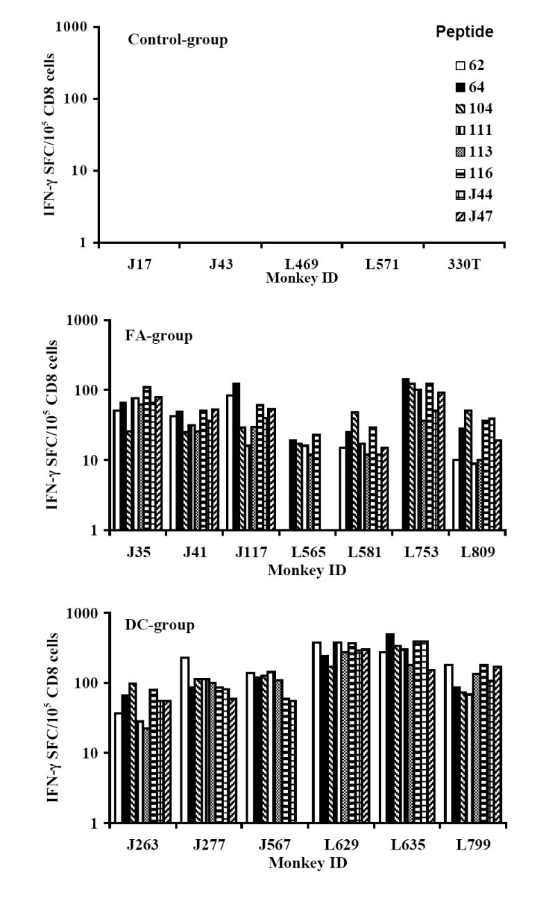
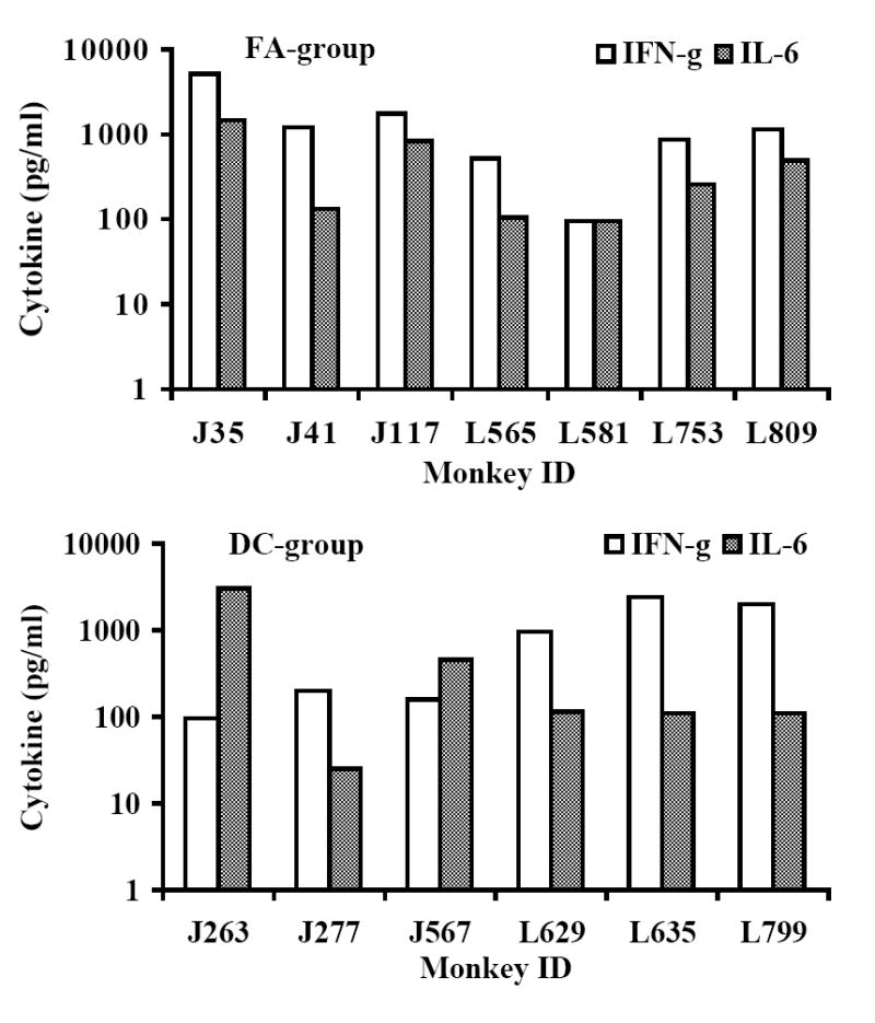
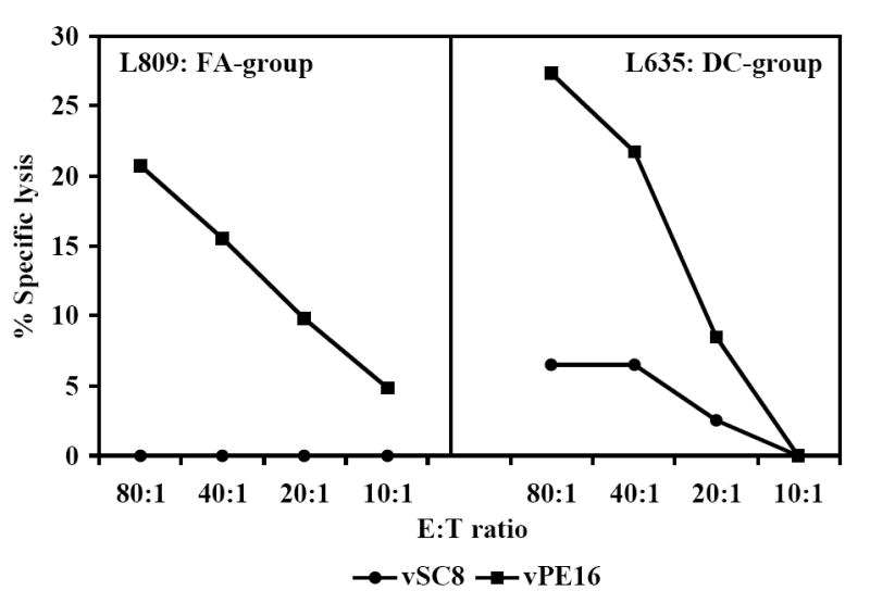
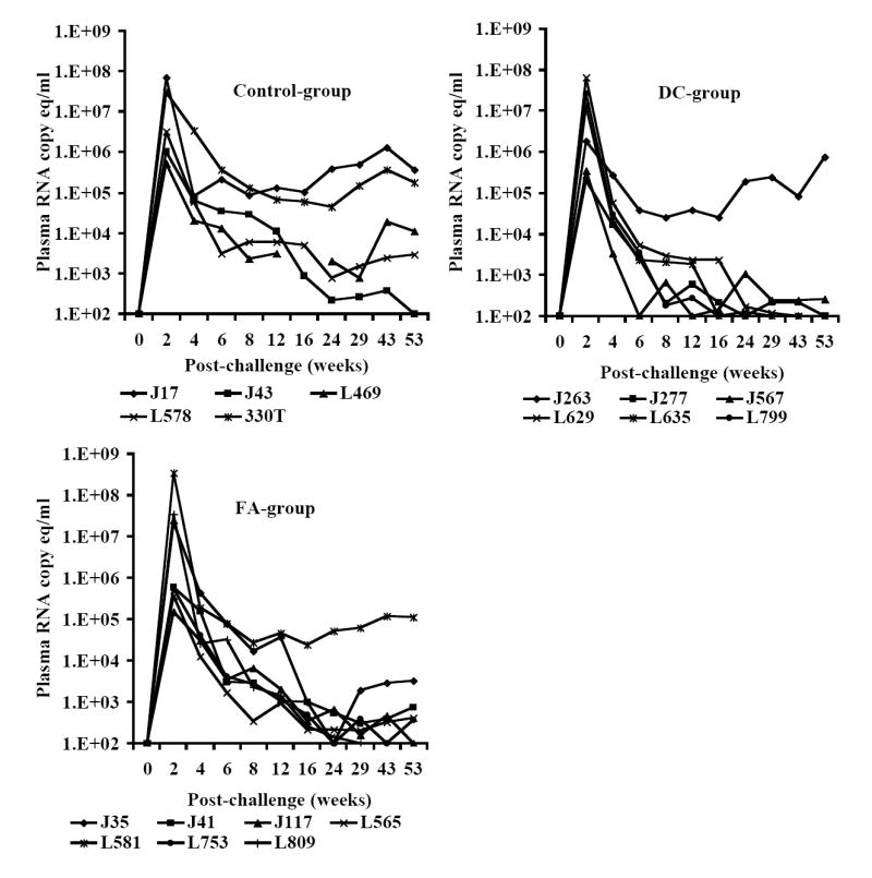
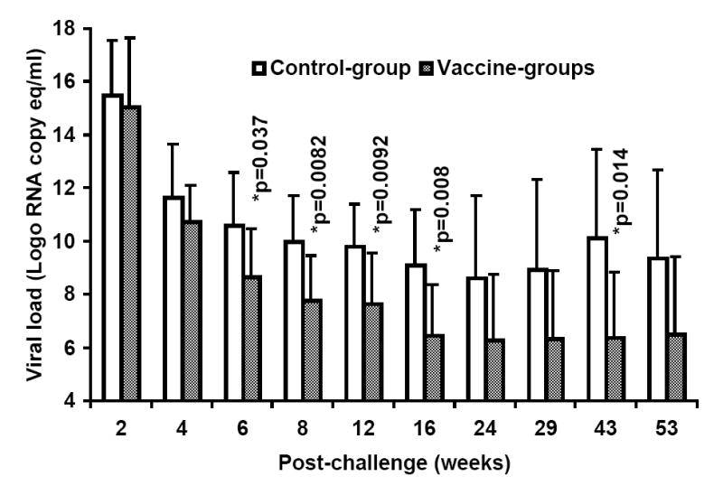
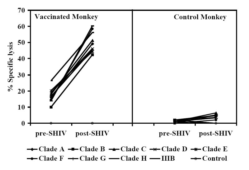
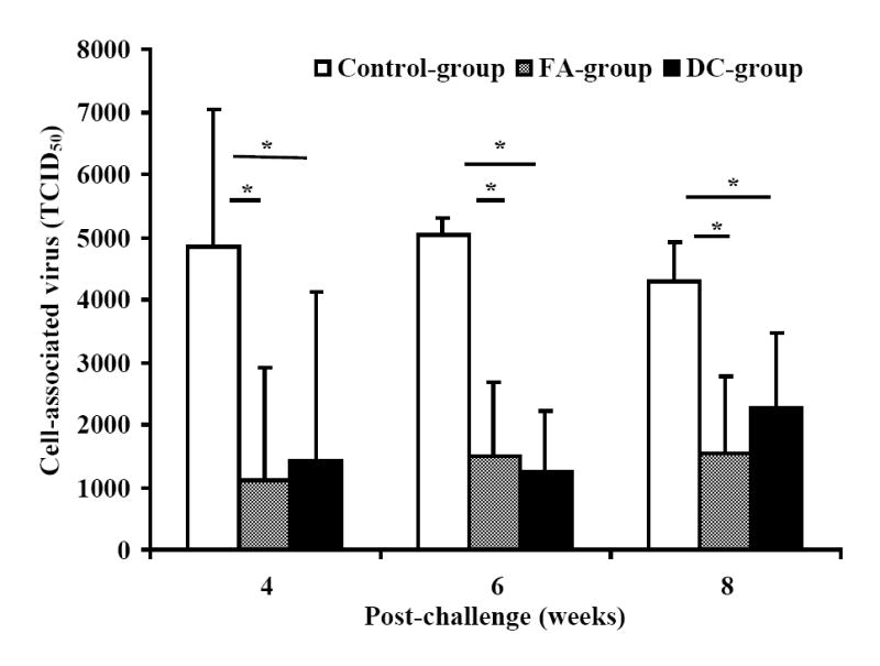
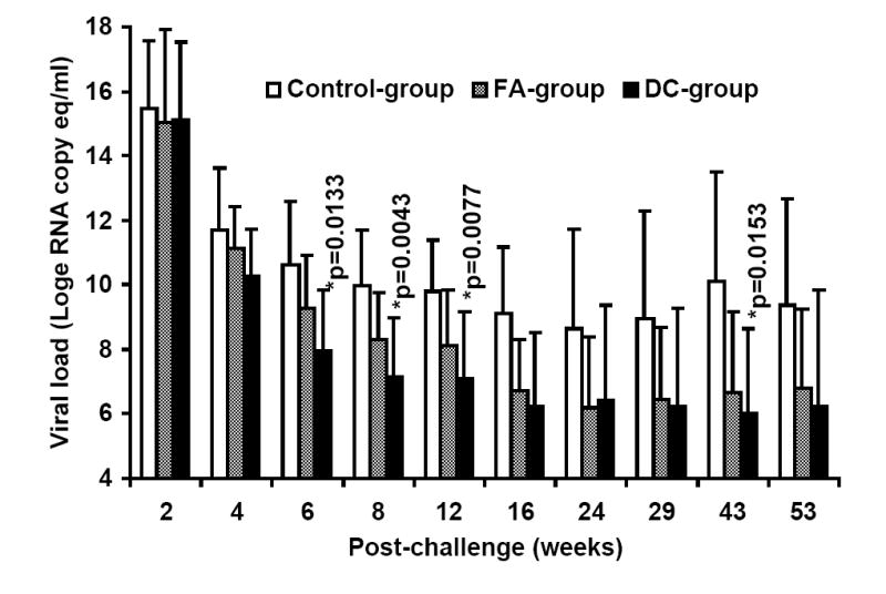
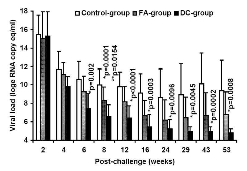
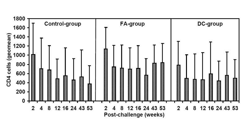
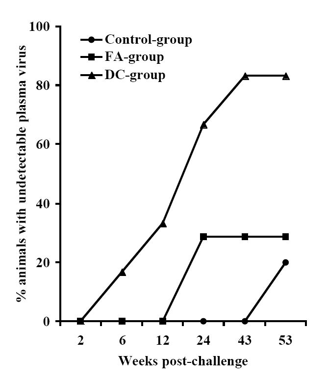
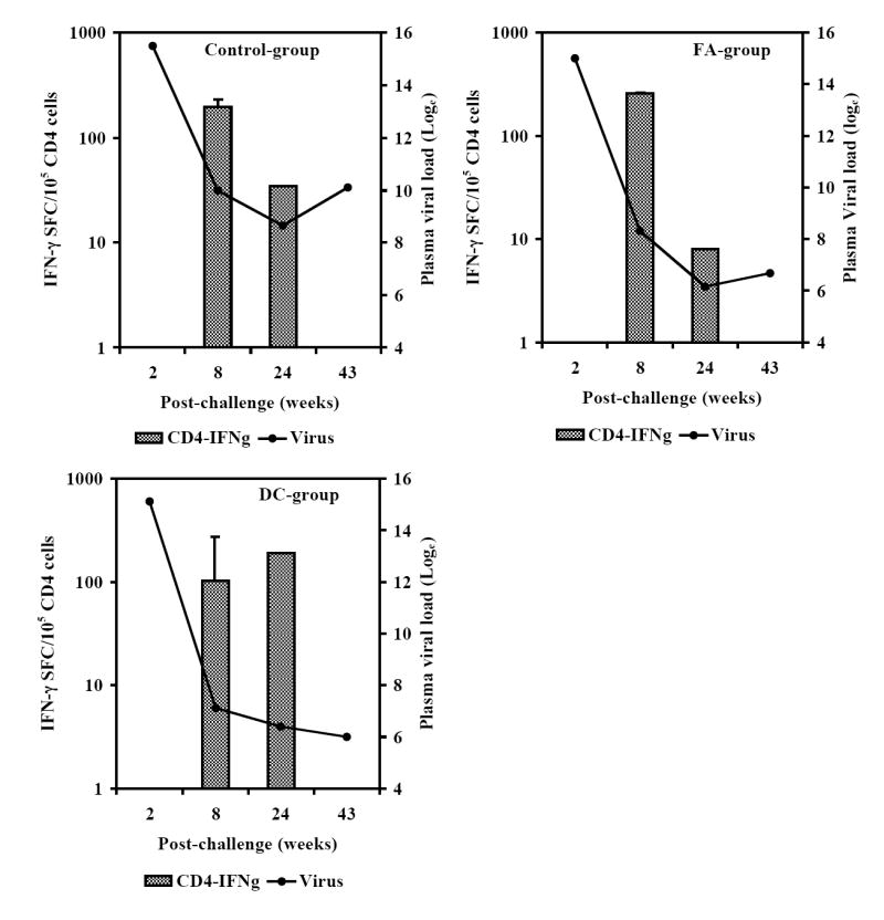
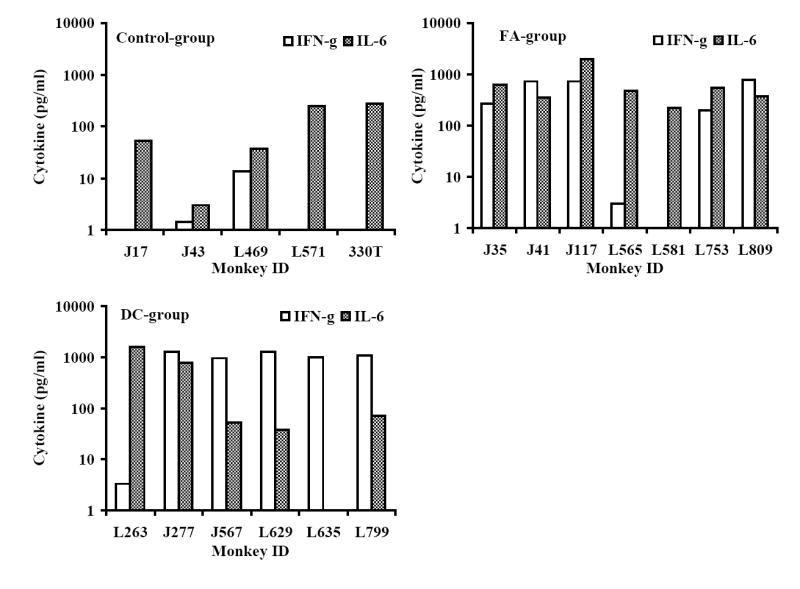
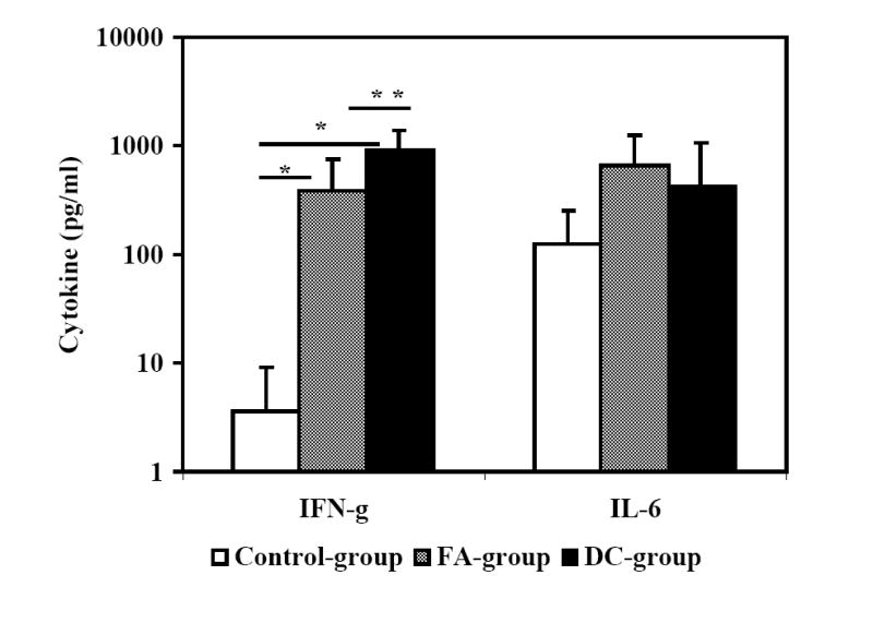
Infection of Indian-origin rhesus macaques by the simian human immunodeficiency virus (SHIV) is considered to be a suitable preclinical model for directly testing efficacy of vaccine candidates based on the HIV-1 envelope. We used this model for prophylactic vaccination with a peptide-cocktail comprised of highly conserved HIV-1 envelope sequences immunogenic/antigenic in macaques and humans. Separate groups of macaques were immunized with the peptide-cocktail by intravenous and subcutaneous routes using autologous dendritic cells (DC) and Freund's adjuvant, respectively. The vaccine elicited antigen specific IFN-gamma-producing cells and T-cell proliferation, but not HIV-neutralizing antibodies. The vaccinated animals also exhibited efficient cross-clade cytolytic activity against target cells expressing envelope proteins corresponding to HIV-1 strains representative of multiple clades that increased after intravenous challenge with pathogenic SHIV(KU2). Virus-neutralizing antibodies were either undetectable or present only transiently at low levels in the control as well as vaccinated monkeys after infection. Significant control of plasma viremia leading to undetectable levels was achieved in majority of vaccinated monkeys compared to mock-vaccinated controls. Monkeys vaccinated with the peptide-cocktail using autologous DC, compared to Freund's adjuvant, and the mock-vaccinated animals, showed significantly higher IFN-gamma production, higher levels of vaccine-specific IFN-gamma producing CD4(+) cells and significant control of plasma viremia. These results support DC-based vaccine delivery and the utility of the conserved HIV-1 envelope peptide-cocktail, capable of priming strong cell-mediated immunity, for potential inclusion in HIV vaccination strategies.


Figures

Fig. 1
Fig. 1
Immunization and challenge scheme for rhesus monkeys in the three different groups: Control-group, FA-group and DC-group where monkeys were either mock-vaccinated or immunized with the HIV-1 envelope peptide-cocktail delivered to rhesus macaques using either the Freund’s adjuvant or autologous DC, respectively. Adult female rhesus macaques (Macaca mulatta) of Indian-origin between the ages of 8-17 years from the specific pathogen-free breeding colony were uniformly distributed in the three study groups.
Fig. 2
Fig. 2
Induction of antigen-specific cellular immune responses by the HIV-1 envelope peptide-cocktail delivered to rhesus macaques using either the Freund’s adjuvant or autologous DC (FA-group and DC-group, respectively). The peak responses in a total of 105 input CD8+ T cells (isolated from the PBMC of the monkeys) in the three different groups in terms of IFN-γ producing cells in response to stimulation for 36 h at 37°C with individual peptides in the peptide-cocktail vaccine were determined by the ELISPOT assay (Fig. 2A). Induction of antigen-specific cytokine production in the monkeys from the FA-group and the DC-group was determined by the CBA assay (Fig. 2B). Monkeys in the Control-group did not show production of either of the cytokines in response to stimulation with the peptide-mix (data not shown). Immunization with the HIV-1 envelope peptide-cocktail primes HIV-1 envelope-specific CTL responses (Fig. 2C). The PBMC obtained from the monkeys in the FA-group and DC-group were stimulated in vitro for 14 days with the mixture of the eight HIV-1 envelope peptides in the peptide-cocktail vaccine (each peptide at 10ug/ml final concentration), and the CTL activity was assayed against autologous B lymphocyte cell lines (B-LCL) infected with recombinant vaccinia virus expressing gp160 from HIV-1IIIB (vPE160), and the control vaccinia virus (vSC8) in the standard 6 h 51Cr-release methodology. Data for a representative monkey each from the FA-group and the DC-group are shown.
Fig. 2
Fig. 2
Induction of antigen-specific cellular immune responses by the HIV-1 envelope peptide-cocktail delivered to rhesus macaques using either the Freund’s adjuvant or autologous DC (FA-group and DC-group, respectively). The peak responses in a total of 105 input CD8+ T cells (isolated from the PBMC of the monkeys) in the three different groups in terms of IFN-γ producing cells in response to stimulation for 36 h at 37°C with individual peptides in the peptide-cocktail vaccine were determined by the ELISPOT assay (Fig. 2A). Induction of antigen-specific cytokine production in the monkeys from the FA-group and the DC-group was determined by the CBA assay (Fig. 2B). Monkeys in the Control-group did not show production of either of the cytokines in response to stimulation with the peptide-mix (data not shown). Immunization with the HIV-1 envelope peptide-cocktail primes HIV-1 envelope-specific CTL responses (Fig. 2C). The PBMC obtained from the monkeys in the FA-group and DC-group were stimulated in vitro for 14 days with the mixture of the eight HIV-1 envelope peptides in the peptide-cocktail vaccine (each peptide at 10ug/ml final concentration), and the CTL activity was assayed against autologous B lymphocyte cell lines (B-LCL) infected with recombinant vaccinia virus expressing gp160 from HIV-1IIIB (vPE160), and the control vaccinia virus (vSC8) in the standard 6 h 51Cr-release methodology. Data for a representative monkey each from the FA-group and the DC-group are shown.
Fig. 3
Fig. 3
A. Control of SHIVKU2 infection in monkeys vaccinated with the HIV-1 envelope peptide-cocktail. Viral loads in plasma samples from individual monkeys in the Control-group as well as the two vaccine groups (FA-group and DC-group) were determined by real-time RT-PCR methodology at several time points over 53 weeks post-challenge with SHIVKU2 (104 TCID50) by the intravenous route. The peak viral loads were observed in the monkeys in all the three groups at week 2 post-challenge and the relative changes subsequently are different in the different groups with majority of the monkeys in the DC-group group showing lower levels compared to that in the Control-group monkeys. Data in Fig. 3B shows significant differences in the mean plasma viral RNA copy numbers for monkeys in the control-group with those in the FA-, and DC-groups combined over the 53 weeks. Log-transformed average viral load changes show significant reduction in viral load (p<0.001) in the vaccinated monkeys at weeks 6, 8, 12, 16 and 43 post-challenge (*).
Fig. 3
Fig. 3
A. Control of SHIVKU2 infection in monkeys vaccinated with the HIV-1 envelope peptide-cocktail. Viral loads in plasma samples from individual monkeys in the Control-group as well as the two vaccine groups (FA-group and DC-group) were determined by real-time RT-PCR methodology at several time points over 53 weeks post-challenge with SHIVKU2 (104 TCID50) by the intravenous route. The peak viral loads were observed in the monkeys in all the three groups at week 2 post-challenge and the relative changes subsequently are different in the different groups with majority of the monkeys in the DC-group group showing lower levels compared to that in the Control-group monkeys. Data in Fig. 3B shows significant differences in the mean plasma viral RNA copy numbers for monkeys in the control-group with those in the FA-, and DC-groups combined over the 53 weeks. Log-transformed average viral load changes show significant reduction in viral load (p<0.001) in the vaccinated monkeys at weeks 6, 8, 12, 16 and 43 post-challenge (*).
Fig. 4
Fig. 4
Cross-clade CTL responses in monkeys vaccinated with the HIV-1 envelope peptide-cocktail. Higher CTL responses, specific to HIV-1 envelope sequences corresponding to 8 different clade HIV-1 isolates, in addition to HIV-1IIIB, were observed in the vaccinated monkey compared to that in a control monkey and the responses in the vaccinated monkey increasedsubstantially after SHIVKU2-challenge (post-SHIV) compared to levels prior to challenge (pre-SHIV). The CTL assay was performed as described in Fig. 2C, and the results are shown for the 25:1 effecter to target ratio for a representative monkey from the control and vaccinated groups.
Fig. 5
Fig. 5
Decrease in cell-associated virus in monkeys vaccinated with the HIV-1 envelope peptide-cocktail. A significant decrease (*, p<0.05) in cell-associated virus at the set point (weeks 4-8) was observed in vaccinated animals (FA-group and DC-group) compared to that in the Control-group. Serial 10-fold dilutions of PBMC (106 cells/ml) from the monkeys in the different groups were co-cultured with indicator C8166 cells for 7 days. At the end of the culture period, the cell-free supernatant from each well was used for determining the amount of infectious virus by incubating 10-fold serial dilutions with fresh indicator MT4 cells and the 50% tissue culture infectious dose (TCID50) was calculated by the standard MTT-dye reduction assay. Results shown are average values for the 50% tissue culture infectious dose (TCID50) for 106 PBMC form monkeys in each group.
Fig. 6
Fig. 6
Delivery of the HIV-1 envelope peptide-cocktail vaccine using DC is more effective than the Freund’s adjuvant for significantly lowering the viral load. The mean plasma viral RNA copy numbers for monkeys in the control-group was compared to that in the vaccinated monkeys (the FA-, and DC-groups) over the 53 weeks. The DC-group, but not the FA-group, showed significantly lower mean plasma viral loads (*) between weeks 6-12 and also at week 43 (Fig.6A). Furthermore, as shown in Fig. 6B, the mean viral load in five monkeys in the DC-group as a sub-group, except J263, that showed similar trends in the viral load changes, showed significant reduction in the virus load past week 4 post-challenge compared to that in the Control-group at all time points (*) and also when compared to the FA-group at week 8 (**). Fig. 6C shows geometric mean values for CD4 cells in the blood for the three different groups with steady decline in the Control-group over the 53 weeks follow-up while stabilization of the CD4 cells was observed in the animals from the two vaccine groups. Fig. 6D shows percentages of monkeys with undetectable plasma viral loads (using 300 RNA copy equivalents/ml as the lower cut-off value for the sensitivity of the assay) in the three different groups; Control-group, FA-group and DC-group.
Fig. 6
Fig. 6
Delivery of the HIV-1 envelope peptide-cocktail vaccine using DC is more effective than the Freund’s adjuvant for significantly lowering the viral load. The mean plasma viral RNA copy numbers for monkeys in the control-group was compared to that in the vaccinated monkeys (the FA-, and DC-groups) over the 53 weeks. The DC-group, but not the FA-group, showed significantly lower mean plasma viral loads (*) between weeks 6-12 and also at week 43 (Fig.6A). Furthermore, as shown in Fig. 6B, the mean viral load in five monkeys in the DC-group as a sub-group, except J263, that showed similar trends in the viral load changes, showed significant reduction in the virus load past week 4 post-challenge compared to that in the Control-group at all time points (*) and also when compared to the FA-group at week 8 (**). Fig. 6C shows geometric mean values for CD4 cells in the blood for the three different groups with steady decline in the Control-group over the 53 weeks follow-up while stabilization of the CD4 cells was observed in the animals from the two vaccine groups. Fig. 6D shows percentages of monkeys with undetectable plasma viral loads (using 300 RNA copy equivalents/ml as the lower cut-off value for the sensitivity of the assay) in the three different groups; Control-group, FA-group and DC-group.
Fig. 7
Fig. 7
Monkeys immunized with autologous DC pulsed with the HIV-1 envelope peptide-cocktail exhibit strong TH1 responses and control of viremia. The numbers of IFN-γ-producing cells in a total of 105 input CD4+ T cells (isolated from the PBMC), in terms of IFN-γ spot forming cells (SFC) were analyzed by the ELISPOT assay at the early and late time points (8 and 24 weeks post-challenge, respectively) as described in the methods section. Fig. 7A shows the average numbers of IFN-γ-producing CD4+ T cells showed a steep decrease between weeks 8 and 24 time points for the monkeys in the Control-group and FA-group where significant drop in viral loads were not observed, but they increased over the same period in the DC-group where the viral loads decreased significantly. Fig. 7B shows the levels of IFN-γ and IL-6 produced by PBMC collected at week 24 post-challenge from the individual monkeys in the three different groups in response to stimulation with the mixture of the peptides in the vaccine-cocktail using the Cytometric Bead Array assay as described in the methods section. Monkeys in the control group predominantly showed production of IL-6 but not IFN-γ. Fig. 7C shows comparison of average values for the levels of IFN-γ and IL-6 produced, in response to stimulation with the mixture of the peptides in the vaccine-cocktail, in the three different groups. Significantly higher amount of IFN-γ production (p<0.05) was observed for monkeys in the FA-group and DC-group compared to that in the Control-group (*), as well as significantly higher amounts in the DC-group compared to the FA-group (**).
Fig. 7
Fig. 7
Monkeys immunized with autologous DC pulsed with the HIV-1 envelope peptide-cocktail exhibit strong TH1 responses and control of viremia. The numbers of IFN-γ-producing cells in a total of 105 input CD4+ T cells (isolated from the PBMC), in terms of IFN-γ spot forming cells (SFC) were analyzed by the ELISPOT assay at the early and late time points (8 and 24 weeks post-challenge, respectively) as described in the methods section. Fig. 7A shows the average numbers of IFN-γ-producing CD4+ T cells showed a steep decrease between weeks 8 and 24 time points for the monkeys in the Control-group and FA-group where significant drop in viral loads were not observed, but they increased over the same period in the DC-group where the viral loads decreased significantly. Fig. 7B shows the levels of IFN-γ and IL-6 produced by PBMC collected at week 24 post-challenge from the individual monkeys in the three different groups in response to stimulation with the mixture of the peptides in the vaccine-cocktail using the Cytometric Bead Array assay as described in the methods section. Monkeys in the control group predominantly showed production of IL-6 but not IFN-γ. Fig. 7C shows comparison of average values for the levels of IFN-γ and IL-6 produced, in response to stimulation with the mixture of the peptides in the vaccine-cocktail, in the three different groups. Significantly higher amount of IFN-γ production (p<0.05) was observed for monkeys in the FA-group and DC-group compared to that in the Control-group (*), as well as significantly higher amounts in the DC-group compared to the FA-group (**).

Similar articles

Cited by

References

    1. Amara RR, Villinger F, Altman JD, Lydy SL, O’Neil SP, Staprans SI, Montefiori DC, Herndon GS, Wyatt MA, Candido NL, Kozyr PL, Earl JM, Smith HL, Ma BD, Grimm ML, Hulsey J Miller, McClure HM, McNicholl JM, Moss B, Robinson HL. Control of a mucosal challenge and prevention of AIDS by a multiprotein DNA/MVA vaccine. Science. 2001;92:69–74. - PubMed
    1. Baba TW, Liska V, Lehmann RH, Vlasak J, Xu W, Ayehunie S, Cavacini LA, Posner MR, Katinger H, Stiegler G, Bernacky BJ, Rizvi TA, Schmidt R, Hill LR, Keeling ME, Lu Y, Wright JE, Chou T, Ruprecht RM. Human neutralizing monoclonal antibodies of the IgG1 subtype protect against mucosal simian-human immunodeficiency virus infection. Nature Med. 2000;6:200–206. - PubMed
    1. Banchereau J, Paczesny S, Blanco P, Bennett L, Pascual V, fay J, Palucka AK. Dendritic cells: controllers of the immune system and a new promise for immunotherapy. Ann N Y Acad Sci. 2003;987:180–187. - PubMed
    1. Barouch DH, Fu TM, Montefiori DC, Lewis MG, Shiver JW, Letvin NL. Vaccine-elicited immune responses prevent clinical AIDS in SHIV (89.6P)-infected rhesus monkeys. Immunol Lett. 2001;79:57–61. - PubMed
    1. Barouch DH, Kunstaman J, Juroda MJ, Schmitz JE, Santra S, Peyerl FW, Krivulka GR, Beaudry K, Lifton MA, Gotgone DA, Montefiori DC, Lewis MG, Wolinsky SM, Letvin NL. Eventual AIDS vaccine failure in a rhesus monkey by viral escape from cytotoxic T lymphocytes. Nature. 2002;415:335–39. - PubMed

Publication types

MeSH terms