

Oxidatively modified RNA in mild cognitive impairment
- PMID: 17920285
- PMCID: PMC2700659
- DOI: 10.1016/j.nbd.2007.07.030
Oxidatively modified RNA in mild cognitive impairment
Abstract
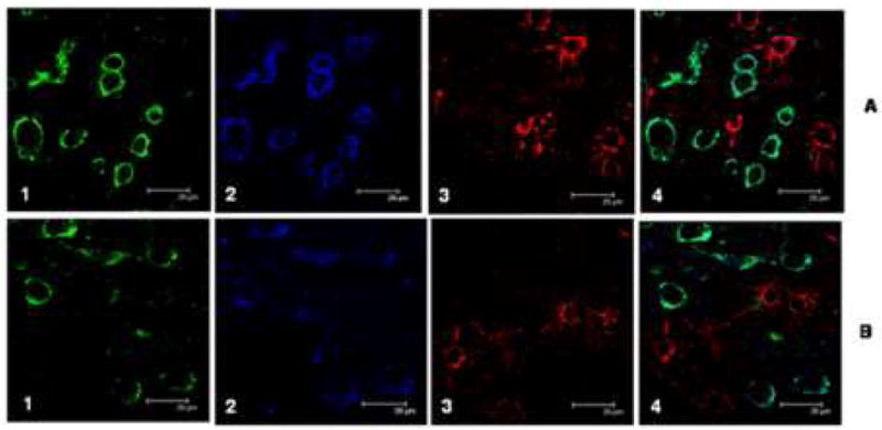
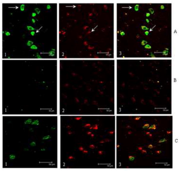
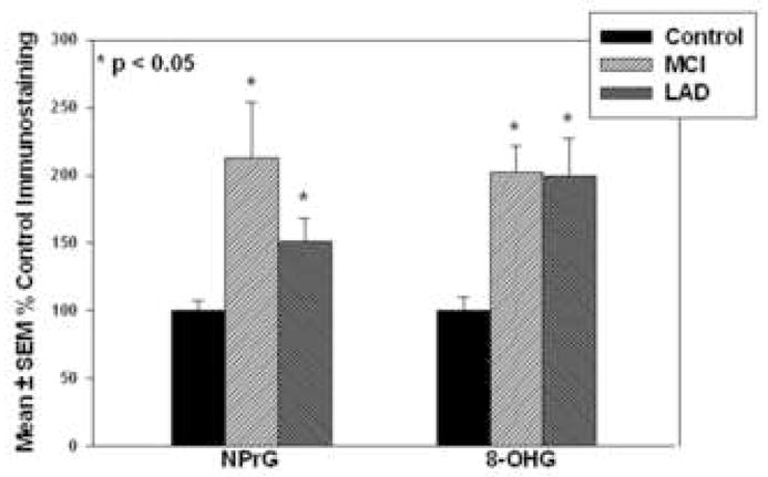
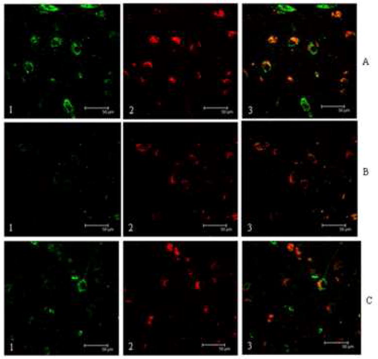
Studies show increased oxidative damage in the brains of subjects with Alzheimer's disease (AD) and mild cognitive impairment (MCI). To determine if RNA oxidation occurs in MCI, sections of hippocampus/parahippocampal gyrus (HPG) from 5 MCI, 5 late stage AD (LAD) and 5 age-matched normal control (NC) subjects were subjected to immunohistochemistry using antibodies against 8-hydroxyguanine (8-OHG) and 1-N2-propanodeoxyguanosine (NPrG). Confocal microscopy showed 8-OHG and NPrG immunostaining was significantly (p<0.05) elevated in MCI and LAD HPG compared with NC subjects and was predominately associated with neurons identified using the MC-1 antibody that recognizes conformational alterations of tau, which are associated with early neurofibrillary tangle formation. Pretreating sections with RNase or DNase-I showed immunostaining for both adducts was primarily associated with RNA. In addition, levels of both adducts in MCI were comparable to those measured in LAD, suggesting RNA oxidation may be an early event in the pathogenesis of neuron degeneration in AD.
Figures
Similar articles
-
Alterations of zinc transporter proteins ZnT-1, ZnT-4 and ZnT-6 in preclinical Alzheimer's disease brain.Brain Pathol. 2010 Mar;20(2):343-50. doi: 10.1111/j.1750-3639.2009.00283.x. Epub 2009 Apr 7. Brain Pathol. 2010. PMID: 19371353 Free PMC article.
-
Elevated zinc transporter-6 in mild cognitive impairment, Alzheimer disease, and pick disease.J Neuropathol Exp Neurol. 2006 May;65(5):489-98. doi: 10.1097/01.jnen.0000229237.98124.91. J Neuropathol Exp Neurol. 2006. PMID: 16772872
-
Oxidatively modified nucleic acids in preclinical Alzheimer's disease (PCAD) brain.Mech Ageing Dev. 2011 Aug;132(8-9):443-8. doi: 10.1016/j.mad.2011.08.003. Epub 2011 Aug 22. Mech Ageing Dev. 2011. PMID: 21878349 Free PMC article.
-
Oxidative DNA damage in mild cognitive impairment and late-stage Alzheimer's disease.Nucleic Acids Res. 2007;35(22):7497-504. doi: 10.1093/nar/gkm821. Epub 2007 Oct 18. Nucleic Acids Res. 2007. PMID: 17947327 Free PMC article. Review.
-
Oxidatively modified proteins in Alzheimer's disease (AD), mild cognitive impairment and animal models of AD: role of Abeta in pathogenesis.Acta Neuropathol. 2009 Jul;118(1):131-50. doi: 10.1007/s00401-009-0517-0. Epub 2009 Mar 14. Acta Neuropathol. 2009. PMID: 19288120 Free PMC article. Review.
Cited by
-
Mitochondrial ATP-synthase in the entorhinal cortex is a target of oxidative stress at stages I/II of Alzheimer's disease pathology.Brain Pathol. 2010 Jan;20(1):222-33. doi: 10.1111/j.1750-3639.2009.00266.x. Epub 2009 Feb 27. Brain Pathol. 2010. PMID: 19298596 Free PMC article.
-
Urinary 8-OxoGsn as a Potential Indicator of Mild Cognitive Impairment in Frail Patients With Cardiovascular Disease.Front Aging Neurosci. 2021 Aug 25;13:672548. doi: 10.3389/fnagi.2021.672548. eCollection 2021. Front Aging Neurosci. 2021. PMID: 34531733 Free PMC article.
-
Amyloid β-peptide (1-42)-induced oxidative stress in Alzheimer disease: importance in disease pathogenesis and progression.Antioxid Redox Signal. 2013 Sep 10;19(8):823-35. doi: 10.1089/ars.2012.5027. Epub 2013 Feb 14. Antioxid Redox Signal. 2013. PMID: 23249141 Free PMC article. Review.
-
Role of a Urinary Biomarker in the Common Mechanism of Physical Performance and Cognitive Function.Front Med (Lausanne). 2022 Feb 18;9:816822. doi: 10.3389/fmed.2022.816822. eCollection 2022. Front Med (Lausanne). 2022. PMID: 35252255 Free PMC article.
-
Organoselenium (Sel-Plex diet) decreases amyloid burden and RNA and DNA oxidative damage in APP/PS1 mice.Free Radic Biol Med. 2009 Jun 1;46(11):1527-33. doi: 10.1016/j.freeradbiomed.2009.03.008. Epub 2009 Mar 19. Free Radic Biol Med. 2009. PMID: 19303433 Free PMC article.
References
-
- Banerjee A, Roach MC, Trcka P, Luduena RF. Increased microtubule assembly in bovine brain tubulin lacking the type III isotype of beta-tubulin. J Biol Chem. 1990;265:1794–9. - PubMed
-
- Beelman CA, Parker R. Degradation of mRNA in eukaryotes. Cell. 1995;81:179–83. - PubMed
-
- Bennett DA, Wilson RS, Schneider JA, Evans DA, Beckett LA, Aggarwal NT, Barnes LL, Fox JH, Bach J. Natural history of mild cognitive impairment in older persons. Neurology. 2002;59:198–205. - PubMed
-
- Calingasan NY, Uchida K, Gibson GE. Protein-bound acrolein: a novel marker of oxidative stress in Alzheimer’s disease. J Neurochem. 1999;72:751–6. - PubMed
-
- Chung FL, Young R, Hecht SS. Formation of cyclic 1,N2-propanodeoxyguanosine adducts in DNA upon reaction with acrolein or crotonaldehyde. Cancer Res. 1984;44:990–5. - PubMed
Publication types
MeSH terms
Substances
Grants and funding
LinkOut - more resources
Full Text Sources
