. 2007 Dec 15;371(2):135-45.
doi: 10.1016/j.ab.2007.08.019. Epub 2007 Aug 22.

Alkaline methanolysis of lipid extracts extends shotgun lipidomics analyses to the low-abundance regime of cellular sphingolipids

Affiliations

Alkaline methanolysis of lipid extracts extends shotgun lipidomics analyses to the low-abundance regime of cellular sphingolipids

Xuntian Jiang et al. Anal Biochem. .

Abstract

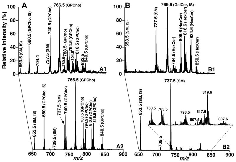
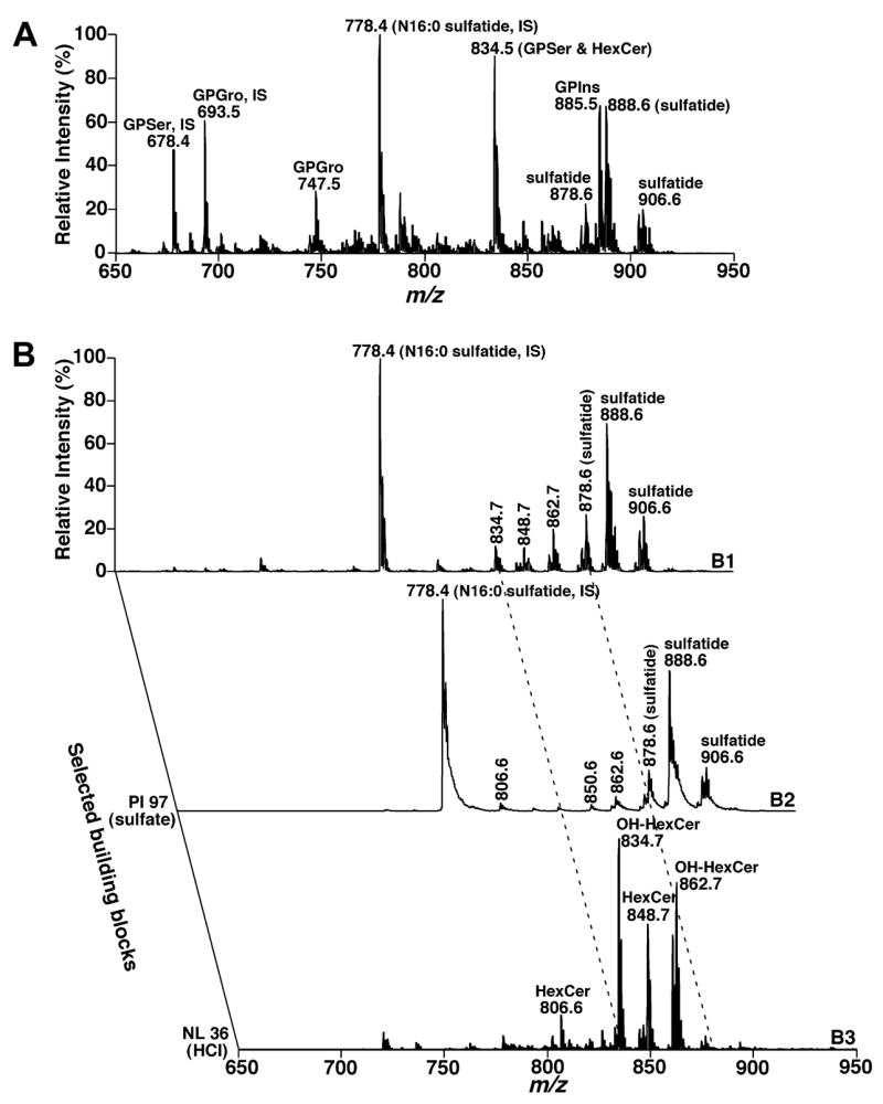
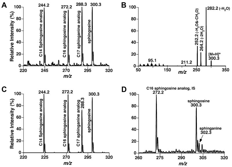
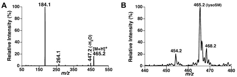
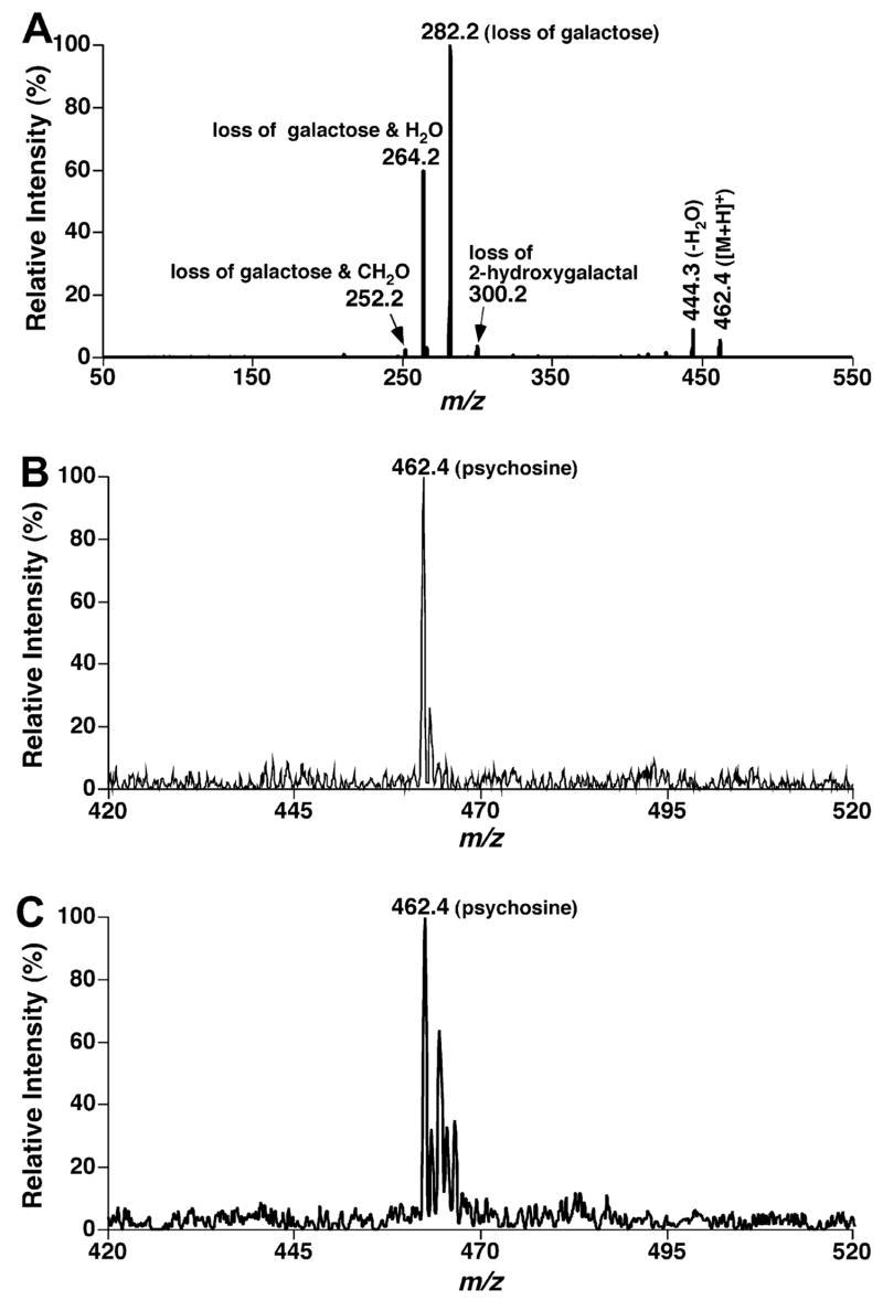
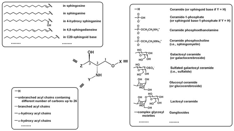
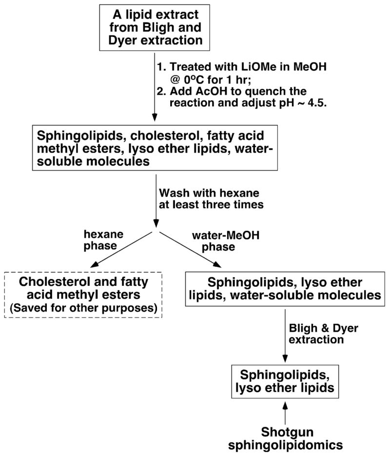
Sphingolipids that contain a sphingoid base are composed of hundreds to thousands of distinct compounds, many of which serve as lipid regulators of biological functions. The global analysis of the large number of low-abundance sphingolipid molecular species has been hampered in many cases by the sphingolipid molecular species being overwhelmed by the quantity of other classes of lipid (e.g., glycerophospholipid) molecular species present, thereby imposing severe restrictions on the dynamic range of their measurement using shotgun lipidomics. Herein, we developed a facile approach in which the sphingolipids of cellular extracts were dramatically enriched by direct alkaline methanolysis of lipid extracts followed by extraction to remove the large majority of other endogenous lipid classes. Through direct infusion of the resultant enriched solution, we identified and quantitated a variety of very-low-abundance sphingolipid classes (e.g., sphingosine, psychosine, and lysosphingomyelin) and molecular species (e.g., sphingomyelin) using electrospray ionization mass spectrometry (i.e., shotgun sphingolipidomics). Accordingly, through utilization of these facile enrichment techniques, direct penetrance into the sphingolipidomes has been greatly extended, facilitating new insights into their metabolism and signaling functions in biological systems.


Figures

Fig. 1
Fig. 1
Shotgun lipidomics analyses of sphingolipid molecular species before and after treatment of a mouse cortex lipid extract with LiOMe in the positive-ion mode in the presence of a small amount of LiOH. The mass spectra were acquired directly from a lipid extract of mouse cortex before (left) and after (right) treatment with LiOMe as illustrated in Scheme 2, respectively. Mass spectra A1 and B1 are the survey scans and mass spectra A2 and B2 were acquired from the neutral loss of 183.1 u (i.e., phosphocholine). IS denotes internal standard. The ion peaks in mass spectrum B2 represent lithiated SM molecular species. Each spectrum displayed is normalized to the base peak in the spectrum.
Fig. 2
Fig. 2
Shotgun lipidomics analyses of sphingolipid molecular species before and after treatment of a mouse cortex lipid extract with LiOMe in the negative-ion mode. The mass spectra in A and B1 were acquired directly from a lipid extract of mouse cortex before and after treatment with LiOMe as illustrated in Scheme 2, respectively. Tandem mass spectrometric analyses of the lipid extract after treatment with LiOMe were acquired by using precursor-ion scanning of 97 Th (i.e., sulfate) (B2) and neutral loss scanning of 36 u (loss of HCl from chlorinated HexCer molecular species) (B3), respectively. IS denotes internal standard. Each spectrum displayed is normalized to the base peak in the spectrum.
Fig. 3
Fig. 3
ESI/MS and MS/MS analyses of sphingosine and its analogs. ESI/MS analysis (A) of an equimolar mixture of sphingosine analogs (0.1 pmol/μl each in 1:1 CHCl3/MeOH in the presence of 0.1% formic acid) was performed in the positive-ion mode. Product ion analysis of sphingosine (B) was performed after collision-induced dissociation. Tandem MS analyses of the equimolar mixture and a lipid extract from mouse spinal cord were also performed by using neutral-loss (NL) scanning of 48 u (i.e., loss of a H2O and a CH2O) (C and D, respectively). IS denotes internal standard.
Fig. 4
Fig. 4
Tandem mass spectrometric analyses of lysosphingomyelin. Product ion analysis of lysosphingomyelin (0.1 pmol/μl in 1:1 CHCl3/MeOH in the presence of 0.1% formic acid) was performed after collision-induced dissociation with a collision gas (nitrogen) pressure at 1.0 mTorr and a collision energy of 22 eV (A). Precursor-ion scanning of 184.1 Th (i.e., protonated phosphocholine) from a lipid extract of mouse spinal cord after treatment with LiOMe was conducted under experimental conditions as described (B).
Fig. 5
Fig. 5
Tandem mass spectrometric analyses of psychosine. Product ion analysis of psychosine (0.1 pmol/μl in 1:1 CHCl3/MeOH in the presence of 0.1% formic acid) was performed after collision-induced dissociation with a collision gas (nitrogen) pressure at 1.0 mTorr and a collision energy of 24 eV (A). Neutral loss scanning of 180 u (i.e., loss of a galactose) (B) and 198 u (i.e., loss of a galactose and a water molecule) (C) from a lipid extract of mouse spinal cord after treatment with LiOMe was conducted under experimental conditions as described.
Scheme 1
Scheme 1
General structure of sphingoid-based lipids. The building block X represents a different polar moiety (linked to the oxygen at the C1 position of the sphingoid base). The building block Y represents fatty acyl chains (acylated to the primary amine at the C2 position of the sphingoid base) with or without the presence of a hydroxyl group which is usually located at the α or ω position. The building block Z represents the aliphatic chains in all possible sphingoid bases, which are carbon–carbon linked to the C3 position of sphingoid bases and vary with the aliphatic chain length, the degree of unsaturation, the presence of a branch, and the presence of an additional hydroxyl group. This illustration has been modified from [7] with permission.
Scheme 2
Scheme 2
Schematic illustration of sample preparation for shotgun sphingolipidomics.

References

    1. Han X, Gross RW. Global analyses of cellular lipidomes directly from crude extracts of biological samples by ESI mass spectrometry: a bridge to lipidomics. J Lipid Res. 2003;44:1071–1079. - PubMed
    1. Lagarde M, Geloen A, Record M, Vance D, Spener F. Lipidomics is emerging. Biochim Biophys Acta. 2003;1634:61. - PubMed
    1. Wenk MR. The emerging field of lipidomics. Nat Rev Drug Discov. 2005;4:594–610. - PubMed
    1. Han X, Gross RW. Shotgun lipidomics: Electrospray ionization mass spectrometric analysis and quantitation of the cellular lipidomes directly from crude extracts of biological samples. Mass Spectrom Rev. 2005;24:367–412. - PubMed
    1. Walker JM, Krey JF, Chen JS, Vefring E, Jahnsen JA, Bradshaw H, Huang SM. Targeted lipidomics: fatty acid amides and pain modulation. Prostaglandins Other Lipid Mediat. 2005;77:35–45. - PubMed

Publication types

LinkOut - more resources