. 2007 Nov 23;374(2):374-83.
doi: 10.1016/j.jmb.2007.08.058. Epub 2007 Aug 31.

Bis-methionyl coordination in the crystal structure of the heme-binding domain of the streptococcal cell surface protein Shp

Affiliations

Bis-methionyl coordination in the crystal structure of the heme-binding domain of the streptococcal cell surface protein Shp

Roman Aranda 4th et al. J Mol Biol. .

Abstract

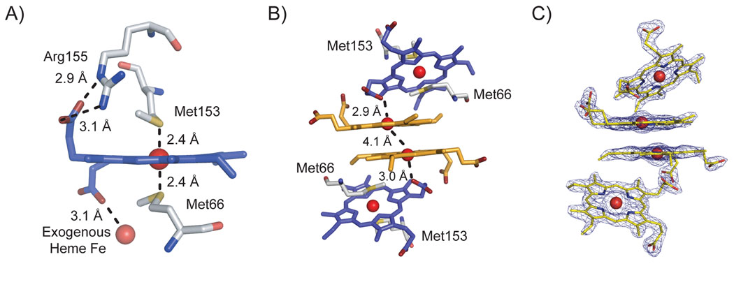
Surface proteins Shr, Shp, and the ATP-binding cassette (ABC) transporter HtsABC are believed to make up the machinery for heme uptake in Streptococcus pyogenes. Shp transfers its heme to HtsA, the lipoprotein component of HtsABC, providing the only experimentally demonstrated example of direct heme transfer from a surface protein to an ABC transporter in Gram-positive bacteria. To understand the structural basis of heme transfer in this system, the heme-binding domain of Shp (Shp(180)) was crystallized, and its structure determined to a resolution of 2.1 A. Shp(180) exhibits an immunoglobulin-like beta-sandwich fold that has been recently found in other pathogenic bacterial cell surface heme-binding proteins, suggesting that the mechanisms of heme acquisition are conserved. Shp shows minimal amino acid sequence identity to these heme-binding proteins and the structure of Shp(180) reveals a unique heme-iron coordination with the axial ligands being two methionine residues from the same Shp molecule. A negative electrostatic surface of protein structure surrounding the heme pocket may serve as a docking interface for heme transfer from the more basic outer cell wall heme receptor protein Shr. The crystal structure of Shp(180) reveals two exogenous, weakly bound hemins, which form a large interface between the two Shp(180) molecules in the asymmetric unit. These "extra" hemins form a stacked pair with a structure similar to that observed previously for free hemin dimers in aqueous solution. The propionates of the protein-bound heme coordinate to the iron atoms of the exogenous hemin dimer, contributing to the stability of the protein interface. Gel filtration and analytical ultracentrifugation studies indicate that both full-length Shp and Shp(180) are monomeric in dilute aqueous solution.


Figures

Figure 1
Figure 1. Shp180 structure
The stereo image of Shp180 in cartoon representation. The hemin is shown in the ball and stick model. The termini and the β-sheets are labeled.
Figure 2
Figure 2. Bis-methionyl coordination in Shp180 and hemin stacking in the crystallization interface
A) Coordination of the protein-bound heme by Met66 and Met153 in Shp180. Distances between the sulfur atom (in yellow) and heme iron (sphere) are listed. Hydrogen bond distances between Arg155 and a propionate are shown and the coordination distance between the propionate and the exogenous heme iron is shown. B) The figure depicts the crystallographic hemin packing between the bound hemes (blue) and the exogenous hemins (orange). Distances between the bound propionate oxygen (red) and the exogenous hemin iron atom (sphere) and the distance between the two exogenous hemin irons are shown. C) Electron density of the protein-bound and exogenous hemins at 1.5σ using the orientation from B). The hemins are displayed in yellow.
Figure 3
Figure 3. Spectroscopic, gel-filtration, and ultracentrifugation analysis of Shp180 and Shp
A) Spectroscopic scan of oxidized Shp180 in solution (Oxi Shp180) and crushed crystals in mother liquor (Crystallized Shp180). An additional scan of the Shp180 crystals corrected for Rayleigh scattering is shown (Corrected Crystallized Shp180). B) Elution of oxidized (Oxi) and reduced (Red) Shp180 and Shp. Molecular weights are listed next to their respective plots. The standard curve used to calculate the molecular weights and correlation coefficient is shown in the upper right. C) Molecular weight distribution results obtained from sedimentation velocity experiments with oxidized Shp180 (blue) and Shp (red), as described in Materials and Methods. Molecular weights peaks are labeled.
Figure 4
Figure 4. Structural comparisons of Shp180 and IsdC proteins
A) Stereo image of the C. trace between Shp180 (blue) and IsdC (red, PDB ID 2O6P). The arrow points to differences between the longer alpha helix Shp180 where Met66 coordinates one side of the heme and the shorter 310-helical lip of IsdC. B) & C) Heme pockets of Shp180 (B) and IsdC (C). The proteins are shown in blue, the residues that coordinate to the heme are labeled and shown in yellow, and the hemes are in gray. D) & E) Electrostatic surface potential of the Shp180 (D) and IsdC (E) proteins. Electrostatic surface potentials were calculated (keV) in the presence of 140 mM monovalent salt concentration at 310 K. The hemins are shown in yellow for both Shp180 and IsdC.

Similar articles

Cited by

References

    1. Mazmanian SK, Skaar EP, Gaspar AH, Humayun M, Gornicki P, Jelenska J, Joachmiak A, Missiakas DM, Schneewind O. Passage of heme-iron across the envelope of Staphylococcus aureus. Science. 2003;299:906–909. - PubMed
    1. Lei B, Smoot LM, Menning HM, Voyich JM, Kala SV, Deleo FR, Reid SD, Musser JM. Identification and characterization of a novel heme-associated cell surface protein made by Streptococcus pyogenes. Infect Immun. 2002;70:4494–4500. - PMC - PubMed
    1. Field LH, Headley VL, Payne SM, Berry LJ. Influence of iron on growth, morphology, outer membrane protein composition, and synthesis of siderophores in Campylobacter jejuni. Infect Immun. 1986;54:126–132. - PMC - PubMed
    1. Wooldridge KG, Williams PH. Iron uptake mechanisms of pathogenic bacteria. FEMS Microbiol Rev. 1993;12:325–348. - PubMed
    1. Weinberg ED. Iron and infection. Microbiol Rev. 1978;42:45–66. - PMC - PubMed

Publication types

Associated data

LinkOut - more resources