

Maintaining transparency: a review of the developmental physiology and pathophysiology of two avascular tissues
- PMID: 17920963
- PMCID: PMC2276117
- DOI: 10.1016/j.semcdb.2007.08.014
Maintaining transparency: a review of the developmental physiology and pathophysiology of two avascular tissues
Abstract
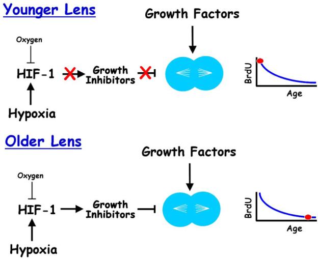
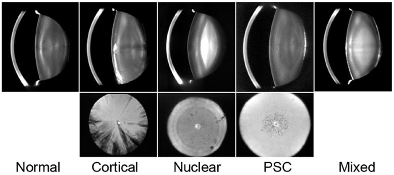
The lens and cornea are transparent and usually avascular. Controlling nutrient supply while maintaining transparency is a physiological challenge for both tissues. During sleep and with contact lens wear the endothelial layer of the cornea may become hypoxic, compromising its ability to maintain corneal transparency. The mechanism responsible for establishing the avascular nature of the corneal stroma is unknown. In several pathological conditions, the stroma can be invaded by abnormal, leaky vessels, leading to opacification. Several molecules that are likely to help maintain the avascular nature of the corneal stroma have been identified, although their relative contributions remain to be demonstrated. The mammalian lens is surrounded by capillaries early in life. After the fetal vasculature regresses, the lens resides in a hypoxic environment. Hypoxia is likely to be required to maintain lens transparency. The vitreous body may help to maintain the low oxygen level around the lens. The hypothesis is presented that many aspects of the aging of the lens, including increased hardening, loss of accommodation (presbyopia), and opacification of the lens nucleus, are caused by exposure to oxygen. Testing this hypothesis may lead to prevention for nuclear cataract and insight into the mechanisms of lens aging. Although they are both transparent, corneal pathology is associated with an insufficient supply of oxygen, while lens pathology may involve excessive exposure to oxygen.
Figures
Similar articles
-
Hypoxia adaptation in the cornea: Current animal models and underlying mechanisms.Animal Model Exp Med. 2021 Nov 28;4(4):300-310. doi: 10.1002/ame2.12192. eCollection 2021 Dec. Animal Model Exp Med. 2021. PMID: 34977481 Free PMC article. Review.
-
A refined model on flow and oxygen consumption in the human cornea depending on the oxygen tension at the interface cornea/post lens tear film during contact lens wear.J Optom. 2022 Apr-Jun;15(2):160-174. doi: 10.1016/j.optom.2020.12.002. Epub 2021 Feb 13. J Optom. 2022. PMID: 33589396 Free PMC article.
-
Lens cells: more than meets the eye.Int J Biochem Cell Biol. 2007;39(10):1754-9. doi: 10.1016/j.biocel.2007.06.021. Epub 2007 Jul 12. Int J Biochem Cell Biol. 2007. PMID: 17707680 Review.
-
Review of the Experimental Background and Implementation of Computational Models of the Ocular Lens Microcirculation.IEEE Rev Biomed Eng. 2016;9:163-76. doi: 10.1109/RBME.2016.2583404. Epub 2016 Jun 21. IEEE Rev Biomed Eng. 2016. PMID: 27337724 Review.
-
The physiological optics of the lens.Prog Retin Eye Res. 2017 Jan;56:e1-e24. doi: 10.1016/j.preteyeres.2016.09.002. Epub 2016 Sep 14. Prog Retin Eye Res. 2017. PMID: 27639549 Review.
Cited by
-
Formation of persistent hyperplastic primary vitreous in ephrin-A5-/- mice.Invest Ophthalmol Vis Sci. 2014 Mar 19;55(3):1594-606. doi: 10.1167/iovs.13-12706. Invest Ophthalmol Vis Sci. 2014. PMID: 24550361 Free PMC article.
-
Corneal neovascularization: updates on pathophysiology, investigations & management.Rom J Ophthalmol. 2019 Jan-Mar;63(1):15-22. Rom J Ophthalmol. 2019. PMID: 31198893 Free PMC article. Review.
-
Hypoxia-regulated activity of PKCepsilon in the lens.Invest Ophthalmol Vis Sci. 2009 Mar;50(3):1271-82. doi: 10.1167/iovs.08-2599. Epub 2008 Nov 7. Invest Ophthalmol Vis Sci. 2009. PMID: 18997087 Free PMC article.
-
Aqueous Humor Circulation in the Era of Minimally Invasive Surgery for Glaucoma.Ann Biomed Eng. 2024 Apr;52(4):898-907. doi: 10.1007/s10439-023-03427-3. Epub 2023 Dec 28. Ann Biomed Eng. 2024. PMID: 38155316
-
Effects of VEGF Inhibitor Conbercept on Corneal Neovascularization Following Penetrating Keratoplasty in Rabbit Model.Clin Ophthalmol. 2020 Jul 31;14:2185-2193. doi: 10.2147/OPTH.S260302. eCollection 2020. Clin Ophthalmol. 2020. PMID: 32801629 Free PMC article.
References
-
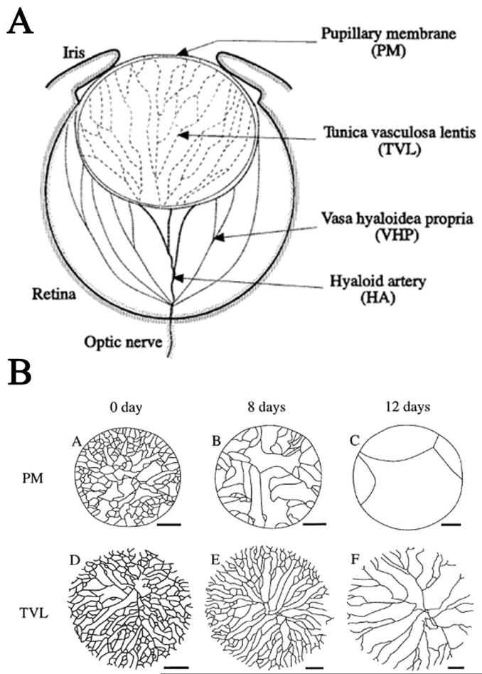
- Ito M, Yoshioka M. Regression of the hyaloid vessels and pupillary membrane of the mouse. Anat Embryol (Berl) 1999;200(4):403–11. - PubMed
-
- Mitchell CA, Risau W, Drexler HC. Regression of vessels in the tunica vasculosa lentis is initiated by coordinated endothelial apoptosis: a role for vascular endothelial growth factor as a survival factor for endothelium. Dev Dyn. 1998;213(3):322–33. - PubMed
-
- Shui Y-B, et al. Vascular Endothelial Growth Factor Expression and Signaling in the Lens. Invest Ophthalmol Vis Sci. 2003;44(9):3911–3919. - PubMed
-
- Gerber HP, et al. VEGF is required for growth and survival in neonatal mice. Development. 1999;126(6):1149–59. - PubMed
Publication types
MeSH terms
Substances
Grants and funding
LinkOut - more resources
Full Text Sources
Medical
