Review
. 2007 Nov;26(6):649-73.
doi: 10.1016/j.preteyeres.2007.07.004. Epub 2007 Aug 11.

Iron homeostasis and toxicity in retinal degeneration

Affiliations
Review

Iron homeostasis and toxicity in retinal degeneration

Xining He et al. Prog Retin Eye Res. 2007 Nov.

Abstract

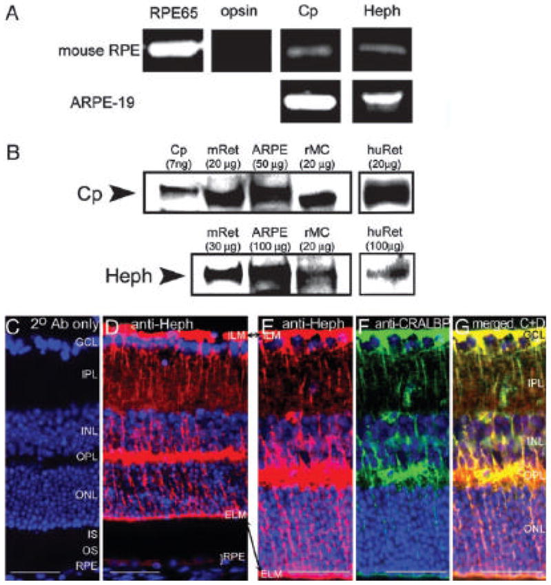
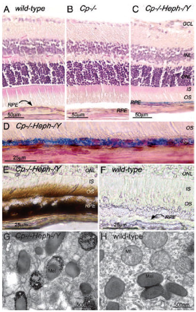
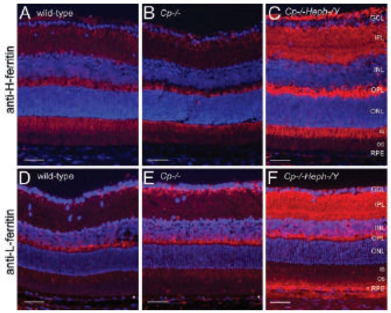
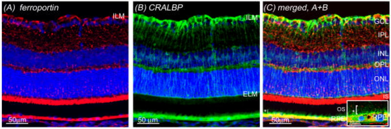
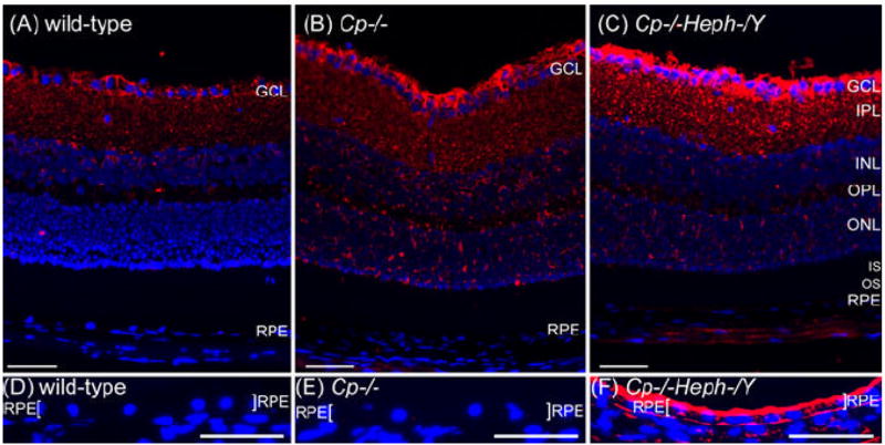
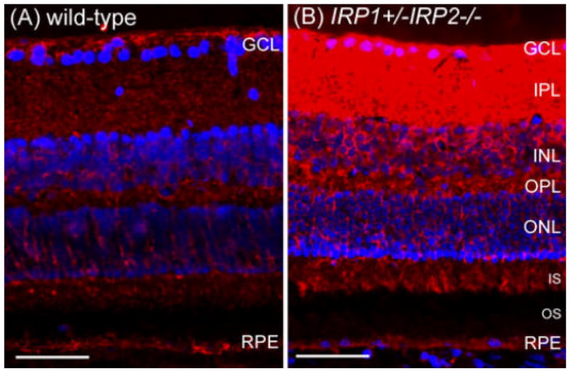
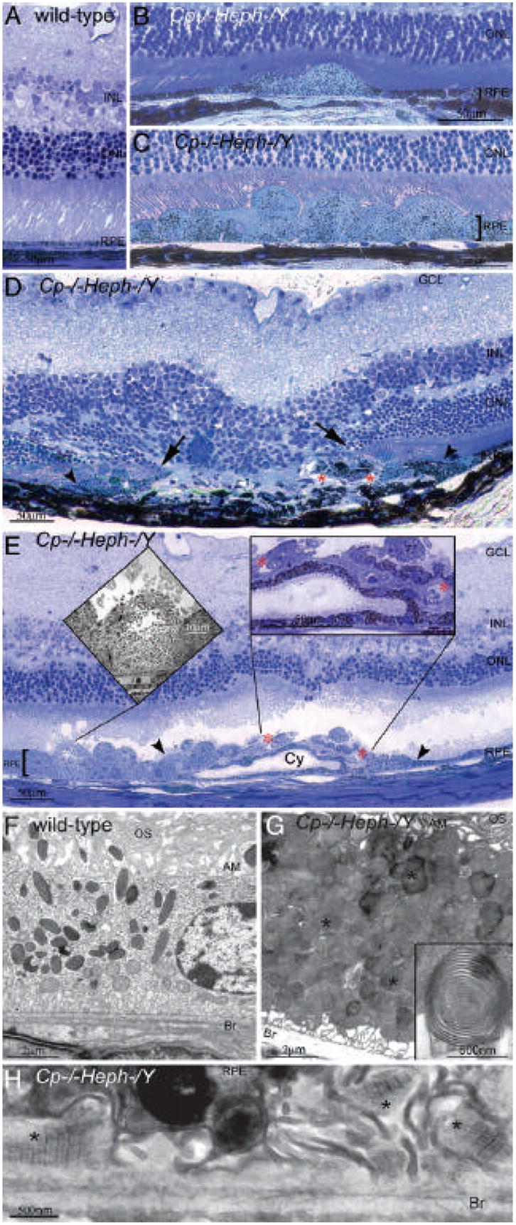
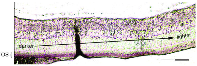
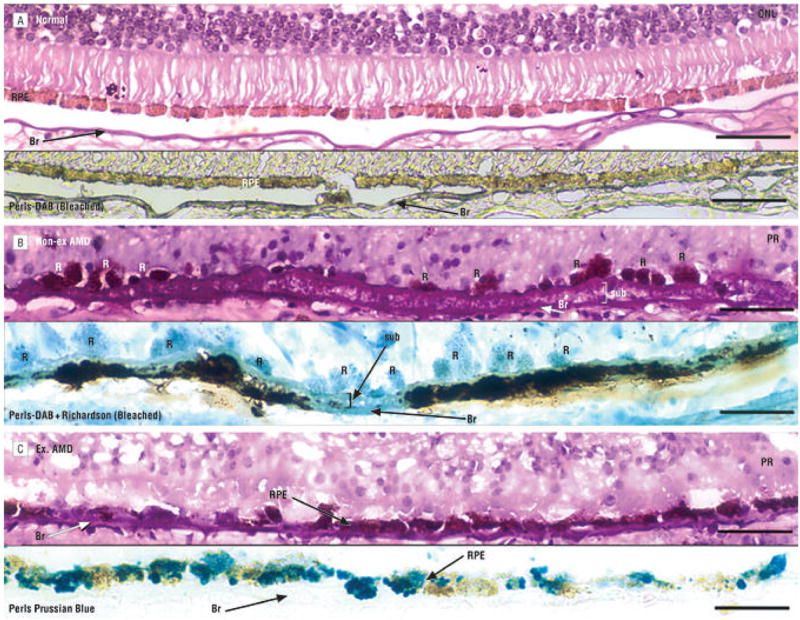
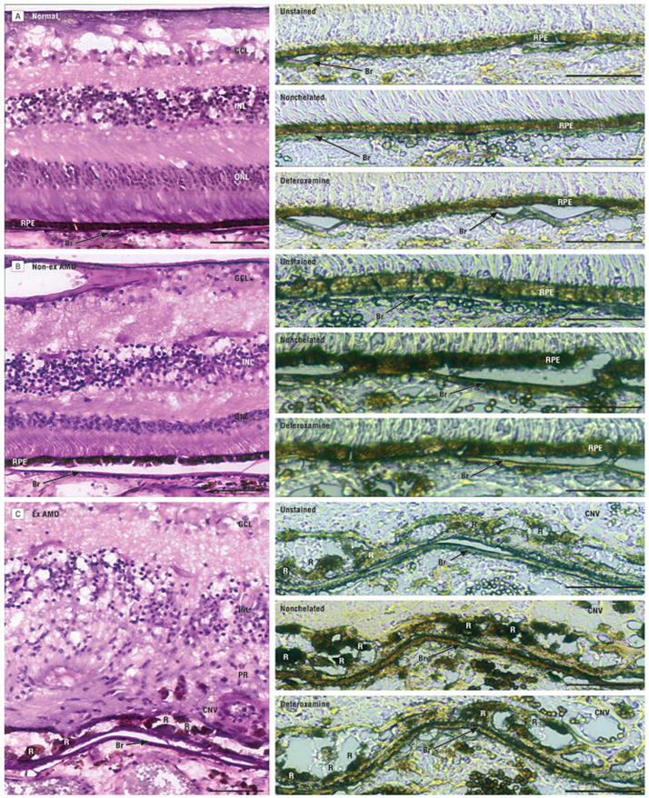
Iron is essential for many metabolic processes but can also cause damage. As a potent generator of hydroxyl radical, the most reactive of the free radicals, iron can cause considerable oxidative stress. Since iron is absorbed through diet but not excreted except through menstruation, total body iron levels buildup with age. Macular iron levels increase with age, in both men and women. This iron has the potential to contribute to retinal degeneration. Here we present an overview of the evidence suggesting that iron may contribute to retinal degenerations. Intraocular iron foreign bodies cause retinal degeneration. Retinal iron buildup resulting from hereditary iron homeostasis disorders aceruloplasminemia, Friedreich's ataxia, and panthothenate kinase-associated neurodegeneration cause retinal degeneration. Mice with targeted mutation of the iron exporter ceruloplasmin have age-dependent retinal iron overload and a resulting retinal degeneration with features of age-related macular degeneration (AMD). Post mortem retinas from patients with AMD have more iron and the iron carrier transferrin than age-matched controls. Over the past 10 years much has been learned about the intricate network of proteins involved in iron handling. Many of these, including transferrin, transferrin receptor, divalent metal transporter-1, ferritin, ferroportin, ceruloplasmin, hephaestin, iron-regulatory protein, and histocompatibility leukocyte antigen class I-like protein involved in iron homeostasis (HFE) have been found in the retina. Some of these proteins have been found in the cornea and lens as well. Levels of the iron carrier transferrin are high in the aqueous and vitreous humors. The functions of these proteins in other tissues, combined with studies on cultured ocular tissues, genetically engineered mice, and eye exams on patients with hereditary iron diseases provide clues regarding their ocular functions. Iron may play a role in a broad range of ocular diseases, including glaucoma, cataract, AMD, and conditions causing intraocular hemorrhage. While iron deficiency must be prevented, the therapeutic potential of limiting iron-induced ocular oxidative damage is high. Systemic, local, or topical iron chelation with an expanding repertoire of drugs has clinical potential.


Figures

Fig. 1
Fig. 1
BALB/c WT immunostained with DMT1 (red). Strong DMT1 label can be seen in the rod bipolar cell bodies, rod bipolar cell termini, horizontal cell bodies, and photoreceptor inner segments. Scale bar represents 50 μm.
Fig. 2
Fig. 2
H- and L- ferritin are present in normal retina in rod bipolar cells. Normal retina labeled for H- ferritin (red; A) and L- ferritin (red; D). Nuclei are counterstained with DAPI (blue). Both ferritin subunits have punctate label in the inner IPL, the region adjacent to the ganglion cell layer (GCL). This punctate label is characteristic of the synaptic terminals of rod bipolar cells, labeled with anti-PKCa (green), which colocalizes (gold) with both H-ferritin (BC) and L- ferritin (EF). This colocalization is best seen in high power images of these synaptic terminals which are positive for both PKCa and L- ferritin (arrowheads, inset F).
Fig. 3
Fig. 3
IRP1+/IRP2/− retinas have increased L-ferritin. Fluorescence photomicrographs of age matched wild type and IRP1+/IRP2/− retinas immunolabeled for L-ferritin (red), counterstained for nuclei with DAPI (blue), and imaged under identical exposure parameters. L- ferritin is increased in the IRP1+/IRP2/− inner retina, including bipolar cell synapses in the IPL, as well as in the OPL and inner segments. Scale bars represent 50 μm.
Fig. 4
Fig. 4
Mitochondrial ferritin is present in normal inner segments and is increased in Cp/− and Cp/Heph-/Y retinas. Fluorescence photomicrographs of age matched wild type, Cp/−, and Cp/Heph-/Y retinas immunolabeled for mitochondrial ferritin (AC) and imaged under identical exposure parameters. Cp/− retinas (B) have increased mitochondrial ferritin (red) in the inner segments (IS) compared to wild type (A), and Cp/Heph-/Y retinas (C) have further increased mitochondrial ferritin. The label in the Cp/− inner segments (E) excludes the inner segment myoid (arrowhead, DF) and colocalizes with a mitochondria-specific antibody (green) to the inner segment ellipsoid (D,F), suggesting mitochondrial localization of MtF. Nuclei are labeled with DAPI (blue). Scale bars represent 50 μm.
Fig. 5
Fig. 5
Cp and Heph mRNA and proteins are present in normal RPE and retina within Müller cells. (A) Bands from ethidium bromide-stained agarose gels corresponding to RT-PCR amplification products of the indicated mRNAs from dissected C57BL/6 murine RPE and cultured ARPE-19 cells. In all cases, the amplification product was of the expected size. (B) Western analysis of Cp and Heph in dissected C57BL/6 murine retinas (mRet) human retinas (huRet), cultured ARPE-19 cells, and cultured rMC-1 cells. Purified human Cp (labeled Cp) was used as control. (C) Control BALB/c retina labeled with a Cy3-conjugated donkey anti-rabbit secondary antibody. (D) Normal BALB/c retina immunolabeled for Heph. (EG) Normal BALB/c retina double- labeled with anti-Heph and anti-CRALBP, a marker for Muller glia. A single red exposure reveals anti-Heph immunoreactivity (E). A single green exposure shows anti-CRALBP immunore-activity (F). Double exposure shows yellow colocalization of Heph and CRALBP (G). Scale bars: 50 μm.
Fig. 6
Fig. 6
Adult (6- month-old) Cp/Heph–/Y RPE and photoreceptors accumulate iron. (AC) 6-month-old WT (A), Cp/− (B), and Cp/Heph–/Y (C) retinas Perls’ stained for iron (blue) and counterstained with hematoxylin/eosin. (D) High magnification of Prussian blue Perls’ label in 6-month-old Cp/Heph–/Y RPE. (E and F) Light photomicrographs of 6- month-old Cp/Heph–/Y (E) and WT (F) retinas after DAB enhancement (brown) of Perls’ stain. (GH) Electron micrographs of RPE from 6- month-old Cp/Heph–/Y (G) and WT (H) eyes. Only the Cp/Heph–/Y RPE (G) contains electrondense vesicles (*) sometimes fused with melanosomes.
Fig. 7
Fig. 7
Cp/Heph–/Y retinas have increased ferritin. Fluorescence photomicrographs of 6-month-old WT, Cp/−, and Cp/Heph–/Y retinas immunolabeled for H-ferritin (AC) and L- ferritin (DF) and imaged by using identical exposure parameters. Scale bars: 50 μm.
Fig. 8
Fig. 8
Ferroportin is present in normal retina at high levels in the Müller cell endfeet and RPE. Normal BALB/c retina immunolabeled with omission of primary antibody (A) localizes to Müller endfeet near the ILM, in photoreceptor inner segments (IS), and in the RPE, as demonstrated by the co-label with CRALBP (green), a marker for Müller cells and RPE (B,C). Ferroportin in the RPE excludes its apical microvilli, the green only label indicated with an asterisk (“*”) in C and in the inset of C, which shows a high power image of RPE co- labeled with ferroportin and CRALBP. Ferroportin is also present in a punctate pattern throughout the inner retina.
Fig. 9
Fig. 9
Cp/Heph-/Y retinas have increased ferroportin. Fluorescence photomicrographs of age matched wild type, Cp/−, and Cp/Heph-/Y retinas immunolabeled for ferroportin (red) and imaged under identical exposure parameters. Nuclei were counterstained with DAPI (blue). Differences between wild type (A) and Cp/− (B) were subtle, but there is a clear increase in ferroportin label in the Müller endfeet of the Cp/Heph-/Y retina (C). The punctate ferroportin label throughout the IPL is also increased in the Cp/Heph-/Y retina. In order to optimally detect differences in ferroportin in the pigmented RPE, it was necessary to pre-bleach sections (DF). Equivalently bleached retinas immunolabeled with ferroportin and imaged with equivalent exposure parameters reveals a robust increase in the Cp/Heph-/Y RPE of ferroportin, which localized to both the apical and basolateral surfaces of the RPE (demarcated with brackets). Scale bars represent 50 μm.
Fig. 10
Fig. 10
IRP1+/IRP2/− retinas have increased ferroportin. Fluorescence photomicrographs of age matched wild type and IRP1+/IRP2/− retinas immunolabeled for ferroportin (red), counterstained for nuclei with DAPI (blue), and imaged under identical exposure parameters. Ferroportin label is increased in the IRP1+/IRP2/− inner retina including Müller endfeet. Scale bars represent 50 μm.
Fig. 11
Fig. 11
Nine- month-old Cp/Heph–/Y mice have retinal degeneration. (A) Light photomicrograph of WT retina. (B and C) Cp/Heph–/Y retina has focal patches of hypertrophic RPE cells in some areas (B) and confluent hypertrophic RP E cells in other areas (C). (D) In an area of RPE hyperplasia (demarcated by arrowheads), Cp/Heph–/Y retinas have local photoreceptor degeneration [demarcated by arrows in the outer nuclear layer (ONL)] and subretinal neovascularization (red *). (E) In an area of hypertrophic, hyperplastic (area demarcated by arrowheads) RPE cells, a necrotic RPE cell also observed by electron microscopy (Left Inset) is present. Within the area of RPE hyperplasia, there is local photoreceptor thinning and subretinal neovascularization (red *) visible as small vessels containing erythrocytes (Right Inset). The hyperplastic RPE have formed a localized cyst (Cy). (F) Electron micrograph of WT RPE. Br, Bruch’s membrane; AM, apical microvilli; OS, photoreceptor outer segments. (G) Electron micrograph of Cp/Heph–/Y RPE overloaded with phagosomes and lysosomes containing photoreceptor outer segments at various stages of digestion. Some of these lysosomes (*) contained multilamellar structures characteristic of outer segment memb ranes (Inset). (H) Electron micrograph of Cp/Heph–/Y deposits between RPE and Bruch’s membrane containing wide-spaced collagen (*). (Scale bars: AE, 50 μm; F and G, 2 μm; H, 500 nm.)
Fig. 12
Fig. 12
Perls’ Prussian blue staining of rabbit eye section injected with autologous blood into the subretinal space. Label can be seen in photoreceptor outer segments with the strongest label at the site of subretinal injection with fading further away from the site of blood accumulation. Scale bar represents 50 μm.
Fig. 13
Fig. 13
Photomicrographs of increased Perls-positive iron in age-related macular degeneration (AMD)–affected retinas compared with healthy retinas. Insets show Perls Prussian blue–stained retinal pigment epithelium (RPE) and Bruch’s membrane (Br) in the same region as the larger figure of an adjacent periodic acid–Schiff (PAS)-hematoxylin–stained section. The PAS-hematoxylin–stained sections were unbleached, while sections shown in the insets were bleached as indicated. ONL indicates outer nuclear layer; PR, disorganized photoreceptors. A, Healthy macula has no Perls-3,3′-diaminobenzidine (DAB) stain after bleaching (inset). The remaining melanin in the RPE is identical to the DAB–hydrogen peroxide–only control (not shown), consisting of incompletely bleached endogenous RPE and not Perls-DAB signal. B, Nonexudative (Non-ex) AMD–affected, geographic atrophic macula with severe photoreceptor loss, RPE atrophy, and sub-RPE deposits. Bruch’s membrane and sub-RPE deposits (sub) are positive for iron (inset). The RPE cells are unpigmented because endogenous RPE melanin has been bleached, but the Richardson (methylene blue/azure II) counterstain identifies RPE cells (Rs). C, Exudative AMD–affected retina adjacent to a fibrovascular scar in the macula. There is severe photoreceptor loss with RPE atrophy and thickened BR. The RPE contains iron detectable by the Perls Prussian blue stain without DAB enhancement (inset). Scale bars indicate 50 μm.
Fig. 14
Fig. 14
Photomicrographs of chelatable and nonchelatable iron accumulation in unbleached age-related macular degeneration (AMD)–affected maculas but not healthy maculas. Large panels show maculas stained with peroidic acid–Schiff (PAS)-hematoxylin, and insets show adjacent sections unstained or stained with Perls-3,3′-diaminobenzidine (DAB) with or without prior deferoxamine chelation. ONL indicates outer nuclear layer; INL, inner nuclear layer; GCL, ganglion cell layer; CNV, choroidal neovascularization. Note that red blood cells are positive for Perls-DAB stain, as expected. A, Healthy macula with minimal Perls-DAB signal observed in the retinal pigment epithelium (RPE) or Bruch’s membrane (Br) of either the chelated or nonchelated sections, which appear only slightly darker than the unstained section. B, Nonexudative AMD–affected macula with signs of early AMD including drusen (not shown), slightly thickened Br, and mild photoreceptor (PR) loss without RPE atrophy. The RPE and Br in the nonchelated section are darker than in the unstained section, owing to increased Perls-DAB stain corresponding to total iron accumulation. Some of the increased iron is chelatable, demonstrated by decreased Perls-DAB stain in the deferoxamine-chelated compared with the nonchelated section. C, Exudative AMD–affected macula with marked PR loss, fibrovascular scarring, sub-RPE deposits, and thickened Br. Choroidal neovascularization is present, with the same vessel in all sections, and few RPE cells (Rs) remain. Iron is increased in the RPE cells and Br and consists of both chelatable and nonchelatable forms. Scale bars indicate 50 μm.

Similar articles

Cited by

References

    1. Abboud S, Haile DJ. A novel mammalian iron-regulated protein involved in intracellular iron metabolism. J Biol Chem. 2000;275:19906–19912. - PubMed
    1. Aisen P, Enns C, Wessling-Resnick M. Chemistry and biology of eukaryotic iron metabolism. Int J Biochem Cell Biol. 2001;33:940–959. - PubMed
    1. Anderson LJ, Wonke B, Prescott E, Holden S, Walker JM, Pennell DJ. Comparison of effects of oral deferiprone and subcutaneous desferrioxamine on myocardial iron concentrations and ventricular function in beta-thalassaemia. Lancet. 2002;360:516–520. - PubMed
    1. Aouad F, Florence A, Zhang Y, Collins F, Henry C, Ward RJ, Crichton R. Evaluation of new iron chelators and their therapeutic potential. Inorg Chim Acta. 2002;339:470–480.
    1. AREDS. A randomized, placebo-controlled, clinical trial of high-dose supplementation with vitamins C and E, beta carotene, and zinc for age-related macular degeneration and vision loss: AREDS report no. 8. Arch Ophthalmol. 2001;119:1417–1436. - PMC - PubMed

Publication types