

Review
. 2007 Oct 1;7 Spec No A(Special issue A):S1-14.
doi: 10.1102/1470-7330.2007.9084.
Distinguishing benign from malignant liver tumours
Affiliations
- PMID: 17921080
- PMCID: PMC2727979
- DOI: 10.1102/1470-7330.2007.9084
Item in Clipboard
Review
Distinguishing benign from malignant liver tumours
Cancer Imaging.
.
Abstract
Liver masses are very common and most are benign. It is therefore important to avoid unnecessary interventions for benign lesions, while at the same time ensuring accurate diagnosis of hepatic malignancies. Many cancer patients, like the general population, have incidental benign liver lesions. In planning treatment for cancer patients, it is critical to avoid inappropriate treatment decisions based on misdiagnosis of a benign lesion as a metastasis or primary liver malignancy. This article describes the salient imaging features of the common benign liver masses and outlines a general approach to distinguishing between benign and malignant hepatic lesions.
Figures
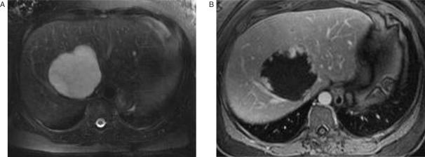
Hemangioma. Unenhanced T2-weighted MR image (A) shows a large hyperintense hepatic mass. Gadolinium enhanced T1-weighted image (B) demonstrates the characteristic nodular enhancement at the periphery of the lesion. Reprinted with permission from Lee et al.[132].
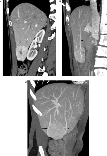
Focal nodular hyperplasia. Contrast-enhanced, arterial phase sagittal CT image (A) shows a well-defined homogeneously enhancing hypervascular mass at the inferior edge of the right lobe of the liver. Note the non-enhancing central scar. A sagittal maximum intensity projection (MIP) image (B) demonstrates early drainage of the mass into a large hepatic vein (arrowheads). An off axis coronal MIP image (C) demonstrates that the mass has two large draining veins (arrows).
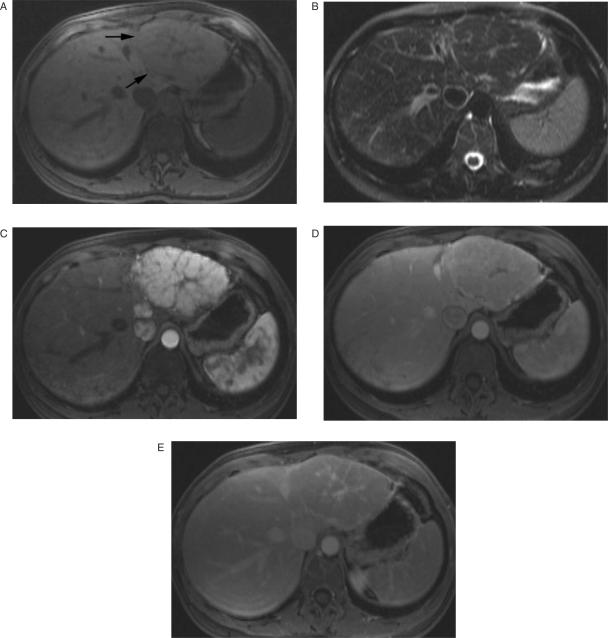
Focal nodular hyperplasia. Unenhanced T1-weighted MR image (A) shows a nearly isointense hepatic mass (arrows) that contains a hypointense central scar. On a T2-weighted image (B) the mass is isointense, & the scar is hyperintense. Arterial phase gadolinium-enhanced image (C) demonstrates marked enhancement of the mass, except for the central scar and fibrous septa radiating from the scar. Portal venous phase image (D) shows rapid contrast enhancement washout of the lesion, which is now isointense with liver parenchyma. On a delayed postcontrast image (E) the mass remains isointense, but the central scar now is hyperintense.
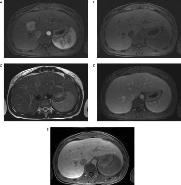
Focal nodular hyperplasia. Arterial phase gadolinium-BOPTA enhanced image (A) demonstrates an intensely enhancing mass in segment 8 of the liver. Note the non-enhancing linear central scar. The mass is isointense on unenhanced T1-weighted (B) and T2-weighted (C) images. Portal venous phase image (D) shows rapid contrast enhancement washout of the lesion, which is now isointense with liver parenchyma. One hour delay image (E) demonstrates persistent enhancement of the mass, which is now hyperintense relative to the normal hepatic parenchyma.
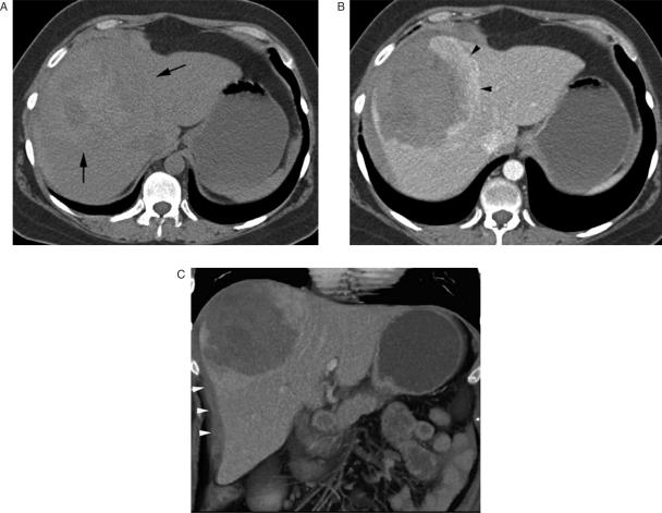
Hepatocellular carcinoma. Arterial phase contrast-enhanced transaxial (A) and coronal (B) CT images shows a large exophytic hypervascular hepatic mass (arrow) that contains a large central scar. Arterial phase gadolinium-BOPTA enhanced MR image (C) shows similar findings. One hour delay image (D) after gadolinium-BOPTA administration demonstrates lack of enhancement of the mass. Note enhancement of the normal hepatic parenchyma (arrows).
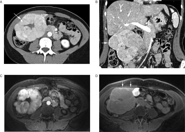
Ruptured hepatocellular adenoma. Precontrast CT image (A) shows a large heterogeneous mass (arrows) near the dome of the liver. Central areas of hyperattenuation represent hemorrhage. Note the high attenuation perihepatic blood. Contrast-enhanced image (B) shows enhancement of the peripheral intact portion of the mass (black arrowheads). The hemorrhagic portion of the mass does not enhance. Note loss of integrity of the liver capsule anterolaterally. Coronal volume rendered image (C) shows the peripherally enhancing mass, ruptured liver capsule, and perihepatic blood (white arrowheads). Reprinted with permission from Lee et al.[132].
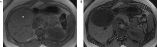
Hepatocellular adenoma. In-phase T1-weighted spoiled gradient-echo MR image (A) shows a large isointense hepatic mass (M). Out-of-phase image (B) shows diffuse decrease in signal intensity within the mass due to the presence of intracellular lipid. Reprinted with permission from Lee et al.[132].
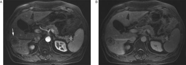
Metastatic colon carcinoma. T1-weighted arterial phase gadolinium-enhanced MR image (A) shows a heterogeneously enhancing mass (arrow) in the right lobe of the liver. On an equilibrium phase image (B) the periphery of the lesion, which demonstrated enhancement during the arterial phase, is now less intense than the center of the lesion. This phenomenon is termed ‘peripheral washout’.
Similar articles
-
Imaging of paediatric liver tumours with pathological correlation.Clin Radiol. 2009 Oct;64(10):1015-25. doi: 10.1016/j.crad.2009.04.014. Epub 2009 Jul 17. Clin Radiol. 2009. PMID: 19748008 Review.
-
[Benign focal hepatic lesions].Radiologe. 2011 Aug;51(8):688-96. doi: 10.1007/s00117-010-2127-1. Radiologe. 2011. PMID: 21809146 Review. German.
-
[Surgical treatment of benign liver tumours--indications and results].Zentralbl Chir. 2009 Apr;134(2):141-4. doi: 10.1055/s-2008-1076871. Epub 2009 Apr 20. Zentralbl Chir. 2009. PMID: 19382044 German.
-
Review article: the evaluation of solitary liver masses.Aliment Pharmacol Ther. 2008 Oct 15;28(8):953-65. doi: 10.1111/j.1365-2036.2008.03805.x. Epub 2008 Jul 16. Aliment Pharmacol Ther. 2008. PMID: 18643922 Review.
-
ACG Clinical Guideline: Focal Liver Lesions.Am J Gastroenterol. 2024 Jul 1;119(7):1235-1271. doi: 10.14309/ajg.0000000000002857. Epub 2024 Jan 26. Am J Gastroenterol. 2024. PMID: 38958301
Cited by
-
Differential diagnoses and diagnostic troubleshooting of upper abdominal masses.Gastroenterol Hepatol (N Y). 2013 Jun;9(6):399-400. Gastroenterol Hepatol (N Y). 2013. PMID: 23935549 Free PMC article. No abstract available.
-
[Benign liver tumors : Diagnostics and treatment].Chirurg. 2019 Dec;90(12):1033-1046. doi: 10.1007/s00104-019-01068-8. Chirurg. 2019. PMID: 31784769 German.
-
[Benign liver tumors : Diagnostics and treatment].Pathologe. 2020 Mar;41(2):181-192. doi: 10.1007/s00292-020-00758-z. Pathologe. 2020. PMID: 32103337 German.
-
Giant Cavernous Hemangioma of the Liver in a Patient with Autosomal Dominant Polycystic Kidney Disease.Am J Case Rep. 2020 Nov 18;21:e927188. doi: 10.12659/AJCR.927188. Am J Case Rep. 2020. PMID: 33206631 Free PMC article.
-
Imaging findings of mimickers of hepatocellular carcinoma.Clin Mol Hepatol. 2015 Dec;21(4):326-43. doi: 10.3350/cmh.2015.21.4.326. Epub 2015 Dec 24. Clin Mol Hepatol. 2015. PMID: 26770920 Free PMC article. Review.
References
-
- Wright TL, Venook AP, Millward-Sadler GH. GH M-S. Hepatic tumours. In: Millward-Sadler GH, Wright R, Arthur MJP, editors. Wright's liver and biliary disease. 3rd. Vol. 2. Philadelphia: WB Saunders; 1992. pp. 1079–21.
-
- Gibney RG, Hendin AP, Cooperberg PL. Sonographically detected hepatic hemangiomas: absence of change over time. AJR Am J Roentgenol. 1987;149:953–7. - PubMed
-
- Mungovan JA, Cronan JJ, Vacarro J. Hepatic cavernous hemangiomas: lack of enlargement over time. Radiology. 1994;191:111–13. - PubMed
-
- Nghiem HV, Bogost GA, Ryan JA, Lund P, Freeny PC, Rice KM. Cavernous hemangiomas of the liver: enlargement over time. AJR Am J Roentgenol. 1997;169:137–40. - PubMed
Publication types
MeSH terms
LinkOut - more resources
Full Text Sources
Medical