

PET/CT and cross sectional imaging of gynecologic malignancy
- PMID: 17921092
- PMCID: PMC2727972
- DOI: 10.1102/1470-7330.2007.9015
PET/CT and cross sectional imaging of gynecologic malignancy
Abstract
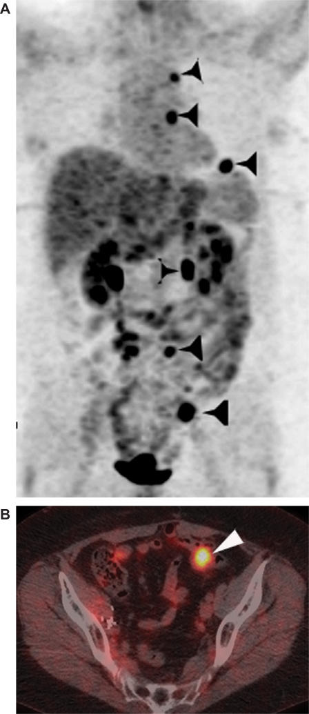
Gynecologic cancers are a common cause of morbidity and mortality in women of all ages. While many gynecologic cancers are staged clinically using the International Federation of Gynecology and Obstetrics (FIGO) staging system, imaging can be a useful adjunct to clinical staging. Cross sectional imaging techniques such as ultrasound (US), computed tomography (CT) and magnetic resonance imaging (MRI) have been used to detect and follow patients with gynecologic cancer. These imaging modalities can show anatomic detail and morphologic changes in the female genitourinary tract to good advantage. Positron emission tomography (PET) differs in that it shows functional information that is not easily obtained by the other cross sectional imaging techniques. The fusion of PET with CT allows anatomic localization of functional abnormalities in the female genital tract and thereby allows the detection of gross disease in many malignant conditions both within and outside the confines of the female pelvis. The utility and limitations of imaging common gynecologic tumors such as cervical, ovarian and endometrial cancer are discussed with particular emphasis on PET/CT imaging.
Figures


Similar articles
-
PET/MRI in Gynecologic Malignancy.Radiol Clin North Am. 2023 Jul;61(4):713-723. doi: 10.1016/j.rcl.2023.02.013. Radiol Clin North Am. 2023. PMID: 37169433 Review.
-
PET/CT in gynecologic malignancies.Radiol Clin North Am. 2013 Sep;51(5):895-911. doi: 10.1016/j.rcl.2013.05.006. Epub 2013 Jul 17. Radiol Clin North Am. 2013. PMID: 24010912 Review.
-
Nuclear Medicine and Molecular Imaging Applications in Gynecologic Malignancies: A Comprehensive Review.Semin Nucl Med. 2024 Mar;54(2):270-292. doi: 10.1053/j.semnuclmed.2024.01.003. Epub 2024 Feb 11. Semin Nucl Med. 2024. PMID: 38342655 Review.
-
PET/MR imaging in gynecologic cancer: tips for differentiating normal gynecologic anatomy and benign pathology versus cancer.Abdom Radiol (NY). 2022 Sep;47(9):3189-3204. doi: 10.1007/s00261-021-03264-9. Epub 2021 Oct 23. Abdom Radiol (NY). 2022. PMID: 34687323 Review.
-
Positron emission tomography-computed tomography imaging for malignancies in women.Radiol Clin North Am. 2013 Nov;51(6):1111-25. doi: 10.1016/j.rcl.2013.07.006. Radiol Clin North Am. 2013. PMID: 24210447 Review.
Cited by
-
Role of F-18 fluorodeoxyglucose positron emission tomography/computed tomography in the detection of recurrence in patients with cervical cancer.Indian J Nucl Med. 2013 Oct;28(4):216-20. doi: 10.4103/0972-3919.121966. Indian J Nucl Med. 2013. PMID: 24379531 Free PMC article.
-
Role of New Functional MRI Techniques in the Diagnosis, Staging, and Followup of Gynecological Cancer: Comparison with PET-CT.Radiol Res Pract. 2012;2012:219546. doi: 10.1155/2012/219546. Epub 2012 Jan 18. Radiol Res Pract. 2012. PMID: 22315683 Free PMC article.
-
Sentinel Lymph Node Biopsy and Complete Lymph Node Dissection for Melanoma.Curr Oncol Rep. 2019 Apr 26;21(6):54. doi: 10.1007/s11912-019-0798-y. Curr Oncol Rep. 2019. PMID: 31028497 Free PMC article. Review.
-
Correlation between histological grade and positron emission tomography parameters in cervical carcinoma.Oncol Lett. 2016 Aug;12(2):1408-1414. doi: 10.3892/ol.2016.4771. Epub 2016 Jun 23. Oncol Lett. 2016. PMID: 27446445 Free PMC article.
-
FDG-PET in gynaecological cancers: recent observations.Eur J Nucl Med Mol Imaging. 2008 Nov;35(11):2133-9. doi: 10.1007/s00259-008-0929-4. Eur J Nucl Med Mol Imaging. 2008. PMID: 18779962 Review. No abstract available.
References
-
- Jemal A, Siegel R, Ward E, Murray T, Xu J, Thun MJ. Cancer statistics 2007. CA Cancer J Clin. 2007;57:43–66. - PubMed
-
- Hricak H, Lacey CG, Sandles LG, et al. Invasive cervical carcinoma: comparison of MR imaging and surgical findings. Radiology. 1988;166:623–31. - PubMed
-
- Subak LL, Hricak H, Powell CB, et al. Cervical carcinoma: computed tomography and magnetic resonance imaging for preoperative staging. Obstet Gynecol. 1995;86:46–50. - PubMed
-
- Matsukuma K, Tsukamoto N, Matsuyama T, Ono M. Nakano H. Pre-operative CT study of lymph nodes in cervical cancer: its correlation with histological findings. Gyncecol Oncol. 1989;33:168–71. - PubMed
-
- Kim SH, Kim SC, Choi BI, Han MC. Uterine cervical carcinoma: evaluation of pelvic lymph node metastatis with MR imaging. Radiology. 1994;190:807–11. - PubMed
Publication types
MeSH terms
Substances
LinkOut - more resources
Full Text Sources
Medical