

Diffusion tensor imaging in patients with essential tremor
- PMID: 17921227
- PMCID: PMC8119092
- DOI: 10.3174/ajnr.A0744
Diffusion tensor imaging in patients with essential tremor
Abstract
Background and purpose: The traditional paradigm has regarded essential tremor (ET) as a benign disorder. However, recent clinical, neuroimaging, and neuropathologic studies suggest that ET may be a progressive neurologic disorder. Based on clinicopathologic findings that cerebellum and its outflow are the key structures in ET and degeneration of gray matter in cerebellum is followed by consequent wallerian degeneration of white matter (WM) fibers, the aim of the present study was to investigate changes in anisotropy in patients with ET.
Materials and methods: Fractional anisotropy (FA) images were generated from DTI data acquired at 1.5T in 10 patients with ET compared with 8 control subjects by using statistical parametric mapping to make voxel-by-voxel comparisons.
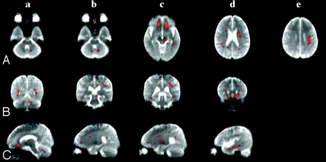
Results: Compared with the control subjects, the patients with ET exhibited significantly reduced FA (P(uncorrected) < .005) in the anterolateral portion of the right pons and decreased FA in the bilateral cerebellum, left retrorubral area of the midbrain, and bilateral deep WM, including the orbitofrontal, lateral frontal, parietal, and temporal WM.
Conclusion: This study demonstrates that structural changes in the WM are extensive in patients with ET, supporting the findings of previous functional neuroimaging and pathologic studies.
Figures
References
-
- Louis ED. Essential tremor. Lancet Neurol 2005;4:100–10 - PubMed
-
- Louis ED, Shungu DC, Chan S, et al. Metabolic abnormality in the cerebellum in patients with essential tremor: a proton magnetic resonance spectroscopic imaging study. Neurosci Lett 2002;333:17–20 - PubMed
-
- Pagan FL, Butman JA, Dambrosia JM, et al. Evaluation of essential tremor with multi-voxel magnetic resonance spectroscopy. Neurology 2003;60:1344–47 - PubMed
-
- Louis ED, Vonsattel JP, Honig LS, et al. Neuropathologic findings in essential tremor. Neurology 2006;66:1756–59 - PubMed
-
- Louis ED, Vonsattel JP, Honig LS, et al. Essential tremor associated with pathologic changes in the cerebellum. Arch Neurol 2006;63:1189–93 - PubMed
Publication types
MeSH terms
LinkOut - more resources
Full Text Sources