. 2007 Oct 16;104(42):16492-7.
doi: 10.1073/pnas.0708025104. Epub 2007 Oct 5.

A glycosyltransferase with a length-controlling activity as a mechanism to regulate the size of polysaccharides

Affiliations

A glycosyltransferase with a length-controlling activity as a mechanism to regulate the size of polysaccharides

Andrés E Ciocchini et al. Proc Natl Acad Sci U S A. .

Abstract

Cyclic beta-1,2-glucans (CbetaG) are osmolyte homopolysaccharides with a cyclic beta-1,2-backbone of 17-25 glucose residues present in the periplasmic space of several bacteria. Initiation, elongation, and cyclization, the three distinctive reactions required for building the cyclic structure, are catalyzed by the same protein, the CbetaG synthase. The initiation activity catalyzes the transference of the first glucose from UDP-glucose to a yet-unidentified amino acid residue in the same protein. Elongation proceeds by the successive addition of glucose residues from UDP-glucose to the nonreducing end of the protein-linked beta-1,2-oligosaccharide intermediate. Finally, the protein-linked intermediate is cyclized, and the cyclic glucan is released from the protein. These reactions do not explain, however, the mechanism by which the number of glucose residues in the cyclic structure is controlled. We now report that control of the degree of polymerization (DP) is carried out by a beta-1,2-glucan phosphorylase present at the CbetaG synthase C-terminal domain. This last activity catalyzes the phosphorolysis of the beta-1,2-glucosidic bond at the nonreducing end of the linear protein-linked intermediate, releasing glucose 1-phosphate. The DP is thus regulated by this "length-controlling" phosphorylase activity. To our knowledge, this is the first description of a control of the DP of homopolysaccharides.


Conflict of interest statement

The authors declare no conflict of interest.

Figures
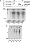
Fig. 1.
Fig. 1.
The C-terminal region of Cgs controls the DP of the CβG. (A) Schematic representation of the wild type and representative truncated and pentapeptide insertion mutants of Cgs. Numbers above the bars indicate the position of residues of Cgs. Dark gray cylinders represent the TMSs. (B) TLC analysis of CβG produced by truncated mutants. (C) TLC analysis of CβG produced by in-frame pentapeptide insertion mutants. pBA24, plasmid expressing wild-type Cgs. * and **, migration of anionic and neutral CβG, respectively.
Fig. 2.
Fig. 2.
Characterization of CβG. HPAEC-PAD elution profiles (A) and MALDI-TOF mass spectrometry analysis (B) of neutral-CβG of A. tumefaciens A1045 strains harboring the indicated plasmid. pBA24, plasmid expressing wild-type Cgs. The numbers in B indicate the DP (in glucose units) corresponding to each m/z value.
Fig. 3.
Fig. 3.
Sequence and site-directed mutagenesis analysis of B. abortus Cgs. (A) Modular organization of Cgs showing the GT-84 (GT family 84) and GH-94 (glycoside phosphorylase family 94) domains, and the conserved domains identified by National Center for Biotechnology Information-Conserved Domain Search (22). CBM-X, putative carbohydrate-binding domain; GH-94 AF, GH-94-associated family domain. Numbers above the bar indicate the position of residues of Cgs. TMSs I–VI are indicated. (B) Multiple sequence alignment of cyclic glucan synthases and the catalytic domain of GH-94 glycoside phosphorylases. The alignment was performed by using the ClustalW program (23) and edited with the Jalview 2.2 program (24). The length of proteins in amino acids is indicated on the left. Cgs, B. abortus cyclic glucan synthase (GenBank accession no. AF047823); ChvB, A. tumefaciens cyclic glucan synthase (GenBank accession no. NP_533395); NdvB, Sinorhizobium meliloti cyclic glucan synthase (GenBank accession no. P20471); ChBP, V. proteolyticus ChBP (GenBank accession no. BAC87867); CBP, Cellvibrio gilvus cellobiose phosphorylase (GenBank accession no. BAA28631); CDP, Clostridium stercorarium cellodextrin phosphorylase (GenBank accession no. AAC45511). (C) Schematic representation of the wild type and site-directed mutants of Cgs. The dark gray cylinders represent the TMSs. (D) TLC analysis of the CβG produced by site-directed mutants. pBA25, plasmid expressing wild-type Cgs. * and **, migration of anionic and neutral CβG, respectively. (E) Bio-Gel P4 chromatography of [14C]glucose-labeled glycopeptides. Glycopeptides were obtained and analyzed as described in Materials and Methods.
Fig. 4.
Fig. 4.
Characterization of the recombinant C-terminal domain of Cgs. (A) Schematic representation of Cgs and the recombinant C-terminal proteins. Dark gray cylinders represent the TMSs. HT, histidine tag. (B) Phosphorylase activity as a function of protein concentration. Phosphorylase activity was determined by measuring the amount of glucose-1-P formed from phosphorolysis of the substrate, as described in Materials and Methods. For each reaction 400 μg of partial acid-hydrolyzed CβG was used as substrate. (C) Analysis of products released from [14C]glucose-labeled glycopeptides by the recombinant C-terminal domain (Cgs-CT) of Cgs. [14C]Glucose-labeled glycopeptides (3,400 cpm) were incubated with Cgs-CT (10 μg), and the reaction products were analyzed by DEAE-Sephadex chromatography. The column was eluted with 1 ml of water and with 1 ml each of 50, 100, 150, 200, 250, and 500 mM NaCl. Light gray columns, glycopeptides without Cgs-CT; dark gray columns, glycopeptides plus Cgs-CT; white columns, glycopeptides plus Cgs-CT in the absence of inorganic phosphate; black columns, [14C]glucose-1-P in the reaction mixture lacking glycopeptides.
Fig. 5.
Fig. 5.
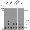
Characterization of 32P radioactive products generated in vitro. Total membranes fractions of A. tumefaciens A1045 strains harboring the indicated plasmid were incubated with inorganic [32P]phosphate, and the radioactive products were analyzed by paper electrophoresis, as indicated in Materials and Methods. Radioactivity was detected by autoradiography. Glucose-1-P was differentiated from glucose 6-phosphate by mild acid hydrolysis (0.1 N HCl for 10 min at 100°C). Plus and minus signs indicate that 5 mM UDP-glucose was added or not to the reaction mixture, or that the sample was subjected or not to mild acid hydrolysis after the incubation. pBA24, plasmid expressing wild-type Cgs; Glc, glucose; Glc 1P, glucose-1-P; UDP-Glc, UDP-glucose; Pi, inorganic phosphate.
Fig. 6.
Fig. 6.
Proposed mechanism for the synthesis of CβG. Cgs itself acts as a protein intermediate and catalyzes the four enzymatic reactions required for the synthesis of CβG: initiation (A), elongation (B), phosphorolysis (C), and cyclization (D). At some point, probably after the glucan chain length reaches >17 glucose residues, the opposing activities of the glucosyltransferase and glucan phosphorylase control the DP. Cyclization puts an end to both reactions and releases the glucan from the protein. Light gray circle, glucose; dark gray circle, glucose at the nonreducing end of the polyglucose chain linked to the protein. Pi, inorganic phosphate. The GT-84 domain between TMSs II and III and the C-terminal GH-94 domain are indicated in light gray.

References

    1. Bohin JP. FEMS Microbiol Lett. 2000;186:11–19. - PubMed
    1. Roset MS, Ciocchini AE, Ugalde RA, Inon de Iannino N. J Bacteriol. 2006;188:5003–5013. - PMC - PubMed
    1. Breedveld MW, Miller KJ. Microbiol Rev. 1994;58:145–161. - PMC - PubMed
    1. Puvanesarajah V, Schell FM, Stacey G, Douglas CJ, Nester EW. J Bacteriol. 1985;164:102–106. - PMC - PubMed
    1. Dylan T, Ielpi L, Stanfield S, Kashyap L, Douglas C, Yanofsky M, Nester E, Helinski DR, Ditta G. Proc Natl Acad Sci USA. 1986;83:4403–4407. - PMC - PubMed

Publication types

MeSH terms

LinkOut - more resources