

Spatial organization of Myxococcus xanthus during fruiting body formation
- PMID: 17921303
- PMCID: PMC2168639
- DOI: 10.1128/JB.01008-07
Spatial organization of Myxococcus xanthus during fruiting body formation
Abstract
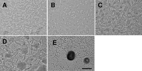
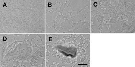
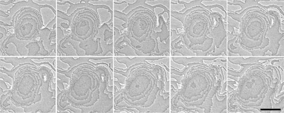
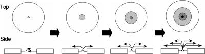
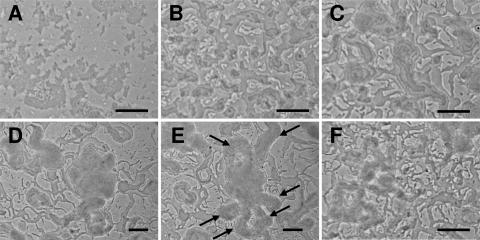
Microcinematography was used to examine fruiting body development of Myxococcus xanthus. Wild-type cells progress through three distinct phases: a quiescent phase with some motility but little aggregation (0 to 8 h), a period of vigorous motility leading to raised fruiting bodies (8 to 16 h), and a period of maturation during which sporulation is initiated (16 to 48 h). Fruiting bodies are extended vertically in a series of tiers, each involving the addition of a cell monolayer on top of the uppermost layer. A pilA (MXAN_5783) mutant produced less extracellular matrix material and thus allowed closer examination of tiered aggregate formation. A csgA (MXAN_1294) mutant exhibited no quiescent phase, aberrant aggregation in phase 2, and disintegration of the fruiting bodies in the third phase.
Figures
Similar articles
-
Characterization of the Exopolysaccharide Biosynthesis Pathway in Myxococcus xanthus.J Bacteriol. 2020 Sep 8;202(19):e00335-20. doi: 10.1128/JB.00335-20. Print 2020 Sep 8. J Bacteriol. 2020. PMID: 32778557 Free PMC article.
-
Aggregation during fruiting body formation in Myxococcus xanthus is driven by reducing cell movement.J Bacteriol. 2007 Jan;189(2):611-9. doi: 10.1128/JB.01206-06. Epub 2006 Nov 10. J Bacteriol. 2007. PMID: 17098901 Free PMC article.
-
Cell behavior and cell-cell communication during fruiting body morphogenesis in Myxococcus xanthus.J Microbiol Methods. 2003 Dec;55(3):829-39. doi: 10.1016/j.mimet.2003.08.007. J Microbiol Methods. 2003. PMID: 14607429 Review.
-
Short-range C-signaling restricts cheating behavior during Myxococcus xanthus development.mBio. 2024 Nov 13;15(11):e0244024. doi: 10.1128/mbio.02440-24. Epub 2024 Oct 18. mBio. 2024. PMID: 39422488 Free PMC article.
-
Cell-cell interactions that direct fruiting body development in Myxococcus xanthus.Curr Opin Genet Dev. 1991 Oct;1(3):363-9. doi: 10.1016/s0959-437x(05)80301-6. Curr Opin Genet Dev. 1991. PMID: 1840894 Review.
Cited by
-
Data-Driven Models Reveal Mutant Cell Behaviors Important for Myxobacterial Aggregation.mSystems. 2020 Jul 14;5(4):e00518-20. doi: 10.1128/mSystems.00518-20. mSystems. 2020. PMID: 32665330 Free PMC article.
-
Local polar order controls mechanical stress and triggers layer formation in Myxococcus xanthus colonies.Nat Commun. 2025 Jan 22;16(1):952. doi: 10.1038/s41467-024-55806-6. Nat Commun. 2025. PMID: 39843452 Free PMC article.
-
Bacterial landlines: contact-dependent signaling in bacterial populations.Curr Opin Microbiol. 2009 Apr;12(2):177-81. doi: 10.1016/j.mib.2009.01.011. Epub 2009 Feb 24. Curr Opin Microbiol. 2009. PMID: 19246237 Free PMC article. Review.
-
Draft Genome Sequence of Myxococcus xanthus Wild-Type Strain DZ2, a Model Organism for Predation and Development.Genome Announc. 2013 May 9;1(3):e00217-13. doi: 10.1128/genomeA.00217-13. Genome Announc. 2013. PMID: 23661486 Free PMC article.
-
Cell density, alignment, and orientation correlate with C-signal-dependent gene expression during Myxococcus xanthus development.Proc Natl Acad Sci U S A. 2021 Nov 9;118(45):e2111706118. doi: 10.1073/pnas.2111706118. Proc Natl Acad Sci U S A. 2021. PMID: 34732578 Free PMC article.
References
-
- Black, W. P., Q. Xu, and Z. Yang. 2006. Type IV pili function upstream of the Dif chemotaxis pathway in Myxococcus xanthus EPS regulation. Mol. Microbiol. 61:447-456. - PubMed
-
- Bonner, P. J., W. P. Black, Z. Yang, and L. J. Shimkets. 2006. FibA and PilA act cooperatively during fruiting body formation of Myxococcus xanthus. Mol. Microbiol. 61:1283-1293. - PubMed
Publication types
MeSH terms
LinkOut - more resources
Full Text Sources
