. 2007 Oct;19(10):3157-69.
doi: 10.1105/tpc.107.051714. Epub 2007 Oct 5.

Arabidopsis cotyledon-specific chloroplast biogenesis factor CYO1 is a protein disulfide isomerase

Affiliations

Arabidopsis cotyledon-specific chloroplast biogenesis factor CYO1 is a protein disulfide isomerase

Hiroshi Shimada et al. Plant Cell. 2007 Oct.

Abstract

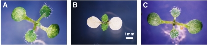
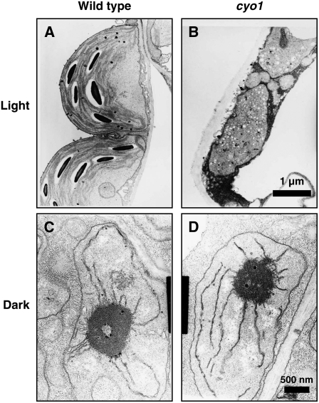
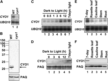
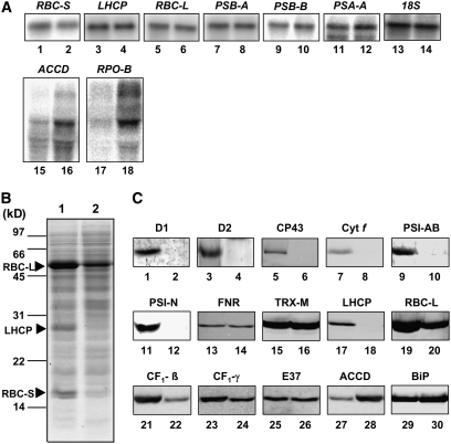
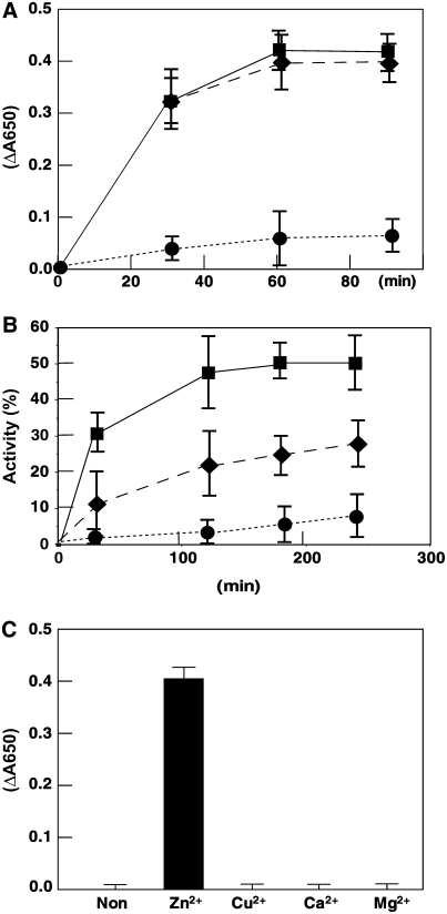
Chloroplast development in cotyledons differs in a number of ways from that in true leaves, but the cotyledon-specific program of chloroplast biogenesis has not been clarified. The cyo1 mutant in Arabidopsis thaliana has albino cotyledons but normal green true leaves. Chloroplasts develop abnormally in cyo1 mutant plants grown in the light, but etioplasts are normal in mutants grown in the dark. We isolated CYO1 by T-DNA tagging and verified that the mutant allele was responsible for the albino cotyledon phenotype by complementation. CYO1 has a C(4)-type zinc finger domain similar to that of Escherichia coli DnaJ. CYO1 is expressed mainly in young plants under light conditions, and the CYO1 protein localizes to the thylakoid membrane in chloroplasts. Transcription of nuclear photosynthetic genes is generally unaffected by the cyo1 mutation, but the level of photosynthetic proteins is decreased in cyo1 mutants. Recombinant CYO1 accelerates disulfide bond reduction in the model substrate insulin and renatures RNase A, indicating that CYO1 has protein disulfide isomerase activity. These results suggest that CYO1 has a chaperone-like activity required for thylakoid biogenesis in cotyledons.


Figures

Figure 1.
Figure 1.
The cyo1 Mutant Plant Has Albino Cotyledons. A 10-d-old wild-type plant (A), a cyo1 mutant plant (B), and a cyo1 mutant plant complemented with the CYO1 genomic DNA clone (C). All were grown under continuous light at 23°C on 1/2 MS agar plate medium with 1.5% sucrose.
Figure 2.
Figure 2.
The Chloroplasts in Cotyledons of cyo1 Mutants Are Small and Abnormal in Shape. Wild-type chloroplast (A) and cyo1 mutant plastid (B) in cotyledons from 7-d-old plants grown under light. The magnification is the same in (A) and (B). Wild-type (C) and cyo1 mutant (D) etioplast in cotyledons from 7-d-old plants grown in the dark. The magnification is the same in (C) and (D).
Figure 3.
Figure 3.
Molecular Characterization of the CYO1 Gene. (A) Structure of CYO1. Boxes and bars represent exons and introns, respectively. (B) Structure of the CYO1 cDNA. The black box and white boxes represent the open reading frame and untranslated region, respectively. (C) to (E) DNA fragments used for the complementation assay. The DNA fragment in (C) contains the ATG codon at 3 bp, whereas the DNA fragments between 5 and 606 bp, shown in (D) and (E), do not. The ATG at 32 bp was mutated to ATT in the DNA fragment shown in (E). 35S represents the cauliflower mosaic virus 35S promoter.
Figure 4.
Figure 4.
Homologs and Putative Topology of CYO1. (A) Alignment of the amino acid sequence of CYO1 (accession number AY059925) and CYO1 homologs from potato (TC133656), tomato (TC180110), Medicago truncatula (ABE80527), soybean (TC228649), poplar (TC44537), pine (TC76401), spruce (TC24344), maize (TC343209), rice (TC332052), and wheat (TC257683). Sequences of potato, tomato, soybean, poplar, pine, spruce, maize, rice, and wheat were drawn from The Institute for Genomic Research Gene Indices (http://tigrblast.tigr.org/tgi/). The sequences of spruce, maize, and wheat are not full-length amino acid sequences. Conserved residues are shown in black, and similar residues are shown in gray. Asterisks indicate Cys residues, the bar shows the zinc finger motif, and the vertical arrow indicates the putative cleavage site of the transit peptide of Arabidopsis CYO1. (B) Putative topology of the zinc finger motif in CYO1. Residues conserved between CYO1 and DnaJ are highlighted. Residues conserved in the zinc finger domain are shown in black, and identical residues between CYO1 and DnaJ are shown in gray. (C) The topology of E. coli DnaJ (Shi et al., 2005).
Figure 5.
Figure 5.
RNA Expression and Immunoblot Analysis of CYO1. (A) Analysis of CYO1 transcripts from 7-d-old wild-type (lane 1) and cyo1 mutant (lane 2) plants grown under light. Ubiquitin10 (UBQ10) was analyzed as a loading control. (B) Immunoblot analysis of CYO1 levels in 7-d-old wild-type (left lane) and cyo1 mutant (right lane) plants grown under light conditions. Fifty micrograms of total protein was loaded in each lane. The arrow indicates CYO1. Plasma Aquaporin (PAQ) was analyzed as a loading control. (C) Effect of light on CYO1 transcript levels. Seven-day-old wild-type Arabidopsis plants grown in continuous dark were illuminated for the times indicated (0 to 12 h; lanes 1 to 6). Lane 7 shows 7-d-old plants grown under continuous light. (D) Effect of light on CYO1 protein levels. Seven-day-old wild-type Arabidopsis plants grown in continuous dark were illuminated for the times indicated (0 to 12 h; lanes 1 to 4). Lane 5 shows 7-d-old plants grown under continuous light. Fifty micrograms of total protein was loaded in each lane. (E) Organ-specific transcripts of CYO1 in continuously illuminated adult plants. Total RNA was isolated from 4-week-old wild-type Arabidopsis for organ-specific quantification of the CYO1 transcript (lanes 2 to 5). (F) Organ-specific CYO1 protein levels. Fifty micrograms of total protein was loaded in each lane.
Figure 6.
Figure 6.
Localization of CYO1 Protein. (A) In vitro chloroplast import and protease protection assay for CYO1. 35S-labeled CYO1 and RBC-S were produced by in vitro translation. The proteins were incubated with isolated pea chloroplasts in the presence of 4.0 mM Mg-ATP for 30 min at room temperature. Chloroplasts were recovered by sedimentation through 40% (v/v) Percoll. The recovered intact chloroplasts were then incubated without (lanes 2, 3, 7, and 8) or with (lanes 4, 5, 9, and 10) thermolysin for 30 min at 4°C. Intact chloroplasts were recovered by centrifugation through 40% (v/v) Percoll and fractionated into total membrane (Pellet; lanes 2, 4, 7, and 9) and soluble (Supernatant [Sup.]; lanes 3, 5, 8, and 10) fractions. CYO1 was detected in the chloroplast fraction (Pellet), as was the control protein. TP represents 10% of in vitro translated products (lanes 1 and 6). (B) Immunoblot analysis of CYO1. Total protein is whole cell protein (lane 1). Intact chloroplasts (lane 2) were lysed, and soluble (Sup.; lane 3) and insoluble (Pellet; lane 4) proteins (10 μg/lane) were separated by 12.5% SDS-PAGE. Each protein fraction was prepared from 7-d-old plants. RBC-L (stroma protein), E37 (chloroplast envelope protein), PAQ (plasma membrane protein), and BiP (ER protein) were analyzed as controls for each fraction. (C) Sonicated thylakoids were prepared from 7-d-old plants and were treated with 0.1 M sodium carbonate (lanes 2 and 3), 1.0% Nonidet P-40 (NP40; lanes 4 and 5), or 2.0 M NaCl (lanes 6 and 7) and then soluble (Sup.; lanes 3, 5 and 7) and insoluble (Pellet; lanes 2, 4, and 6) fractions were separated at 12,000g. LHCP and FNR were used as marker proteins for intrinsic and extrinsic thylakoid membrane proteins, respectively. (D) Sonicated thylakoids were prepared from 7-d-old plants and were treated with thermolysin (0, 10, or 100 μg/mL) for 60 min on ice. FNR and PSI-N were used as stroma- and lumen-exposed peripheral thylakoid protein markers, respectively. (E) Intact thylakoids of 7-d-old plants were solubilized by treatment with 1% n-dodecyl-β-d-maltoside and separated by 5 to 14% blue native gel electrophoresis. After electrophoresis, the gel was incubated in 0.1% SDS containing transfer buffer (100 mM Tris, 192 mM Gly, and 5% methanol) for 10 min at room temperature and blotted to a membrane (left panel). The membrane was reacted with antibodies against CYO1 (right panel). Without additional staining, protein complexes were detected by bound Coomassie blue dye and chlorophylls. Bands corresponding to various photosynthetic complexes are indicated (Asakura et al., 2004) (see also http://www.hos.ufl.edu/clineweb/BNgel.htm). Ferritin (880 and 440 kD) and BSA (132 and 66 kD) were used as molecular mass marker proteins. Arrowheads show the bands of CYO1. Closed arrowheads show that CYO1 bands comigrate with PSI/LHCI and PSII/LHCII, and the open arrowhead shows the CYO1 band in an unidentified thylakoid protein complex.
Figure 7.
Figure 7.
RNA Expression and Immunoblot Analysis of Chloroplast Proteins. (A) Total RNA was prepared from 7-d-old wild-type (lane 1) and cyo1 mutant (lane 2) plants. One microgram of total RNA was loaded, and transcript levels were measured in wild-type (odd-numbered lanes) and cyo1 mutant (even-numbered lanes) plants for the following nuclear genes: RBC-S, LHCP, and 18S (18S rRNA); the following chloroplast genes transcribed by PEP: RBC-L, PSB-A (D1), PSB-B (CP47), and PSA-A (PsaA); and the following chloroplast genes transcribed by NEP: ACCD and RPO-B. (B) Twenty micrograms of total protein of 7-d-old wild-type (lane 1) and cyo1 mutant (lane 2) plants was loaded. The SDS-PAGE gel (20 μg of protein per lane) was stained with Coomassie blue. The arrowheads show the major bands of RBC-L, RBC-S, and LHCP. (C) Immunoblot analysis of photosynthesis and control proteins (20 μg of protein per lane) in 7-d-old wild-type (odd-numbered lanes) and cyo1 mutant (even-numbered lanes) plants. D1, D2, and CP43 are proteins of PSII, Cyt f is a protein of cytochrome b6f, PSI-AB and PSI-N are proteins of PSI, FNR is ferredoxin NADP+ oxidoreductase, TRX-M is thioredoxin m, RBC-L is the large subunit of Rubisco, CF1-β and CF1-γ are subunits of chloroplast ATPase, E37 is a chloroplast envelope protein, ACCD is acetyl-CoA carboxylase, and BiP is an ER protein.
Figure 8.
Figure 8.
CYO1 Has Reductase and Oxidase Activity. (A) Purified Δ30CYO1 catalyzes the reduction of insulin. The reaction was initiated by adding DTT into 0.1 M potassium phosphate, pH 6.6, containing 0.13 mM bovine insulin in the absence (circles) or presence of 1.0 μM Δ30CYO1 (diamonds) or 1.0 μM DnaJ (squares). The reduction of insulin and the resulting precipitation of the B chain was monitored by following the optical density at 650 nm. Data represent means ± sd of three independent experiments. (B) Effect of CYO1 on refolding of reduced and denatured RNase A. Refolding of denatured and reduced RNase A (40 μM) was initiated by 10-fold dilution in 50 mM Tris-HCl, pH 8.0, containing 0.1 M NaCl and 0.3 mM DTT at 30°C in the absence (circles) or presence of 1.0 μM Δ30CYO1 (diamonds) or 1.0 μM DnaJ (squares). At the indicated time points, an aliquot containing 40 μmol of RNase A was withdrawn from the reaction to assay RNase A activity. Activity is expressed as a percentage of native RNase A activity and represents the average ± sd of three independent experiments. (C) Effect of metal on CYO1 activity. Purified Δ30CYO1 was denatured with 6.0 M guanidine hydrochloride, and then the proteins were renatured with dialyzing buffer containing 5.0 mM ZnCl2, CuCl2, CaCl2, MgCl2, or no divalent metal ion. The dialyzed proteins were centrifuged, and the supernatant was used for enzymatic assays. The reduction of insulin and the resulting precipitation of the B chain were monitored by following the optical density at 650 nm. Data represent means ± sd of three independent experiments.

Similar articles

Cited by

References

    1. Albrecht, V., Ingenfeld, A., and Apel, K. (2006). Characterization of the snowy cotyledon 1 mutant of Arabidopsis thaliana: The impact of chloroplast elongation factor G on chloroplast development and plant vitality. Plant Mol. Biol. 60 507–518. - PubMed
    1. Anfinsen, C.B., and Scheraga, H.A. (1975). Experimental and theoretical aspects of protein folding. Adv. Protein Chem. 29 205–300. - PubMed
    1. Aronsson, H., and Jarvis, P. (2002). A simple method for isolating import-competent Arabidopsis chloroplasts. FEBS Lett. 529 215–220. - PubMed
    1. Asakura, Y., Hirohashi, T., Kikuchi, S., Belcher, S., Osborne, E., Yano, S., Terashima, I., Barkan, A., and Nakai, M. (2004). Maize mutants lacking chloroplast FtsY exhibit pleiotropic defects in the biogenesis of thylakoid membranes. Plant Cell 16 201–214. - PMC - PubMed
    1. Awai, K., Marechal, E., Block, M.A., Brun, D., Masuda, T., Shimada, H., Takamiya, K., Ohta, H., and Joyard, J. (2001). Two types of MGDG synthase genes, found widely in both 16:3 and 18:3 plants, differentially mediate galactolipid syntheses in photosynthetic and nonphotosynthetic tissues in Arabidopsis thaliana. Proc. Natl. Acad. Sci. USA 98 10960–10965. - PMC - PubMed

Publication types

MeSH terms

Substances