. 2007 Dec;145(4):1681-91.
doi: 10.1104/pp.107.107805. Epub 2007 Oct 5.

Role of the PsbI protein in photosystem II assembly and repair in the cyanobacterium Synechocystis sp. PCC 6803

Affiliations

Role of the PsbI protein in photosystem II assembly and repair in the cyanobacterium Synechocystis sp. PCC 6803

Marika Dobáková et al. Plant Physiol. 2007 Dec.

Abstract

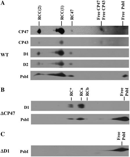
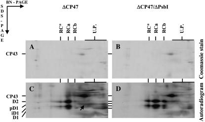
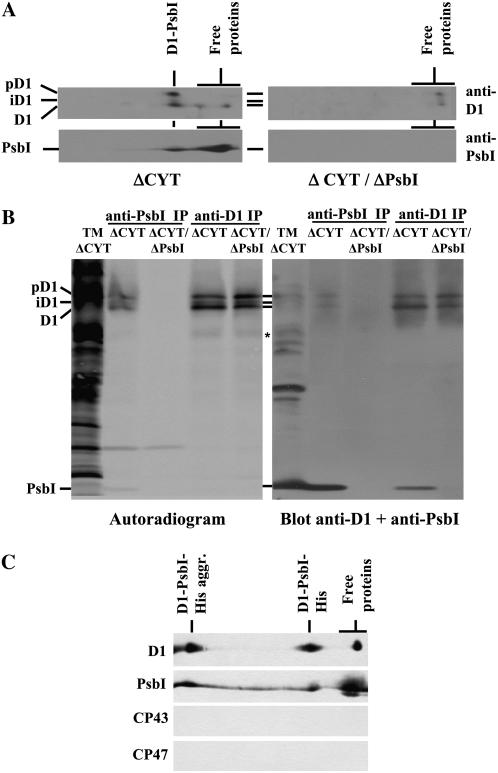
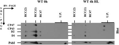
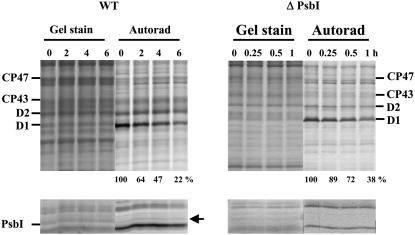
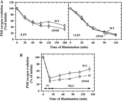
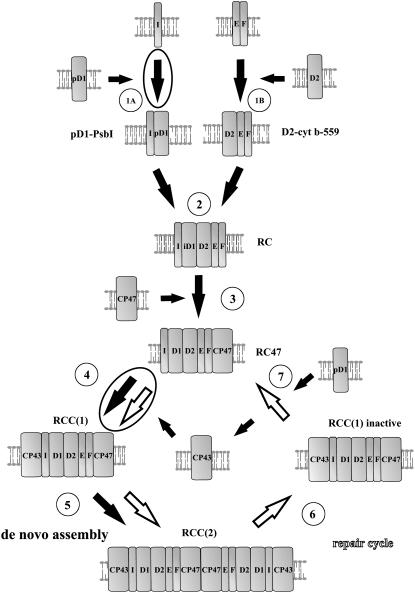
The involvement of the PsbI protein in the assembly and repair of the photosystem II (PSII) complex has been studied in the cyanobacterium Synechocystis sp. PCC 6803. Analysis of PSII complexes in the wild-type strain showed that the PsbI protein was present in dimeric and monomeric core complexes, core complexes lacking CP43, and in reaction center complexes containing D1, D2, and cytochrome b-559. In addition, immunoprecipitation experiments and the use of a histidine-tagged derivative of PsbI have revealed the presence in the thylakoid membrane of assembly complexes containing PsbI and either the precursor or mature forms of D1. Analysis of PSII assembly in the psbI deletion mutant and in strains lacking PsbI together with other PSII subunits showed that PsbI was not required for formation of PSII reaction center complexes or core complexes, although levels of unassembled D1 were reduced in its absence. However, loss of PsbI led to a dramatic destabilization of CP43 binding within monomeric and dimeric PSII core complexes. Despite the close structural relationship between D1 and PsbI in the PSII complex, PsbI turned over much slower than D1, whereas high light-induced turnover of D1 was accelerated in the absence of PsbI. Overall, our results suggest that PsbI is an early assembly partner for D1 and that it plays a functional role in stabilizing the binding of CP43 in the PSII holoenzyme.


Figures

Figure 1.
Figure 1.
Immunoblots of thylakoid membrane proteins from Synechocystis 6803 wild type (WT; A), psbB deletion mutant ΔCP47 (B), and psbA deletion mutant ΔD1 (C) after their separation by 2D BN/SDS-PAGE. Thylakoid proteins were separated by 2D BN-PAGE, blotted onto PVDF membrane, and immunodecorated using antibodies raised against D1, D2, CP47, CP43, and PsbI. Designation of complexes: RCC(2) and RCC(1), dimeric and monomeric PSII core complexes, respectively; RC47, PSII core complex lacking CP43; RC*, RCa, and RCb, RC complexes. One microgram of Chl was loaded for each sample.
Figure 2.
Figure 2.
Pulse-chase analysis of wild type and the psbI deletion strain ΔPsbI. Cells of wild type (A, C, E, and G) and ΔPsbI (B, D, F, and H) were radiolabeled at 500 μmol photons m−2 s−1 and 23°C with a mixture of [35S]Met/Cys for 10 min (pulse) and then CAP (1 mg mL−1) was added and cells incubated at the same temperature at 125 μmol photons m−2 s−1 for another 20 min (chase). Labeled cells were used for isolation of thylakoids, which were analyzed by 2D BN/SDS-PAGE. A and B, Coomassie Blue-stained gels of proteins after the pulse. C and D, Autoradiograms of the same samples with the putative D1-PsbI complex designated by arrow in C. E and F, Autoradiograms of proteins after pulse chase. G and H, Low-Mr region with PsbI detected by immunoblotting (blot) and putative D1-PsbI complex designated by an arrow. Designations of proteins are as described in the legend to Figure 1. RCC-RC47 designates dimeric PSII core complex lacking only one CP43 copy and RC47(2) the dimeric PSII core lacking both CP43 copies. U.P., Unassembled proteins. α- and β-subunits of ATP synthase (designated by αβ*) were used as internal standards during quantification of D1-stained and -labeled bands (see Supplemental Table S2). The arrows in C and G show the complex of PsbI and pD1 in wild type. Six micrograms of Chl was loaded for each sample.
Figure 3.
Figure 3.
Analysis of thylakoid membrane proteins in the psbB deletion mutant ΔCP47 and the double deletion mutant ΔCP47/ΔPsbI. Cells of the Synechocystis 6803 strains were radiolabeled at 500 μmol photons m−2 s−1 and 29°C with [35S]Met/Cys for 30 min and their thylakoid proteins were separated by 2D BN/SDS-PAGE. Designations of proteins are as described in the legend to Figure 1; the arrow indicates the complex of PsbI and pD1. Six micrograms of Chl was loaded for each sample.
Figure 4.
Figure 4.
Identification of a D1-PsbI precomplex. A, Thylakoid membrane proteins (1 μg of Chl) of the ΔCYT and ΔCYT/ΔPsbI strains were separated by 2D BN/SDS-PAGE, transferred onto PVDF membrane, and detected by antibodies against D1 and PsbI. B, Pulse-labeled thylakoid proteins (5 μg of Chl) from the ΔCYT and ΔCYT/ΔPsbI strains were immunoprecipitated using antibodies specific for D1 (anti-D1 IP) or PsbI (anti-PsbI IP) and the immunoprecipitates together with thylakoids from ΔCYT (TM) were analyzed by SDS-PAGE, blotted onto PVDF membrane, autoradiographed (left, autoradiogram), and then probed with antibodies against both PsbI and D1 proteins (right, blot anti-D1 + anti-PsbI). *, A putative 23-kD D1 synthesis intermediate. C, Immunoblots of D1, PsbI, CP43, and CP47 after 2D BN/SDS-PAGE of the protein fraction bound to nickel-affinity column loaded with solubilized thylakoids of the PsbI-His/ΔPsbI/ΔCYT strain. D1-PsbI-His aggr., Aggregates of D1 and PsbI-His.
Figure 5.
Figure 5.
Immunoblots of thylakoid membrane proteins from the Synechocystis 6803 wild-type strain before and after 4-h exposure to high light. Cells of wild type were exposed to high irradiance (1,000 μmol photons m−2 s−1) for 4 h in the presence of the protein synthesis inhibitor lincomycin (100 μg mL−1). Thylakoid membrane proteins (1 μg of Chl) were separated by 2D BN/SDS-PAGE, transferred onto PVDF membrane, and probed with antibodies against the D1, D2, CP43, CP47, and PsbI proteins. Designations of proteins are as described in the legend to Figure 1. To allow direct comparison of protein bands, thylakoids from control and photoinhibited cells were analyzed on a single gel and blot.
Figure 6.
Figure 6.
Degradation of the PSII proteins in the wild-type and ΔPsbI strains under high irradiance monitored by radioactive pulse-chase labeling. Cells of both strains were subjected to 250 μmol photons m−2 s−1 of white light for 20 min in the presence of [35S]Met/Cys. Then the cells were washed, supplemented with unlabeled Met/Cys, and subjected to 500 μmol photons m−2 s−1 of white light for 6 h. Thylakoids were isolated, analyzed by SDS-PAGE, the gel was stained (Gel stain), and the radioactive labeling of the proteins was visualized using a PhosphorImager (Autorad). Quantification of radioactivity in the D1 band was performed by ImageQuant software with samples of each strain equally loaded on Chl basis (2 μg of Chl; see Gel stain) in a single gel. The radioactivity incorporated into the D1 band of each strain during pulse was taken as 100%; numbers show means of three measurements; sd did not exceed 7%. The low-Mr region of the gel is shown in the bottom sections and the stable band of PsbI is designated by an arrow.
Figure 7.
Figure 7.
PSII repair under high and low irradiance in cells of wild type and ΔPsbI. A, Cells of wild type (circles) and ΔPsbI (squares) were illuminated with 500 μmol photons m−2 s−1 of white light for 180 min in the absence (left) or for 120 min in the presence (right) of 100 μg mL−1 lincomycin and during illumination PSII oxygen-evolving activity was assayed in whole cells. B, Cells of wild type (circles) and ΔPsbI (squares) were illuminated at 2,000 μmol photons m−2 s−1 for 20 min (PI), then the cells were transferred to low irradiance of 50 μmol photons m−2 s−1 and incubated for an additional 120 min (REC). During these light regimes, PSII oxygen-evolving activity was assayed in whole cells. Values in the plots represent means of three measurements ± sd. Initial values for wild type and ΔPsbI were in the range of 372 ± 27 and 258 ± 13 μmol O2 mg Chl−1 h−1, respectively.
Figure 8.
Figure 8.
Illustration of function of PsbI during PSII assembly and repair. De novo assembly of PSII (large black arrows) starts with association of PsbI (I) with pD1 (step 1A) forming pD1-PsbI precomplex on one side, and association of Cyt b-559 protein subunits α and β (E and F) with the D2 protein forming Cyt b-559-D2 precomplex on the other side (step 1B). Assembly continues by formation of the RC complex from both precomplexes (step 2). Then CP47 (step 3) and CP43 (step 4) are attached and resulting PSII core monomers RCC(1) form the PSII core dimer [RCC(2), step 5]. PSII repair cycle (white arrows) starts with inactivation of the D1 protein and monomerization (step 6). Then it continues by detachment of CP43 and selective replacement of the D1 protein (step 7). The final two steps, the CP43 attachment and PSII dimerization, seem to be common with the de novo assembly pathway. PsbI is important for processes of formation of pD1-PsbI precomplex (step 1A) and attachment of CP43 to RC47 (step 4) as indicated by encircled arrows. For simplicity, other small and extrinsic PSII subunits were omitted. pD1, Unprocessed forms of D1; iD1, partially processed forms of D1.

Similar articles

Cited by

References

    1. Anbudurai PR, Mor TS, Ohad I, Shestakov SV, Pakrasi HB (1994) The CtpA gene encodes the C-terminal processing protease for the D1 protein of the photosystem-II reaction-center complex. Proc Natl Acad Sci USA 91 8082–8086 - PMC - PubMed
    1. Barber J (2006) Photosystem II: an enzyme of global significance. Biochem Soc Trans 34 619–631 - PubMed
    1. Bumba L, Tichý M, Dobáková M, Komenda J, Vácha F (2005) Localization of the PsbH subunit in photosystem II from the Synechocystis 6803 using the His-tagged Ni-NTA nanogold labeling. J Struct Biol 152 28–35 - PubMed
    1. Burke E, Barik S (2003) Megaprimer PCR: application in mutagenesis and gene fusion. Methods Mol Biol 226 525–532 - PubMed
    1. Eaton-Rye JJ, Vermaas WFJ (1991) Oligonucleotide-directed mutagenesis of psbB, the gene encoding CP47, employing a deletion mutant strain of the cyanobacterium Synechocystis sp. PCC 6803. Plant Mol Biol 17 1165–1177 - PubMed

Publication types

MeSH terms

Substances

LinkOut - more resources