. 2007 Dec;6(12):2406-18.
doi: 10.1128/EC.00222-07. Epub 2007 Oct 5.

Endocytosis in the shiitake mushroom Lentinula edodes and involvement of GTPase LeRAB7

Affiliations

Endocytosis in the shiitake mushroom Lentinula edodes and involvement of GTPase LeRAB7

Ming Tsung Lee et al. Eukaryot Cell. 2007 Dec.

Abstract

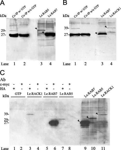
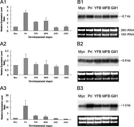
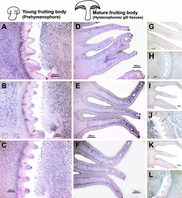
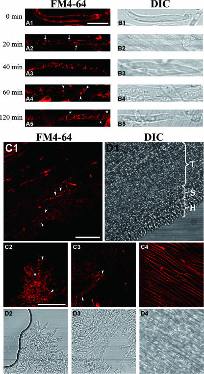
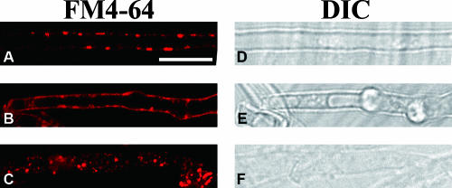
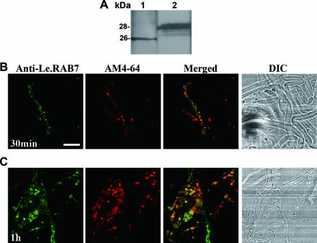
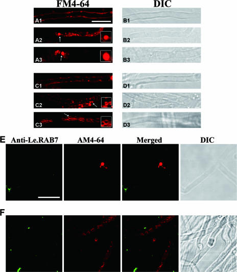
Endocytosis is the process by which substrates enter a cell without passing through the plasma membrane but rather invaginate the cell membrane and form intracellular vesicles. Rab7 regulates endocytic trafficking between early and late endosomes and between late endosomes and lysosomes. LeRab7 in Lentinula edodes is strongly homologous to Rab7 in Homo sapiens. Receptors for activated C kinase-1 (LeRACK1) and Rab5 GTPase (LeRAB5) were isolated as interacting partners of LeRab7, and the interactions were confirmed by in vivo and in vitro protein interaction assays. The three genes showed differential expression in the various developmental stages of the mushroom. In situ hybridization showed that the three transcripts were localized in regions of active growth, such as the outer region of trama cells, and the subhymenium of the hymenophore of mature fruiting bodies and the prehymenophore of young fruiting bodies. The existence of endocytosis in the mycelium and hymenophores was confirmed by the internalization of FM4-64. LeRAB7 was partially colocalized with the AM4-64 and was located in the late endocytic pathway. This is the first report of the presence of endocytosis in homobasidiomycetes. LeRAB7, LeRAB5, and LeRACK1 may contribute to the growth of L. edodes and cell differentiation in hymenophores.


Figures

FIG. 1.
FIG. 1.
In vitro Co-IP protein interaction assay. LeRAB7 was tagged with c-Myc epitope, whereas LeRAB5 and LeRACK1 were tagged with an HA epitope. Each panel shows an autoradiogram from the SDS-PAGE analysis after in vitro transcription/translation and immunoprecipitation. The in vitro-translated proteins were biotinylated and visualized by binding with alkaline phosphatase, followed by chemiluminescent detection. The band intensities of LeRAB5 and LeRACK1 were weaker than that of LeRAB7, which may be due to different translation efficiencies on the different vectors. (A) In vitro Co-IP between LeRAB7 and LeRAB5. Lanes 3 and 4 show the in vitro-translated LeRAB5 and LeRAB7. The predicted products and their respective sizes are indicated by arrows. Lanes 1 and 2 represent the Co-IP reactions of LeRAB5 and LeRAB7 with or without GTP incubation before the mixing of the two proteins and precipitation with the c-myc antibody. LeRAB5 and LeRAB7 were coimmunoprecipitated in lane 1 (with GTP incubation), but only LeRAB7 was pulled out in lane 2 (without GTP incubation). (B) In vitro Co-IP between LeRAB7 and LeRACK1. Lanes 3 and 4 show the in vitro-translated LeRAB7 and LeRACK1. The predicted products and their respective sizes are indicated by arrows. Lanes 1 and 2 represent the Co-IP reactions of LeRAB7 with or without GTP incubation before the mixing of the two proteins. The protein mixtures were immunoprecipitated with c-myc antibody. LeRACK1 and LeRAB7 were coimmunoprecipitated in lane 1 (with GTP incubation), but only LeRAB7 was pulled out in lane 2 (without GTP incubation). (C) The immunoprecipitation of the in vitro-translated proteins and GTP was performed by using either c-myc antibody or HA antibody, as indicated (lanes 1 to 8). The predicted products and their respective sizes are indicated by arrows. The in vitro-translated LeRAB7, LeRAB5, and LeRACK1 are shown in lanes 9, 10, and 11, respectively. GTP was not pulled out by the c-myc or the HA antibody. LeRAB7 was pulled out by the c-myc antibody, whereas LeRAB5 and LeRACK1 were pulled out the by HA antibody.
FIG. 2.
FIG. 2.
Temporal expression profiles of LeRab7, LeRab5, and LeRACK1 at different developmental stages in L. edodes. (A1 to A3) Quantitative real-time RT-PCR of the transcripts at six developmental stages. (A1) LeRab7; (A2) LeRab5; (A3) LeRACK1. The RNA amounts were normalized to the level of LePma and are shown as the expression relative to that in the mycelium. The gene expression level at the mycelium stage was taken as one. The results were calculated as the average values of three individual RNA samples, and each PCR was repeated twice. The error bars indicate standard deviations (n = 4), with P < 0.001 in the LeRab7 and LeRACK1 assay and P < 0.005 in the LeRab5 assay. (B1 to B3) Northern blotting of the transcripts at the five developmental stages. (B1) LeRab7; (B2) LeRab5; (B3) LeRACK1. Equal amounts of total RNA were loaded and confirmed by equal intensities of rRNA before the gels were blotted. Myc, mycelium; Pri, primordium; YFB, young fruiting body; MFB, mature fruiting body; Gill1, gill tissue (hymenophore) before sporulation; Gill2, gill tissue after sporulation.
FIG. 3.
FIG. 3.
Distributions of the LeRab7, LeRab5, LeRACK1 transcripts in young fruiting bodies and mature fruiting bodies as determined by in situ RNA-RNA hybridization. Fixed longitudinal ultrathin sections of the prehymenophores of the young fruiting bodies and the hymenophores of the mature fruiting bodies were hybridized with LeRab7 (A and B), LeRab5 (E and F), and LeRACK1 (I and J) digoxigenin-labeled RNA antisense probes, respectively. The signals were detected by alkaline phosphatase-labeled anti-digoxigenin Fab fragments and subsequent nitroblue tetrazolium-5-bromo-4-chloro-3-indolylphosphate (BCIP) staining. Positive signals are indicated by purple Sense probes of LeRab7 (C and D), LeRab5 (G and H), and LeRACK1 (K and L) were used as negative controls to show the specificity of reactions. Magnification, ×4. Bar, 200 μm.
FIG. 4.
FIG. 4.
Time course of FM4-64 dye internalization in the mycelium and the hymenophore of a fruiting body of L. edodes. The samples in panels A1 to A5 were taken out at the time points indicated after loading with 12 μM FM4-64 at 0°C. (A1 to A3) The FM4-64 was taken up by mycelial cells and detected on the plasma membrane (A1), followed by internalization through punctuated structures (arrows) (A2), and an increase in the number and size of the circular, endosomal-like structures (A3). (A4) The dye was transported into vacuolar and hollow organelles (arrowheads) after 1 h. (A5) After 2 h, the dye was retained in large vacuolar structures (asterisks), which marked the final point of internalization. (B1 to B5) DIC images of the various time points. Bars, 10 μm. (C1) FM4-64 internalization in the hymenophore of a mature fruiting body. The images were captured after the loading of the dye at 0°C, and chasing followed after 40 min at 25°C. The appearance of punctuated structures is indicated by arrowheads. (D1) The trama (T), subhymenium (S), and hymenium (H) were observed in the DIC micrograph. (C2 to C4) Dye internalization was seen in the hymenium (C2) and subhymenium (C3) but not in the trama cells (C4). (D2 to D4) DIC images of the respective structures. Bar, 50 μm.
FIG. 5.
FIG. 5.
Effects of drugs on FM4-64 internalization. The images were collected after the loading of the dye at 0°C followed by chasing after 40 min at 25°C in the presence of a mixture of sodium azide and sodium fluoride (30 mM each) (A), cytochalasin D (20 μM) (B), and benomyl at 3 μg/ml (C). Bar, 10 μm.
FIG. 6.
FIG. 6.
Double labeling of mycelial cells with anti-LeRAB7 and AM4-64. (A) Characterization of the LeRAB7 antiserum. The anti-LeRAB7 detection of LeRAB7 was performed on a total protein extract of L. edodes at the mycelium stage (lane 1) and during the in vitro transcription/translation of c-myc-tagged LeRAB7 (lane 2). A single main band was observed in both lanes. (B and C) The mycelial cells were loaded with 12 μM AM4-64 at 0°C, followed by chasing at 25°C, and were fixed with 4.5% paraformaldehyde at the 30-min (B) and 60-min (C) time points. The cells were then immunostained with LeRAB7 antiserum. At the 30-min time point, the anti-LeRAB7 partly colocalized with the AM4-64. The extent of the colocalization was higher at the 60-min time point than at the 30-min time point. The yellow appearance of the merged images indicates the colocalization of the antibody and dye. Bar, 50 μm.
FIG. 7.
FIG. 7.
Effects of BFA and wortmannin on FM4-64 internalization and LeRAB7 labeling in mycelial cells. (A1 to A3) The cells were incubated with 17.5 μM of BFA for 2 h before loading with 12 μM FM4-64 at 0°C. Samples were taken at 0 min (A1), 20 min (A2), and 1 h (A3) after chasing at 25°C. (C1 to C3) The cells were incubated with 16.5 μM wortmannin for 2 h before loading with 12 μM FM4-64 at 0°C. (B1 to B3) Samples were taken at 0 min (B1), 20 min (B2), and 1 h (B3) after chasing at 25°C. Panels B1 to B3 and panels D1 to D3 are DIC images of the corresponding treatments and time points. The insets are enlarged images of the BFA- and wortmannin-induced structures, which are indicated by arrows. (E and F) Double labeling of mycelial cells with anti-LeRAB7 and AM4-64 after treatment with 17.5 μM BFA (E) and 16.5 μM wortmannin (F). The cells were loaded with 12 μM AM4-64 at 0°C, followed by chasing at 25°C, and were fixed after 1 h. They were then immunostained by LeRAB7 antiserum. No colocalization was observed in either treatment. Bar, 10 μm.

Similar articles

Cited by

References

    1. Armstrong, J., M. W. Craighead, R. Watson, S. Ponnambalam, and S. Bowden. 1993. Schizosaccharomyces pombe ypt5: a homologue of the rab5 endosome fusion regulator. Mol. Biol. Cell 4:583-592. - PMC - PubMed
    1. Atkinson, H. A., A. Daniels, and N. D. Read. 2002. Live-cell imaging of endocytosis during conidial germination in the rice blast fungus, Magnaporthe grisea. Fungal Genet. Biol. 37:233-244. - PubMed
    1. Ayscough, K. R. 2005. Defining protein modules for endocytosis. Cell 123:305-320. - PubMed
    1. Baluska, F., A. Hlavacka, J. Samaj, K. Palme, D. G. Robinson, T. Matoh, D. W. McCurdy, D. Menzel, and D. Volkmann. 2002. F-actin-dependent endocytosis of cell wall pectins in meristematic root cells. Insights from brefeldin A-induced compartments. Plant Physiol. 130:422-431. - PMC - PubMed
    1. Brabec, M., D. Blaas, and D. Fuchs. 2006. Wortmannin delays transfer of human rhinovirus serotype 2 to late endocytic compartments. Biochem. Biophys. Res. Commun. 348:741-749. - PubMed

Publication types

LinkOut - more resources