. 2007 Oct 29;18(16):1657-61.
doi: 10.1097/WNR.0b013e3282f0e42d.

Dp71, utrophin and beta-dystroglycan expression and distribution in PC12/L6 cell cocultures

Affiliations

Dp71, utrophin and beta-dystroglycan expression and distribution in PC12/L6 cell cocultures

Ramses Ilarraza-Lomeli et al. Neuroreport. .

Abstract

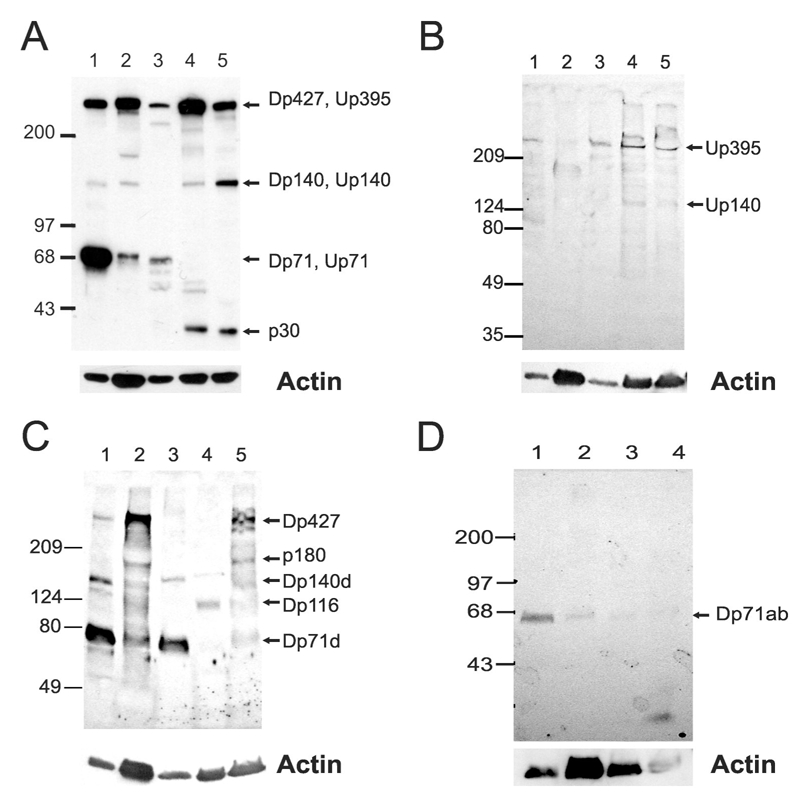
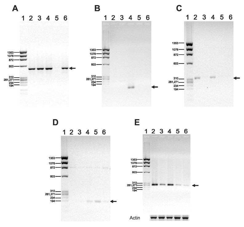
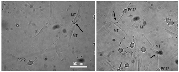
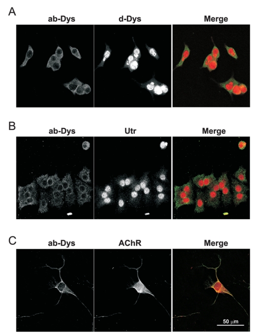
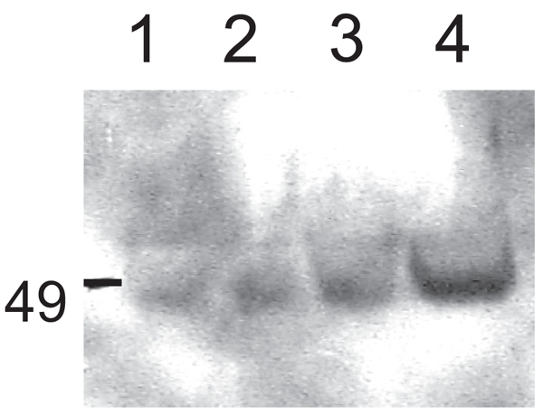
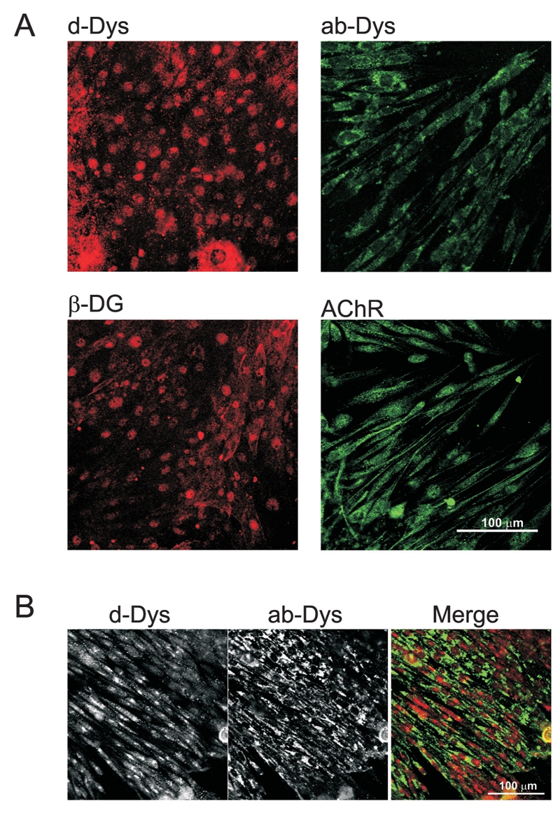
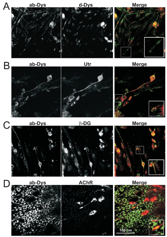
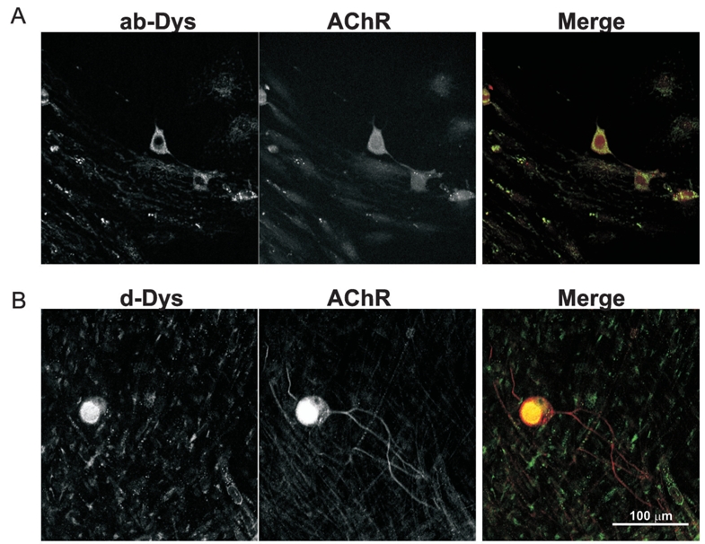
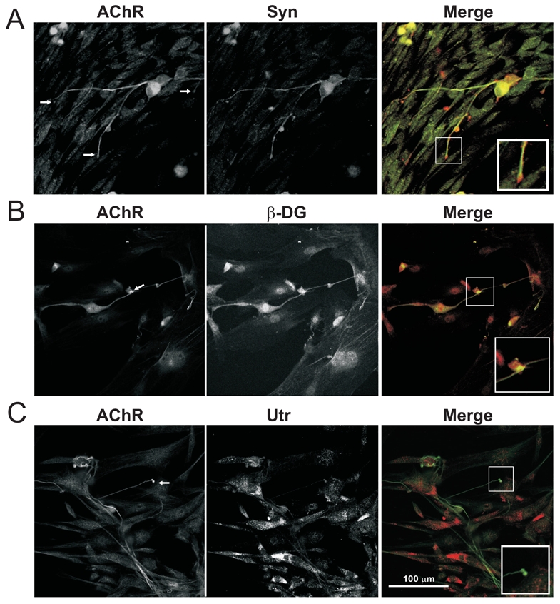
Function of dystrophin Dp71 isoforms is unknown but seems related to neurite outgrowth and synapse formation. To evaluate Dp71 role in myoneural synapses, we established a coculture model using PC12 cells and L6 myotubes and analyzed expression and localization of Dp71 and related proteins, utrophin and beta-dystroglycan, in PC12 cells. Confocal microscopy showed Dp71d isoform in PC12 nuclei, golgi-complex-like and endoplasmic reticulum-like structures, whereas Dp71ab concentrates at neurite tips and cytoplasm, colocalizing with beta-dystroglycan, utrophin, synaptophysin and acetylcholine receptors. Evidences suggest that Dp71ab isoform, unlike Dp71d, may take part in neurite-related processes. This is the first work on Dp and members of Dp-associated protein complex roles in a cell-line based coculturing system, which may be useful in determining Dp71 isoforms associations.


Figures

Fig. 1
Fig. 1
Expression of dystrophin and utrophin in L6 myoblasts and myotubes. Western blots were performed as indicated in materials and methods, using 3–10% gradient PAGE-SDS (A, B and C) or 10% (D) gels, and H5A3 pan-dystrophin-utrophin (A), K7 anti-utrophin (B), H4 anti-d- (C) and 5F3 anti-ab-dystrophin (D) antibodies. A, B and C. Lanes: 1, rat brain; 2, rat muscle; 3, undifferentiated PC12 cells; 4, L6 myoblasts; and, 5, L6 myotubes. D. Lanes: 1, rat brain; 2, mouse muscle; 3, L6 myoblasts; and, 4, L6 myotubes. Proteins detected by the antibodies are indicated by arrows. Actin was used as a loading control. Molecular weights are shown on the left.
Fig. 2
Fig. 2
RT-PCR using oligonucleotides for Dp427 (A), Dp260 (B), Dpl40 (C), Dp116 (D) and Dp71 (E). Total RNA was subjected to RT-PCR assays with oligonucleotides Dp427-5 and Dp427-3 for Dp427, Dp260-5 and Dp260-3 for Dp260, Dpl40-5 and Dpl40-3 for Dpl40, Dpll6U and Dpll6L for Dp116 and Primer 5 and 247 for Dp71. Expected product sizes are identified with arrows, and are 547 bp for Dp427, 141 bp for Dp260, 367 bp for Dp 140, 180 bp for Dp116 and 282 for Dp71. Lanes 1 through 6 are, respectively, molecular size markers, rat brain, muscle and eye, L6 myoblasts and myotubes. RT-PCR assays were performed as described in material and methods. Actin (E, bottom) was used as an amplification control. Molecular sizes are indicated on the left.
Fig. 3
Fig. 3
Co-cultures of PC12 cells differentiated in the presence of L6 myotubes. PC12 cells were layered on coverslips containing L6 myotubes and were differentiated with NGF, as described in materials and methods. Arrows indicate contacts between PC12 neurite distal growth cone and L6 myotube sarcolemma. Size bar (100 μm) is shown at the bottom.
Fig. 4
Fig. 4
Indirect immunofluorescence of differentiated PC12 cells. PC12 cells attached to coverslips and cultured for 7 days with NGF, were treated with primary antibodies against abdystrophins (mainly Dp71ab, left) and d-dystrophins (mainly Dp71d) or utrophin (middle), as described in material and methods. Left images correspond to the green channel, as middle images correspond to the red channel. Merge images are shown on the right. A. Subcellular distribution of Dp71ab (green) and Dp71d (red). B. Localization of Dp71ab (green) and utrophin (red). C. Localization of Dp71 (green) and AChR (red) in differentiated PC12 cells. Size bar (100 μm) is shown at the bottom (C, merge).
Fig. 5
Fig. 5
AChR is present in PC12 nuclear extracts. Western blots of PC12 nuclear and cytoplasmic extracts was performed using 100 μg of nuclear, cytoplasmic and crude extracts in 10% PAGE-SDS gels, transferred to Hybond-N filters and treated with ECL Western blot kit, as specified by manufacturer. The 49 kDa band corresponding to α-subunit of AChR is indicated on the left. Lanes: 1, nuclear extracts; 2, cytoplasmic extracts; 3, crude extracts; and 4, brain crude extracts.
Fig. 6
Fig. 6
Distribution of dystrophins, β-DG and AChR in differentiated L6 cells. Coverslips containing L6 differentiated cells were subject to indirect immunofluorescence assays to identify the subcellular localization of d- and ab-dystrophins, as well as β-DG and AChR. A. Distribution of d-dystrophins (upper left), ab-dystrophins (upper right), β-dystroglycan (lower left) and AChR (lower right). B. Colocalization of ab- (green) and d-(red) dystrophins in L6 cells. Size bars (100 μm) for each section are shown (A, AChR, bottom right; B, merge, bottom right).
Fig. 7
Fig. 7
Subcellular localization of dystrophins, utrophin, β-dystroglycan and synaptophysin in co-cultures of PC12 and L6 cells. Coverslips containing co-cultures of PC12 cells differentiated in the presence of L6 myotubes, were co-treated with 5F3 and H4, K7, LG5 or anti-synaptophysin antibodies. A. Colocalization of ab- and d-dystophins. d- and ab- dystrophins were tested for colocalization in PC12-L6 co-cultures. B. Subcellular localization of ab-dystrophins and utrophin. C. Localization of ab-dystrophins and β-dystrolycan. D. Colocalization of abdystrophins and synaptophysin. For all purposes, ab-dystrophin image corresponds to the green channel (left), while H4, K7, LG5 and anti-AChR detection correspond to the red channel (middle). Merge images are on the right. Inserts (bottom right, merge) correspond to 2X magnifications of the area indicated by squares in the merge image. Size bar (100 μm) is shown at the bottom (D, merge).
Fig. 8
Fig. 8
Colocalization of ab- (A, left) and d-dystrophins (B, left) with AChR (middle) in co-cultured PC12 cells. When codetecting ab-dystrophins and AChR, Alexa 594 linked α-bungarotoxin was used to identify AChR, and for d-dystrophins, the anti-AChR antibody was the choice. For homogeneity purposes, in the merge images (right), channels have been switched to show AChR in red, either with anti-AChR or α-bungarotoxin, while d- and ab-dystrophins are in green. Size bar (100 μm) is shown at the bottom (B, merge).
Fig. 9
Fig. 9
Coimmunofluorescence of AChR with synaptophysin, β-dystroglycan and utrophin in co-cultures of PC12 and L6 cells. AChR localization, in green, was assessed altogether with synaptophysin (A), β-dystroglycan (B) and utrophin (C), all of the latter in red. Merge images are shown on the right. Inserts (bottom right, merge) correspond to 2X magnifications of the area indicated by squares in the merge image. Size bar (100 μm) is shown at the bottom (C, merge).

Similar articles

Cited by

References

    1. Acosta R, et al. Dystrophin Dp71 is required for neurite outgrowth in PC12 cells. Exp Cell Res. 2003;296:265–275. - PubMed
    1. Adams ME. Two forms of mouse syntrophin, a 58 kd dystrophin-associated protein, differ in primary structure and tissue distribution. Neuron. 1993;11:531–540. - PubMed
    1. Apel ED. Rapsyn is required for MuSK signalling and recruits synaptic components to a MuSK-containing scaffold. Neuron. 1997;18:623–635. - PubMed
    1. Avila AM. Differential regulation of nicotinic acetylcholine receptors in PC12 cells by nicotine and nerve growth factor. Mol Pharmacol. 2003;64:974–986. - PubMed
    1. Bardoni A. Loss of Dp140 regulatory sequences is associated with cognitive impairment in dystrophinopathies. Neuromuscul Disord. 2000;10:194–199. - PubMed

Publication types

MeSH terms