. 2007 Dec;152(8):1172-84.
doi: 10.1038/sj.bjp.0707501. Epub 2007 Oct 8.

Phosphatase and tensin homologue deleted on chromosome ten (PTEN) as a molecular target in lung epithelial wound repair

Affiliations

Phosphatase and tensin homologue deleted on chromosome ten (PTEN) as a molecular target in lung epithelial wound repair

J-P Lai et al. Br J Pharmacol. 2007 Dec.

Abstract

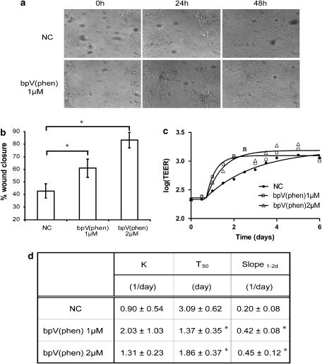
Background and purpose: Epithelial injury contributes to lung pathogenesis. Our work and that of others have identified the phosphoinositide-3 kinase (PI3K)/Akt pathway as a vital component of survival in lung epithelia. Therefore, we hypothesized that pharmacological inhibition of PTEN, a major suppressor of this pathway, would enhance wound closure and restore lung epithelial monolayer integrity following injury.

Experimental approach: We evaluated the ability of two bisperoxovanadium derivatives, bpV(phen) and bpV(pic), in differentiated primary human airway epithelia and BEAS2B cultures for their ability to inhibit PTEN, activate the PI3K/Akt pathway and restore epithelial monolayer integrity following mechanical injury.

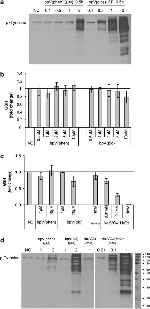
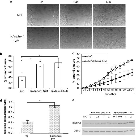
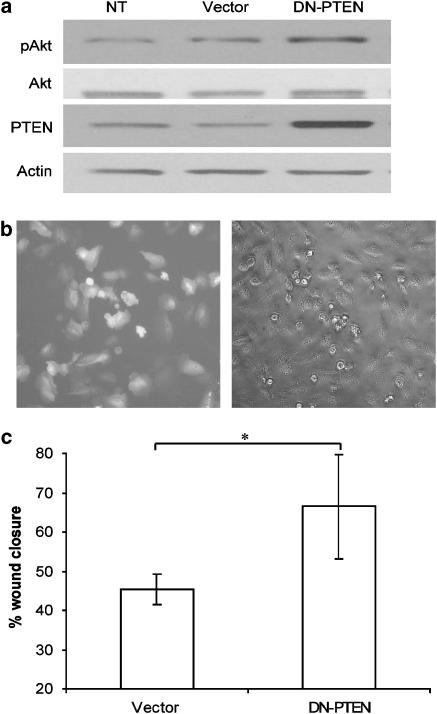
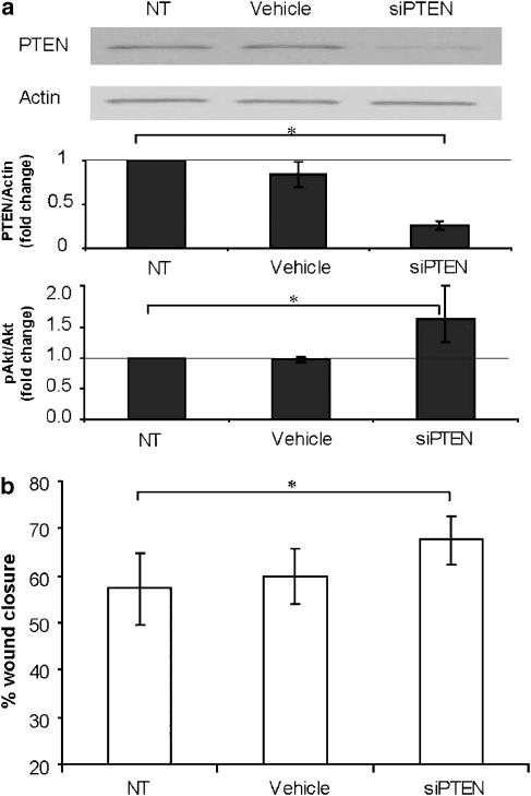
Key results: BpV(phen) and bpV(pic) induced Akt phosphorylation in primary and BEAS2B cells in a dose and time dependent manner. Minimal toxicity was observed as measured by lactate dehydrogenase (LDH) release. To verify that Akt phosphorylation is specifically induced by PTEN inhibition, the PTEN positive cell line, DU145, and two PTEN negative cell lines, LNCaP and PC3, were examined. PTEN positive cells demonstrated a dose responsive increase in Akt phosphorylation whereas PTEN negative cells showed no response indicating that bpV(phen) directly suppresses PTEN without affecting auxiliary pathways. Next, we observed that exposure to either compound resulted in accelerated wound closure following mechanical injury. Similar effects were observed after transfection with a dominant negative isoform of PTEN and PTEN specific siRNA.

Conclusions and implications: From these studies, we conclude that PTEN is a valid target for future studies directed at restoring epithelial barrier function after lung injury.


Figures

Figure 1
Figure 1
Bisperoxovanadium compounds induced Akt phosphorylation. (a) PTEN (phosphatase and tensin homologue deleted on chromosome ten) was constitutively expressed in the lung epithelium. Endogenous PTEN, Akt and phosphorylated Akt protein levels were determined by western analysis in primary human upper airway epithelial cells (hUAECs) and BEAS2B cells, and compared to the PTEN functional cell line, DU145, and a PTEN-null cell line, LNCaP. (b) Potassium bisperoxo (1,10-phenanthroline) oxovanadate (bpV(phen)) induced a dose-dependent increase in Akt phosphorylation in hUAECs within 30 min. Quantitative analysis by densitometry is shown in the bottom panel following comparison of the ratio of phosphorylated Akt to total Akt. PC is the positive control. (c) Similar results are shown in BEAS2B cells following stimulation with bpV(phen) (0.1–10 μM). (d) Direct comparison of bpV(phen) and di-potassium bisperoxo (picolinato) oxovanadate in BEAS2B cells demonstrates that both compounds induced Akt phosphorylation at submicromolar concentrations. The western blots are representative of four experiments; other data are mean±s.d.; n=4.
Figure 2
Figure 2
Prolonged exposure to bisperoxovanadium compounds is associated with minimal cytotoxicity. Cytotoxicity and cell viability were simultaneously determined by lactate dehydrogenase (LDH) and water-soluble tetrazolium salt assays, respectively, following prolonged exposure to potassium bisperoxo (1,10-phenanthroline) oxovanadate (bpV(phen)) and di-potassium bisperoxo (picolinato) oxovanadate (bpV(pic)). In view of the previous results, we evaluated a dosage range between 1 and 5 μM for bpV(phen) and 0.5–5 μM for bpV(pic). (a) Minimal cytotoxicity or alteration in viability was observed following 24-h exposure in BEAS2B cells. (b) Similar results were observed following 48-h exposure to both compounds. The upper right inset compares each compound to complete cell lysis and LDH release with 2% Triton (100% cell death). (c) Again, no detectable toxicity was determined in primary human upper airway epithelial cells under submersion conditions following 96-h exposure to both compounds as measured by LDH release. (d) The same cultures were further evaluated following prolonged exposure to bpV(phen) and demonstrate sustained phosphorylation of Akt above baseline levels in BEAS2B cells. Data are mean±s.d.; n=3 with a representative western blot.
Figure 3
Figure 3
Evaluation of the specificity of bisperoxovanadium compounds for PTEN (phosphatase and tensin homologue deleted on chromosome ten). (a) A dose-dependent increase in Akt phosphorylation was observed with potassium bisperoxo (1,10-phenanthroline) oxovanadate (bpV(phen)) in DU145 cells, a PTEN functional cell line. The dosage range evaluated was between 1 and 5 μM for 0.5 h. Phosphorylated Akt and total Akt levels are shown following western analysis (top panel) and densitometry (bottom panel). (b) We then determined if a similar dose-dependent increase in Akt phosphorylation would be observed with bpV(phen) over an identical dose range in the PTEN-null LNCaP or (c) PC3 cell lines. As before, phosphorylated Akt and total Akt levels were evaluated by western analysis (top panel) and densitometry (bottom panel). In contrast to DU145 cells, bpV(phen) did not induce Akt phosphorylation in PTEN-null cells. The results shown are mean±s.d.; n=3, with representative western blots.
Figure 4
Figure 4
Bisperoxovanadium compounds are specific for PTEN (phosphatase and tensin homologue deleted on chromosome ten) in lung epithelia. (a) BEAS2B cultures were exposed to potassium bisperoxo (1,10-phenanthroline) oxovanadate (bpV(phen)) and di-potassium bisperoxo (picolinato) oxovanadate (bpV(pic)) at a dosage range between 0.1 and 2 μM for 0.5 h. Whole-cell lysates were resolved on a polyacrylamide gel and then evaluated for phosphotyrosine content by western analysis. By direct comparison, bpV(phen) was superior to bpV(pic) in maintaining target specificity over a broader dosage range. (b) In similar experiments, we evaluated the potential for a dose-dependent increase in glutathione content, indicative of a lack of PTEN specificity, over a broader dosage range (0.5–10 μM) for bpV(phen) and bpV(pic) in BEAS2B cultures. Cell lysates were collected after 0.5-h exposure and measured for glutathione content. Again, by direct comparison, bpV(phen) was superior to bpV(pic) in maintaining target specificity over a broader dosage range. (c) As a positive control, we also compared bpV(phen) and bpV(pic) to the less-specific parent compound Na3VO4 following activation with H2O2 (0.01–1 mM) for 0.5 h. H2O2-activated Na3VO4 resulted in a substantial decrease in glutathione content, indicative of broad substrate inhibition. (d) Similarly, a dose-responsive increase in phospho-tyrosine residues as measured by anti-phosphotyrosine staining was observed under the same experimental conditions. Data are mean±s.d.; n=3, with representative Western blots.
Figure 5
Figure 5
Bisperoxovanadium compounds enhance wound closure in the lung epithelium. (a) A 600 μM scrape wound was made in confluent BEAS2B monolayers immediately followed by addition of PTEN (phosphatase and tensin homologue deleted on chromosome ten) inhibitors to cultures. Microscopic images of cultures were obtained over a 48-h period utilizing time-lapse photomicroscopy of live cell cultures. Images shown are of non-treated control or potassium bisperoxo (1,10-phenanthroline) oxovanadate (bpV(phen))-treated cultures at 0 h (left panel), 24 h (middle panel) and 48 h (right panel). Similar images were obtained with both compounds in primary lung epithelial cultures (shown later in Figure 8). (b) Quantitative analysis of wound closure following treatment with PTEN inhibitors in BEAS2B cells demonstrates that 24-h exposure to both bisperoxovanadium compounds resulted in accelerated closure of the lesion when compared to untreated cultures. Wound widths were initially measured immediately after making the lesion (0 h) (100% distance) and then at 24 h after treatment to determine the distance remaining between wound edge margins. Multiple time points were analysed, but only the 24-h data are shown. The percentage of wound closure was then calculated and presented graphically. Both bpV(phen) (1 μM) and di-potassium bisperoxo (picolinato) oxovanadate (bpV(pic)) (0.5 μM) induced a statistically significant enhancement in wound closure when compared to untreated cultures. Data are mean±s.d.; n=3; *P<0.05 vs NC; Student's t-test. (c) Time course of wound closure with or without exposure to bpV(phen) (1 μM) demonstrates accelerated wound closure at early time points, suggesting that cell migration plays a role in wound healing in this model. Multiple time points were analysed every 2 h for 24 h. The percentage of wound closure was calculated as described previously. Data are mean±s.d.; n=3. (d) A migration assay was also utilized to evaluate cell behaviour following exposure to bpV(phen) (1 μM). BEAS2B cells were plated on the apical of migration chamber with or without treatment and incubated at 37 °C to allow migration. Cells that migrated onto the basolateral surface were then dissociated at designated time points and counted. Data are mean±s.d.; n=3; *P<0.05 vs NC; Student's t-test. (e) A simultaneous dose-dependent increase in phospho-glycogen synthase kinase-3 (GSK3), a downstream target of Akt, was observed following 24-h treatment with bpV(phen) or bpV(pic) in BEAS2B cultures. Phospho-GSK3 and total GSK3 levels were analysed by western analysis.
Figure 6
Figure 6
Overexpression of dominant negative (DN) PTEN (phosphatase and tensin homologue deleted on chromosome ten) enhances wound closure in lung epithelia. To determine further if PTEN plays a critical role in wound closure, we transiently overexpressed a DN form of PTEN and examined wound closure rate. (a) Overexpression of DN-PTEN resulted in an increase in PTEN levels and a simultaneous increase in phosphorylated Akt levels in BEAS2B cultures. Total Akt and β-actin levels were unaffected. (b) A transfection efficiency of 70% or greater was routinely achieved as shown by expression of the green fluorescent protein reporter plasmid at 24 h after transfection. (c) Quantitative assessment of wound closure is shown following transfection with the DN-PTEN plasmid. Scrape wounds were made 24 h after transfection and then images were analysed 24 h later. A statistically significant enhancement in wound closure was observed at 24 h in DN-PTEN-transfected cultures when compared to sham-transfected cultures (empty vector). Data are mean±s.d.; n=3; *P<0.05 vs vector; Student's t-test.
Figure 7
Figure 7
Loss of function with PTEN (phosphatase and tensin homologue deleted on chromosome ten) siRNA knockdown enhances wound closure in lung epithelia. To confirm the role of PTEN as a suppressor in lung epithelial wound repair, we utilized siRNA directed at PTEN in scrape wound cultures. (a) Confirmation of successful siRNA-mediated suppression of PTEN protein expression was first done in BEAS2B cultures 48 h after transfection. PTEN protein levels were determined by western analysis (top panel) and measured by densitometry and standardized to β-actin (middle panel). The level of phosphorylated Akt was also evaluated in the same cultures and presented as the ratio of phosphorylated Akt to total Akt (bottom panel). (b) We then evaluated wound closure in cultures following siRNA-mediated PTEN knockdown. Scrape wounds were made 48 h after transfection and images were analysed 24 h later. Similar to previous experiments, a statistically significant enhancement in wound closure was observed following PTEN suppression when compared to untreated or vehicle-treated cultures. Data are mean±s.d.; n=3; *P<0.05 as shown; Student's t-test.
Figure 8
Figure 8
Bisperoxovanadium compounds enhance wound closure in primary human upper airway epithelial cells (hUAECs). (a) A 600 μM scrape wound was made in confluent primary, submersed, cell cultures immediately followed by addition of PTEN (phosphatase and tensin homologue deleted on chromosome ten) inhibitors to cultures. Microscopic images of cultures were obtained over a 48-h period. Images are shown of non-treated control or potassium bisperoxo (1,10-phenanthroline) oxovanadate (bpV(phen))-treated cultures at 0 h (left panel), 24 h (middle panel) and 48 h (right panel). (b) Quantitative analysis of wound closure following treatment with PTEN inhibitors in primary cells under submersion conditions demonstrates that 24-h exposure to both bisperoxovanadium compounds resulted in accelerated closure of the lesion when compared to untreated cultures. The results shown were obtained following measurement of bpV(phen)-treated cultures. Similar results were observed with di-potassium bisperoxo (picolinato) oxovanadate (bpV(pic)) at the same doses (not shown). Again, a statistically significant difference in wound closure was observed following treatment with PTEN inhibitors. Data are mean±s.d.; n=3; *P<0.05 vs NC; analysis of variance. (c) Serial measurements of trans-epithelial electrical resistance (TEER) was also monitored out to 6 days after a mechanical wound was generated in fully differentiated primary hUAECs grown on transwell inserts. In parallel, we compared cultures without any treatment to those receiving bpV(phen) at a dose of 1 and 2 μM. (d) Metric analysis of TEER recovery is expressed by K, TEER recovery rate constant, T50, time to 50% recovery as well as slope1−2d, the linear section of the curve. Data are mean±s.d.; n=3; *P<0.05 vs NC; analysis of variance.

Comment in

Similar articles

Cited by

References

    1. Bao S, Wang Y, Sweeney P, Chaudhuri A, Doseff AI, Marsh CB, et al. Keratinocyte growth factor induces Akt kinase activity and inhibits Fas-mediated apoptosis in A549 lung epithelial cells. Am J Physiol Lung Cell Mol Physiol. 2005;288:L36–L42. - PubMed
    1. Butler M, McKay RA, Popoff IJ, Gaarde WA, Witchell D, Murray SF, et al. Specific inhibition of PTEN expression reverses hyperglycemia in diabetic mice. Diabetes. 2002;51:1028–1034. - PubMed
    1. Cairns P, Okami K, Halachmi S, Halachmi N, Esteller M, Herman JG, et al. Frequent inactivation of PTEN/MMAC1 in primary prostate cancer. Cancer Res. 1997;57:4997–5000. - PubMed
    1. Cantley LC. The phosphoinositide 3-kinase pathway. Science. 2002;296:1655–1657. - PubMed
    1. Cerovac Z, Ban J, Morinville A, Yaccato K, Shaver A, Maysinger D. Activation of MAPK by potassium bisperoxo(1,10-phenanthroline)oxovanadate (V) Neurochem Int. 1999;34:337–344. - PubMed

Publication types

MeSH terms

Substances