

Agonist-dependent cannabinoid receptor signalling in human trabecular meshwork cells
- PMID: 17922024
- PMCID: PMC2095100
- DOI: 10.1038/sj.bjp.0707495
Agonist-dependent cannabinoid receptor signalling in human trabecular meshwork cells
Abstract
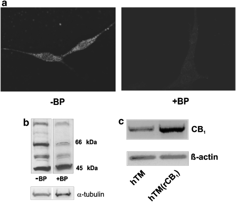
Background and purpose: Trabecular meshwork (TM) is an ocular tissue involved in the regulation of aqueous humour outflow and intraocular pressure (IOP). CB1 receptors (CB1) are present in TM and cannabinoid administration decreases IOP. CB1 signalling was investigated in a cell line derived from human TM (hTM).
Experimental approach: CB1 signalling was investigated using ratiometric Ca2+ imaging, western blotting and infrared In-Cell Western analysis.
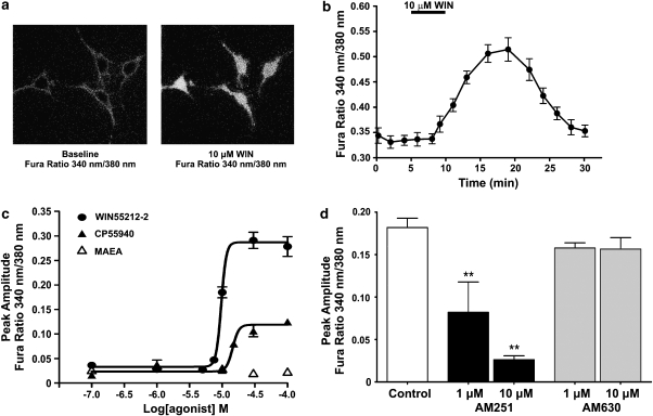
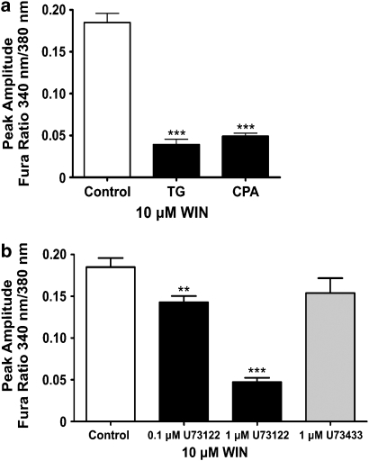
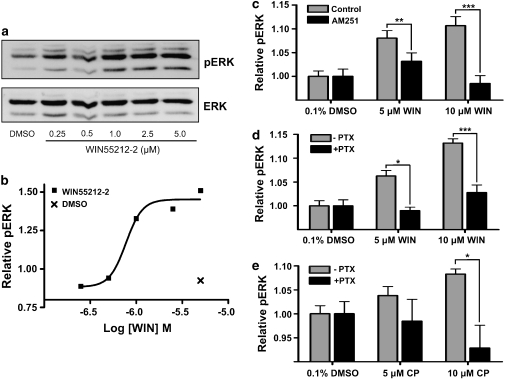
Key results: WIN55212-2, a synthetic aminoalkylindole cannabinoid receptor agonist (10-100 microM) increased intracellular Ca2+ in hTM cells. WIN55,212-2-mediated Ca2+ increases were blocked by AM251, a CB1 antagonist, but were unaffected by the CB2 antagonist, AM630. The WIN55,212-2-mediated increase in [Ca2+]i was pertussis toxin (PTX)-insensitive, therefore, independent of Gi/o coupling, but was attenuated by a dominant negative Galpha(q/11) subunit, implicating a Gq/11 signalling pathway. The increase in [Ca2+]i was dependent upon PLC activation and mobilization of intracellular Ca2+ stores. A PTX-sensitive increase in extracellular signal-regulated kinase (ERK1/2) phosphorylation was also observed in response to WIN55,212-2, indicative of a Gi/o signalling pathway. CB1-Gq/11 coupling to activate PLC-dependent increases in Ca2+ appeared to be specific to WIN55,212-2 and were not observed with other CB1 agonists, including CP55,940 and methanandamide. CP55940 produced PTX-sensitive increases in [Ca2+]i at concentrations>or=15 microM, and PTX-sensitive increases in ERK1/2 phosphorylation.
Conclusions and implications: This study demonstrates that endogenous CB1 couples to both Gq/11 and Gi/o in hTM cells in an agonist-dependent manner. Cannabinoid activation of multiple CB1 signalling pathways in TM tissue could lead to differential changes in aqueous humour outflow and IOP.
Figures

Similar articles
-
The cannabinoid agonist WIN55,212-2 increases intracellular calcium via CB1 receptor coupling to Gq/11 G proteins.Proc Natl Acad Sci U S A. 2005 Dec 27;102(52):19144-9. doi: 10.1073/pnas.0509588102. Epub 2005 Dec 19. Proc Natl Acad Sci U S A. 2005. PMID: 16365309 Free PMC article.
-
Cannabidiol displays unexpectedly high potency as an antagonist of CB1 and CB2 receptor agonists in vitro.Br J Pharmacol. 2007 Mar;150(5):613-23. doi: 10.1038/sj.bjp.0707133. Epub 2007 Jan 22. Br J Pharmacol. 2007. PMID: 17245363 Free PMC article.
-
Anandamide-mediated CB1/CB2 cannabinoid receptor--independent nitric oxide production in rabbit aortic endothelial cells.J Pharmacol Exp Ther. 2007 Jun;321(3):930-7. doi: 10.1124/jpet.106.117549. Epub 2007 Mar 22. J Pharmacol Exp Ther. 2007. PMID: 17379772
-
Stimulation of cannabinoid (CB1) and prostanoid (EP2) receptors opens BKCa channels and relaxes ocular trabecular meshwork.Exp Eye Res. 2005 May;80(5):697-708. doi: 10.1016/j.exer.2004.12.003. Epub 2005 Jan 4. Exp Eye Res. 2005. PMID: 15862177
-
Endothelial atypical cannabinoid receptor: do we have enough evidence?Br J Pharmacol. 2014 Dec;171(24):5573-88. doi: 10.1111/bph.12866. Br J Pharmacol. 2014. PMID: 25073723 Free PMC article. Review.
Cited by
-
Immunolocalization of cannabinoid receptor type 1 and CB2 cannabinoid receptors, and transient receptor potential vanilloid channels in pterygium.Mol Med Rep. 2017 Oct;16(4):5285-5293. doi: 10.3892/mmr.2017.7246. Epub 2017 Aug 14. Mol Med Rep. 2017. PMID: 28849159 Free PMC article.
-
Cannabinoids Stimulate the TRP Channel-Dependent Release of Both Serotonin and Dopamine to Modulate Behavior in C. elegans.J Neurosci. 2019 May 22;39(21):4142-4152. doi: 10.1523/JNEUROSCI.2371-18.2019. Epub 2019 Mar 18. J Neurosci. 2019. PMID: 30886012 Free PMC article.
-
Biased Agonism of Three Different Cannabinoid Receptor Agonists in Mouse Brain Cortex.Front Pharmacol. 2016 Nov 4;7:415. doi: 10.3389/fphar.2016.00415. eCollection 2016. Front Pharmacol. 2016. PMID: 27867358 Free PMC article.
-
Role of ionotropic cannabinoid receptors in peripheral antinociception and antihyperalgesia.Trends Pharmacol Sci. 2009 Feb;30(2):79-84. doi: 10.1016/j.tips.2008.10.008. Epub 2008 Dec 11. Trends Pharmacol Sci. 2009. PMID: 19070372 Free PMC article. Review.
-
Physical and functional interaction between CB1 cannabinoid receptors and beta2-adrenoceptors.Br J Pharmacol. 2010 Jun;160(3):627-42. doi: 10.1111/j.1476-5381.2010.00681.x. Br J Pharmacol. 2010. PMID: 20590567 Free PMC article.
References
-
- Beilin M, Neumann R, Belkin M, Green K, Bar-Ilan A. Pharmacology of the intraocular pressure (IOP) lowering effect of systemic dexanabinol (HU-211), a non-psychotropic cannabinoid. J Ocul Pharmacol Ther. 2000;6:217–230. - PubMed
-
- Berridge M. Inositol trisphosphate and calcium signalling. Nature. 1993;361:315–325. - PubMed
-
- Bisogno T, Delton-Vandenbroucke I, Milone A, Lagarde M, Di Marzo V. Biosynthesis and inactivation of N-arachidonoylethanolamine (anandamide) and N-docosahexaenoylethanolamine in bovine retina. Arch Biochem Biophys. 1999;370:300–307. - PubMed
-
- Bonhaus D, Chang L, Kwan J, Martin G. Dual activation and inhibition of adenylyl cyclase by cannabinoid receptor agonists: evidence for agonist-specific trafficking of intracellular responses. J Pharmacol Exp Ther. 1998;287:884–888. - PubMed
-
- Chen J, Matias I, Dinh T, Lu T, Venezia S, Nieves A, et al. Finding of endocannabinoids in human eye tissues: implications for glaucoma. Biochem Biophys Res Commun. 2005;330:1062–1067. - PubMed
Publication types
MeSH terms
Substances
LinkOut - more resources
Full Text Sources
Other Literature Sources
Miscellaneous
