

Structure and organization of phycobilisomes on membranes of the red alga Porphyridium cruentum
- PMID: 17922299
- PMCID: PMC2173912
- DOI: 10.1007/s11120-007-9264-z
Structure and organization of phycobilisomes on membranes of the red alga Porphyridium cruentum
Abstract
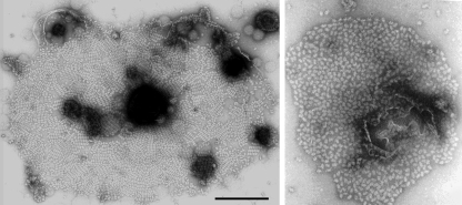
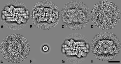
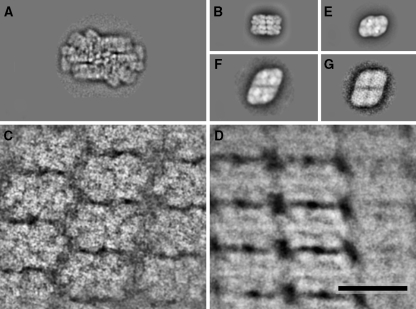
In the present work, electron microscopy and single particle averaging was performed to investigate the supramolecular architecture of hemiellipsoidal phycobilisomes from the unicellular red alga Porphyridium cruentum. The dimensions were measured as 60 x 41 x 34 nm (length x width x height) for randomly ordered phycobilisomes, seen under high-light conditions. The hemiellipsoidal phycobilisomes were found to have a relatively flexible conformation. In closely packed semi-crystalline arrays, observed under low-light conditions, the width is reduced to 31 or 35 nm, about twice the width of the phycobilisome of the cyanobacterium Synechocystis sp. PCC 6803. Since the latter size matches the width of dimeric PSII, we suggest that one PBS lines up with one PSII dimer in cyanobacteria. In red algae, a similar 1:1 ratio under low-light conditions may indicate that the red algal phycobilisome is enlarged by a membrane-bound peripheral antenna which is absent in cyanobacteria.
Figures
References
-
- {'text': '', 'ref_index': 1, 'ids': [{'type': 'DOI', 'value': '10.1007/s11120-004-2143-y', 'is_inner': False, 'url': 'https://doi.org/10.1007/s11120-004-2143-y'}, {'type': 'PubMed', 'value': '15977057', 'is_inner': True, 'url': 'https://pubmed.ncbi.nlm.nih.gov/15977057/'}]}
- Adir N (2005) Elucidation of the molecular structures of components of the phycobilisome: reconstructing a giant. Photosynth Res 85:15–32 - PubMed
-
- {'text': '', 'ref_index': 1, 'ids': [{'type': 'DOI', 'value': '10.1023/B:PRES.0000015399.43503.95', 'is_inner': False, 'url': 'https://doi.org/10.1023/b:pres.0000015399.43503.95'}, {'type': 'PubMed', 'value': '16228392', 'is_inner': True, 'url': 'https://pubmed.ncbi.nlm.nih.gov/16228392/'}]}
- Aspinwall CL, Sarcina M, Mullineaux CW (2004) Phycobilisome mobility in the cyanobacterium Synechococcus sp PCC7942 is influenced by the trimerisation of Photosystem I. Photosynth Res 79:179–187 - PubMed
-
- {'text': '', 'ref_index': 1, 'ids': [{'type': 'DOI', 'value': '10.1007/BF00117661', 'is_inner': False, 'url': 'https://doi.org/10.1007/bf00117661'}, {'type': 'PubMed', 'value': '24271608', 'is_inner': True, 'url': 'https://pubmed.ncbi.nlm.nih.gov/24271608/'}]}
- Bald D, Kruip J, Rögner M (1996) Supramolecular architecture of cyanobacterial thylakoid membranes: how is the phycobilisome connected with the photosystems? Photosynth Res 49:103–118 - PubMed
-
- {'text': '', 'ref_index': 1, 'ids': [{'type': 'DOI', 'value': '10.1039/b300063j', 'is_inner': False, 'url': 'https://doi.org/10.1039/b300063j'}, {'type': 'PubMed', 'value': '12803076', 'is_inner': True, 'url': 'https://pubmed.ncbi.nlm.nih.gov/12803076/'}]}
- Barber J, Morris EP, da Fonseca PCA (2003) Interaction of the allophycocyanin core complex with photosystem II. Photochem Photobiol Sci 2:536–541 - PubMed
-
- {'text': '', 'ref_index': 1, 'ids': [{'type': 'PubMed', 'value': '9165067', 'is_inner': True, 'url': 'https://pubmed.ncbi.nlm.nih.gov/9165067/'}]}
- Betz M (1997) One century of protein crystallography: the phycobiliproteins. Biol Chem 378:167–176 - PubMed
Publication types
MeSH terms
Substances
LinkOut - more resources
Full Text Sources
