. 2007 Dec;5(12):2494-502.
doi: 10.1111/j.1538-7836.2007.02785.x. Epub 2007 Oct 8.

Role of the transmembrane domain of glycoprotein IX in assembly of the glycoprotein Ib-IX complex

Affiliations

Role of the transmembrane domain of glycoprotein IX in assembly of the glycoprotein Ib-IX complex

S-Z Luo et al. J Thromb Haemost. 2007 Dec.

Abstract

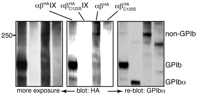
Background: The glycoprotein (GP) Ib-IX complex is critically involved in platelet adhesion to von Willebrand factor and in the initial step of platelet activation. How this complex is assembled is not clear. We previously showed that the transmembrane (TM) domains of the GPIbalpha and GPIbbeta subunits interact and participate in complex assembly.

Objectives and methods: Here, we have investigated the role of the TM and cytoplasmic domains of GPIX in assembly of the GPIb-IX complex, by analyzing the mutational effects on complex expression and assembly in transiently transfected Chinese hamster ovary cells.

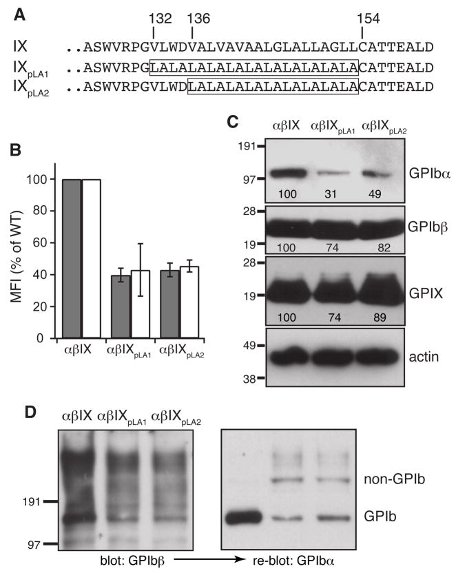
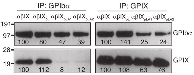
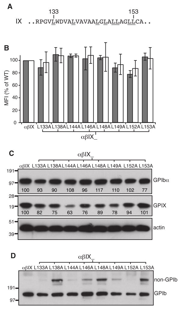
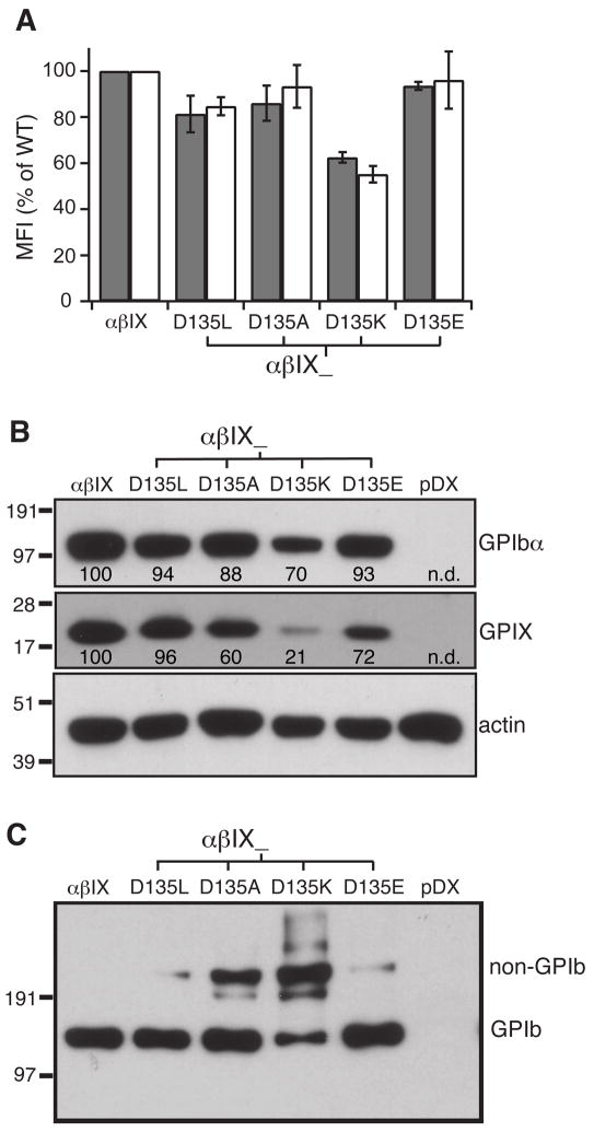
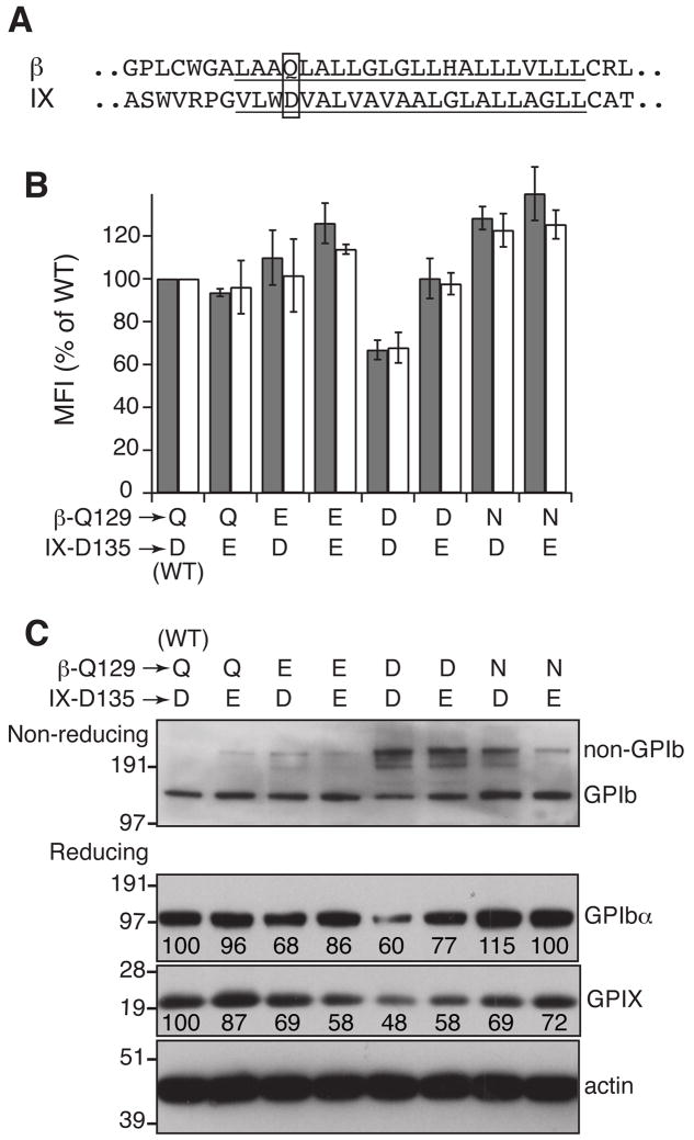
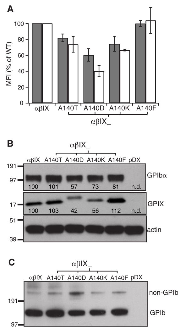
Results: Replacing the cytoplasmic domain of GPIX with a poly-alanine sequence had little effect on surface expression and structural integrity of the GPIb-IX complex. In contrast, replacing the GPIX TM domain (residues 132-153) with a poly-leucine-alanine sequence markedly disrupted complex formation of GPIX with GPIbalpha, interfered with GPIb formation, and decreased surface expression of the host complex. We further analyzed the contributions of a number of GPIX TM residues to complex formation by mutagenesis and found significant roles for Asp135 and several Leu residues.

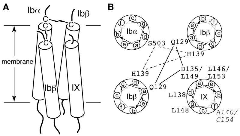
Conclusions: The TM domain, rather than the cytoplasmic domain, of GPIX plays an important role in expression and assembly of the GPIb-IX complex by interacting with its counterparts of GPIb. These TM domains may form a parallel four-helical bundle structure in the complex.


Figures

Fig. 1
Fig. 1. Effect of domain deletion in GPIX on expression and assembly of the GPIb-IX complex in transiently transfected CHO cells
(A) Illustration of mutant GPIX constructs. (B) Surface expression levels of GPIbα (gray bar) and GPIX (white bar) in mutant cells. The vector containing wild type or mutant GPIX cDNA was co-transfected transiently with wild type GPIbα and GPIbβ cDNAs into CHO cells. After 2 days, the cells were harvested, stained by antibodies against GPIbα (AK2) or GPIX (FMC25), and analyzed by flow cytometry. The cells were identified by the subunits transfected. “α” stands for GPIbα, “β” GPIbβ, and “IX” GPIX. The subscript denotes the mutation within the subunit. The mean fluorescence intensity (MFI) were normalized such that the level in wild-type CHOαβIX cells is 100% and that in sham vector transfected CHOpDX cells 0%. The data are calculated from 3 independent experiments and presented as the mean ± s.d.. (C) Overall expression of GPIbα and GPIX in transfected CHO cells. Cell lysates were resolved in reducing SDS gels and transferred to the PVDF membrane. The membrane was probed separately with antibodies against GPIbα (WM23), GPIbβ (Gi27), GPIX (polyclonal), and actin. Band intensities for each complex subunit were quantitated and expressed as a percentage of the wild type level. The numbers below the bands are the average of 3 independent experiments, and the deviation is usually 10%. (D) Effect of domain deletion in GPIX on formation of the GPIbα-GPIbβ disulfide bonds (i.e. GPIb formation). Cell lysates were resolved in non-reducing SDS gels and transferred to PVDF membrane. The membrane was probed with Gi27 first, stripped and re-probed with WM23. The blots are representatives of 2 independent experiments. The membrane was also probed directly with WM23 in 3 additional independent experiments (not shown). The GPIb and non-GPIb bands are indicated on the right.
Fig. 2
Fig. 2. The non-GPIb complex contains GPIbα and GPIbβ
Cell lysates were resolved in a non-reducing 5% Tris-glycine SDS gel and probed with anti-HA antibody for HA-tagged GPIbβ (two exposures shown) and re-probed with WM23 for GPIbα. The blots are representatives of 3 independent experiments. The anti-HA antibody was used instead of Gi27 for slightly better GPIbβ blotting. Addition of the HA epitope tag to the C-terminus of GPIbβ did not affect GPIbα blotting (not shown).
Fig. 3
Fig. 3. Mutational effect on complex expression and assembly by replacement of the GPIX TM domain with pLA sequences
(A) TM sequences in wild type and domain replacement GPIX constructs. The pLA sequence in each mutant is boxed. (B) Surface expression levels of GPIbα (gray) and GPIX (white) in domain replacement cells. Measurement and quantitation followed the description in Fig 1B. (C) Overall expression of GPIbα and GPIX. Cell lysates were resolved in reducing SDS gels. The relative band intensity is the average of 3 independent experiments. (D) GPIb formation in domain replacement cells. Cell lysates were resolved in non-reducing SDS gels and probed as indicated. The blots are representatives of 2 independent experiments.
Fig. 4
Fig. 4. Replacement of the GPIX TM domain with pLA sequences, but not the pL sequence, weakened the interaction between GPIb and GPIX
Cell lysates were co-immunoprecipitated with antibodies AK2 or FMC25, resolved in a reducing SDS gel, and immunoblotted with WM23 or anti-IX polyclonal antibody. Intensities of the GPIbα and GPIX bands were quantitated by densitometry and expressed as a percentage of the wild type level. The numbers below the bands are the average of 3 independent experiments.
Fig. 5
Fig. 5. Certain Leu residues in the GPIX TM domain hampered GPIb formation but not surface expression of the GPIb-IX complex in transfected CHO cells
(A) Sequence of the GPIX TM domain. All Leu residues are underlined with 2 residue numbers marked on top. (B) Surface expression levels of GPIbα (gray) and GPIX (white) in Leu mutant cells. Measurement and quantitation followed the description in Fig 1B. (C) Overall expression of GPIbα and GPIX. Cell lysates were resolved in reducing SDS gels and immunoblotted with antibodies as indicated. The relative band intensity is the average of 3 independent experiments. (D) GPIb formation in Leu mutant cells. Cell lysates were resolved in non-reducing SDS gels and immunoblotted with WM23. The blot shown represents 4 independent experiments.
Fig. 6
Fig. 6. Asp135 in the GPIX TM domain is important to complex assembly and expression
(A) Surface expression levels of GPIbα (gray) and GPIX (white), as well as (B) overall expression of GPIbα and GPIX, in Asp135 mutant cells were measured as described earlier. The relative intensity marked under each band is the average of 3 independent experiments. (C) GPIb formation in Asp135 mutant cells. Cell lysates were resolved in non-reducing SDS gels and immunoblotted with WM23. The blot shown represents 3 independent experiments.
Fig. 7
Fig. 7. Probing the interaction between Asp135 of GPIX and Gln129 of GPIbβ
(A) The TM sequences (underlined) of GPIbβ and GPIX. Residues Gln129 of GPIbβ and Asp135 of GPIX are boxed. (B,C) Effects of mutating Gln129 and Asp135 to certain polar residues on surface expression levels of GPIbα (gray bar) and GPIX (white bar), GPIb formation as well as overall expression of the complex. The data shown were either the average or a representative of 3–4 independent experiments.
Fig. 8
Fig. 8. Ala140 in the GPIX TM domain is not critical to assembly and surface expression of the GPIb-IX complex
Ala140 was mutated to Thr, Asp, Lys, or Phe, and the mutational effects on the surface (A) and overall (B) expression level of GPIbα (gray bar) and GPIX (white bar), as well as (C) the effects on GPIb formation were characterized. For some reason, GPIXA140D migrated at a pace different from the other variants. The data shown were either the average or a representative of 4 independent experiments.
Fig. 9
Fig. 9. A four-helical bundle model for the TM domains of the GPIb-IX complex
(A) The side view of the TM bundle. Each TM helix is shown as a rod traversing the membrane bilayer. (B) The helical wheel diagram of the TM bundle. The possible interactions among the polar residues are marked by lines. The residues in GPIX that play an role in modulating GPIb formation, as well as A140 and Cys154 (Italic), are marked.

References

    1. Berndt MC, Shen Y, Dopheide SM, Gardiner EE, Andrews RK. The vascular biology of the glycoprotein Ib-IX-V complex. Thromb Haemost. 2001;86:178–88. - PubMed
    1. Kanaji T, Russell S, Ware J. Amelioration of the macrothrombocytopenia associated with the murine Bernard-Soulier syndrome. Blood. 2002;100:2102–7. - PubMed
    1. Hoffmeister KM, Felbinger TW, Falet H, Denis CV, Bergmeier W, Mayadas TN, von Andrian UH, Wagner DD, Stossel TP, Hartwig JH. The clearance mechanism of chilled blood platelets. Cell. 2003;112:87–97. - PubMed
    1. Lopez JA, Chung DW, Fujikawa K, Hagen FS, Papayannopoulou T, Roth GJ. Cloning of the alpha chain of human platelet glycoprotein Ib: a transmembrane protein with homology to leucine-rich alpha 2-glycoprotein. Proc Natl Acad Sci USA. 1987;84:5615–9. - PMC - PubMed
    1. Lopez JA, Chung DW, Fujikawa K, Hagen FS, Davie EW, Roth GJ. The alpha and beta chains of human platelet glycoprotein Ib are both transmembrane proteins containing a leucine-rich amino acid sequence. Proc Natl Acad Sci USA. 1988;85:2135–9. - PMC - PubMed

Publication types

MeSH terms