. 2008 Mar;123(3):398-410.
doi: 10.1111/j.1365-2567.2007.02705.x. Epub 2007 Oct 6.

Neuropeptides activate human mast cell degranulation and chemokine production

Affiliations

Neuropeptides activate human mast cell degranulation and chemokine production

Marianna Kulka et al. Immunology. 2008 Mar.

Abstract

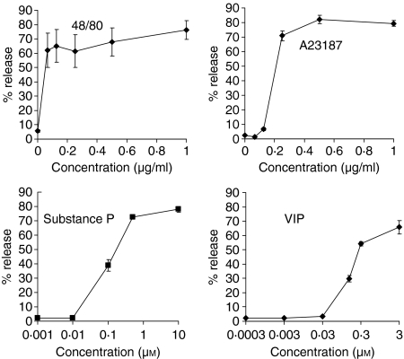
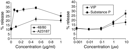
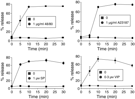
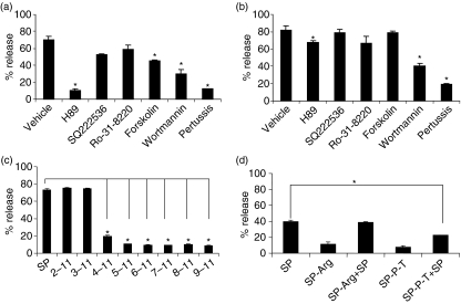
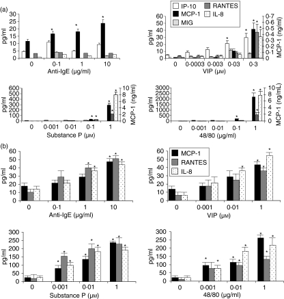
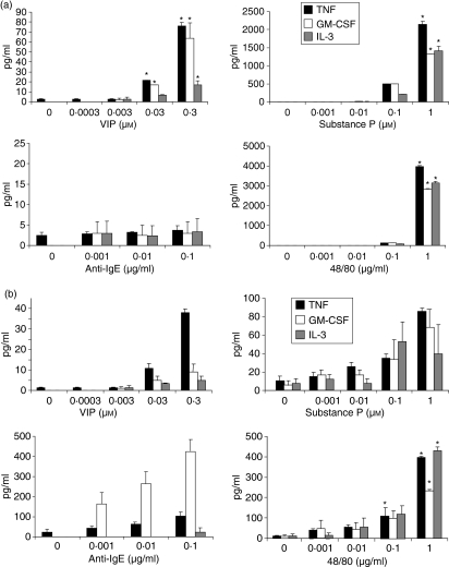
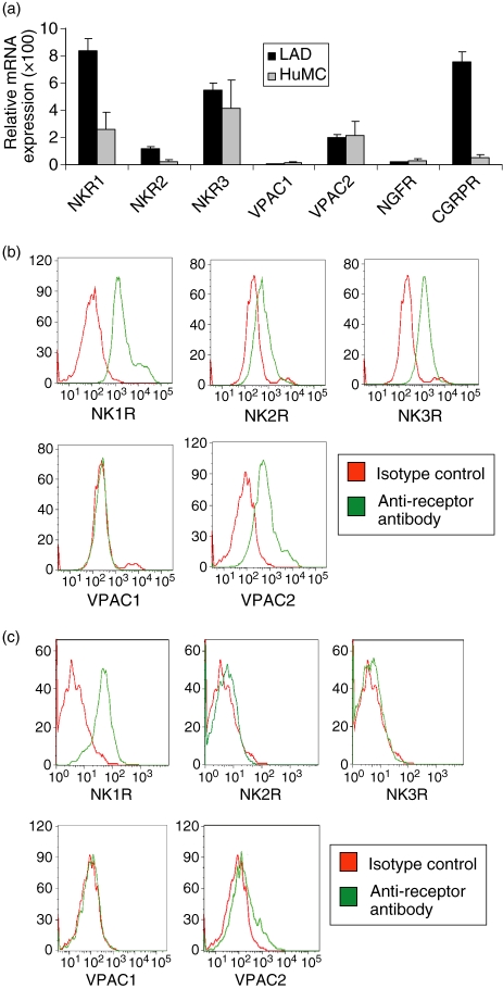
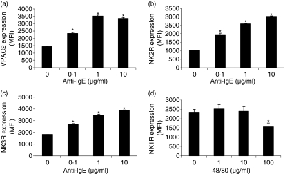
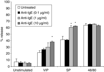
During neuronal-induced inflammation, mast cells may respond to stimuli such as neuropeptides in an FcepsilonRI-independent manner. In this study, we characterized human mast cell responses to substance P (SP), nerve growth factor (NGF), calcitonin gene-related peptide (CGRP) and vasoactive intestinal polypeptide (VIP) and compared these responses to human mast cell responses to immunoglobulin E (IgE)/anti-IgE and compound 48/80. Primary cultured mast cells, generated from CD34(+) progenitors in the presence of stem cell factor and interleukin-6 (IL-6), and human cultured mast cells (LAD2) were stimulated with these and other stimuli (gastrin, concanavalin A, radiocontrast media, and mannitol) and their degranulation and chemokine production was assessed. VIP and SP stimulated primary human mast cells and LAD cells to degranulate; gastrin, concanavalin A, radiocontrast media, mannitol, CGRP and NGF did not activate degranulation. While anti-IgE stimulation did not induce significant production of chemokines, stimulation with VIP, SP or compound 48/80 potently induced production of monocyte chemoattractant protein-1, inducible protein-10, monokine induced by interferon-gamma (MIG), RANTES (regulated on activation, normal, T-cell expressed, and secreted) and IL-8. VIP, SP and compound 48/80 also activated release of tumour necrosis factor, IL-3 and granulocyte-macrophage colony-stimulating factor, but not IL-4, interferon-gamma or eotaxin. Human mast cells expressed surface neurokinin 1 receptor (NK1R), NK2R, NK3R and VIP receptor type 2 (VPAC2) but not VPAC1 and activation of human mast cells by IgE/anti-IgE up-regulated expression of VPAC2, NK2R, and NK3R. These studies demonstrate the pattern of receptor expression and activation of mast cell by a host of G-protein coupled receptor ligands and suggest that SP and VIP activate a unique signalling pathway in human mast cells. These results are likely to have direct relevance to neuronally induced inflammatory diseases.


Figures

Figure 1
Figure 1
Neuropeptides activate LAD2 human mast cell degranulation. LAD2 cells were activated with compound 48/80, A23187, substance P and VIP for 30 min and β-hexosaminidase release was measured (n = 3, P < 0·01).
Figure 2
Figure 2
Neuropeptides activate CD34+ progenitor-derived human mast cell degranulation. Human mast cells were cultured from CD34+ progenitor cells as described in Materials and methods. Cultured mast cells were activated by compound 48/80, A23187, substance P and VIP for 30 min and β-hexosaminidase release was measured (n = 3, P < 0·01). c.p.s., counts per second.
Figure 3
Figure 3
Time course of degranulation. LAD2 cells were stimulated with compound 48/80 (a), A23187 (b), substance P and VIP (at 1 µm and 0·3 µm, respectively; circles) or untreated (squares) for indicated times and β-hexosaminidase release was measured (n = 3, P < 0·01).
Figure 4
Figure 4
Mechanisms of neuropeptide activation. (a, b) Activation by neuropeptides is sensitive to inhibitors of G proteins and PI3 kinase. (a) LAD2 cells were pretreated with vehicle (0·1% DMSO), H89 (1 µm), SQ222536 (10 µm), Ro-31–8220 (100 nm), forskolin (10 µm), wortmannin (10 µm) or pertussis toxin (3·5 nm), stimulated with either VIP (0·3 µm) or (b) SP (1 µm) for 30 min and β-hexosaminidase release was measured (n = 3, P < 0·01; P-value calculated relative to vehicle alone). (c) Truncated substance P peptides activate human mast cell degranulation. LAD2 cells were activated with 1 µg/ml of indicated peptides (see also Table 2) for 30 min and β-hexosaminidase release was measured (n = 3, P < 0·01; P-value calculated relative to entire SP peptide). (d) NK1R peptide inhibitors block substance P activation of human mast cells. LAD2 cells were pretreated with 1 µg/ml of inhibitor (SP-Arg or SP-P-T, see Table 2) for 30 min, then activated with substance P (0·1 µm) for 30 min and β-hexosaminidase release was measured (n = 3, P < 0·01; P-value calculated relative to SP alone).
Figure 5
Figure 5
Neuropeptides activate LAD2 cells to produce chemokines. (a) LAD2 cells were stimulated with IgE/anti-IgE (see legend, Fig. 4), substance P, VIP, or compound 48/80 for 24 hr and IP-10, MCP-1, MIG, RANTES and IL-8 production was measured in cell-free supernatants (n = 3, P < 0·01). (b) HuMC were stimulated as above and IP-10, MCP-1, MIG, RANTES and IL-8 production was measured in cell free supernatants (n = 3, P < 0·01).
Figure 6
Figure 6
Neuropeptides activate LAD2 cells to produce cytokines. (a) LAD2 cells were stimulated with IgE/anti-IgE (see legend, Fig. 4), substance P, VIP, or compound 48/80 for 24 hr and TNF, GM-CSF and IL-3 production were measured in cell free supernatants (n = 3, P < 0·01). (b) HuMC were stimulated as above and TNF, GM-CSF and IL-3 production were measured in cell-free supernatants (n = 3, P < 0·01).
Figure 7
Figure 7
LAD2 cells express receptors for VIP and substance P. (a) Receptor mRNA expression was measured by real-time PCR analysis of LAD2 and HuMC total RNA. (b) Surface expression of NK1R, NK2R, NK3R, VPAC1 and VPAC2 by LAD2 cells (b) and HuMC (c) was measured by flow cytometry.
Figure 8
Figure 8
Stimulation with anti-IgE or compound 48/80 modifies neuropeptide receptor expression by LAD2 cells. (a–c) LAD2 cells were stimulated with IgE/anti-IgE or for 24 hr and expression of VPAC2, NK2R and NK3R was measured by flow cytometry. (d) LAD2 cells were stimulated with compound 48/80 for 24 hr and expression of NK1R was measured by flow cytometry. Expression is presented as mean fluorescence intensity (MFI; n = 3, P < 0·01).
Figure 9
Figure 9
Preincubation of human mast cells with IgE/anti-IgE renders them more sensitive to SP and VIP stimulation. Sensitized LAD2 cells were preincubated with anti-IgE (10 µg/ml) for 24 hr, then stimulated with either SP (1 µm), VIP (0·3 µm) or compound 48/80 (1 µg/ml) and degranulation was measured. (n = 3, P < 0·01; P-value calculated relative to agonist alone).

Similar articles

Cited by

References

    1. Bienenstock J, MacQueen G, Sestini P, Marshall JS, Stead RH, Perdue MH. Mast cell/nerve interactions in vitro and in vivo. Am Rev Respir Dis. 1991;143:S55–S58. - PubMed
    1. Singh LK, Pang X, Alexacos N, Letourneau R, Theoharides TC. Acute immobilization stress triggers skin mast cell degranulation via corticotropin releasing hormone, neurotensin, and substance P. A link to neurogenic skin disorders. Brain Behav Immun. 1999;13:225–39. - PubMed
    1. Ferry X, Brehin S, Kamel R, Landry Y. G protein-dependent activation of mast cell by peptides and basic secretagogues. Peptides. 2002;23:1507–15. - PubMed
    1. Janiszewski J, Bienenstock J, Blennerhassett MG. Picomolar doses of substance P trigger electrical responses in mast cells without degranulation. Am J Physiol – Cell Physiol. 1994;267:C138–C145. - PubMed
    1. Okayama Y, Ono Y, Nakazawa T, Church MK, Mori M. Human skin mast cells produce TNF-alpha by substance P. Int Arch Allergy Immunol. 1998;117:48–51. - PubMed

Publication types

MeSH terms