

Chromogenic in situ hybridisation for the assessment of HER2 status in breast cancer: an international validation ring study
- PMID: 17922920
- PMCID: PMC2242665
- DOI: 10.1186/bcr1776
Chromogenic in situ hybridisation for the assessment of HER2 status in breast cancer: an international validation ring study
Abstract
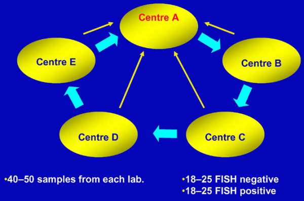
Introduction: Before any new methodology can be introduced into the routine diagnostic setting it must be technically validated against the established standards. To this end, a ring study involving five international pathology laboratories was initiated to validate chromogenic in situ hybridisation (CISH) against fluorescence in situ hybridisation (FISH) and immunohistochemistry (IHC) as a test for assessing human epidermal growth factor receptor 2 (HER2) status in breast cancer.
Methods: Each laboratory performed CISH, FISH and IHC on its own samples. Unstained sections from each case were also sent to another participating laboratory for blinded retesting by CISH ('outside CISH').
Results: A total of 211 invasive breast carcinoma cases were tested. In 76 cases with high amplification (HER2/CEP17 ratio >4.0) by FISH, 73 cases (96%) scored positive (scores >or= 6) by 'outside CISH'. For FISH-negative cases (HER2/CEP17 ratio <2.0), 94 of 100 cases (94%) had CISH scores indicating no amplification (score <or= 5), and only three cases were positive by CISH; in the three remaining cases, no CISH result could be obtained. For cases with low-level amplification using FISH (HER2/CEP17 ratio 2.0-4.0), 20 of 35 had CISH scores indicating gene amplification. Inter-laboratory concordance was also very high: 95% for normal HER2 copy number (1-5 copies); and 92% for cases with HER2 copy numbers >or= 6. CISH intra-laboratory concordance with IHC was 92% for IHC-negative cases (IHC 0/1+) and 91% for IHC 3+ cases. Among IHC 2+ cases, CISH was 100% concordant with samples showing high amplification by FISH, and 94% concordant with FISH-negative samples.
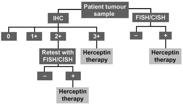
Conclusion: These results show that CISH inter- and intra-laboratory concordance to FISH and IHC is very high, even in equivocal IHC 2+ cases. Therefore, we conclude that CISH is a methodology that is a viable alternative to FISH in the HER2 testing algorithm.
Figures
References
-
- Hynes NE, Stern DF. The biology of erbB-2/neu/HER-2 and its role in cancer. Biochim Biophys Acta. 1994;1198:165–184. - PubMed
-
- Nagai MA, Pacheco MM, Oshima CT, Brentani MM. c-erbB-2 DNA amplification and mRNA expression in human primary breast tumors and its relationship to other prognostic factors. Cancer Biother. 1993;8:29–35. - PubMed
-
- Pauletti G, Godolphin W, Press MF, Slamon DJ. Detection and quantitation of HER-2/neu gene amplification in human breast cancer archival material using fluorescence in situ hybridization. Oncogene. 1996;13:63–72. - PubMed
Publication types
MeSH terms
Substances
LinkOut - more resources
Full Text Sources
Medical
Research Materials
Miscellaneous
