

Early postnatal development of corpus callosum and corticospinal white matter assessed with quantitative tractography
- PMID: 17923457
- PMCID: PMC8134187
- DOI: 10.3174/ajnr.a0751
Early postnatal development of corpus callosum and corticospinal white matter assessed with quantitative tractography
Abstract
Background and purpose: The early postnatal period is perhaps the most dynamic phase of white matter development. We hypothesized that the early postnatal development of the corpus callosum and corticospinal tracts could be studied in unsedated healthy neonates by using novel approaches to diffusion tensor imaging (DTI) and quantitative tractography.
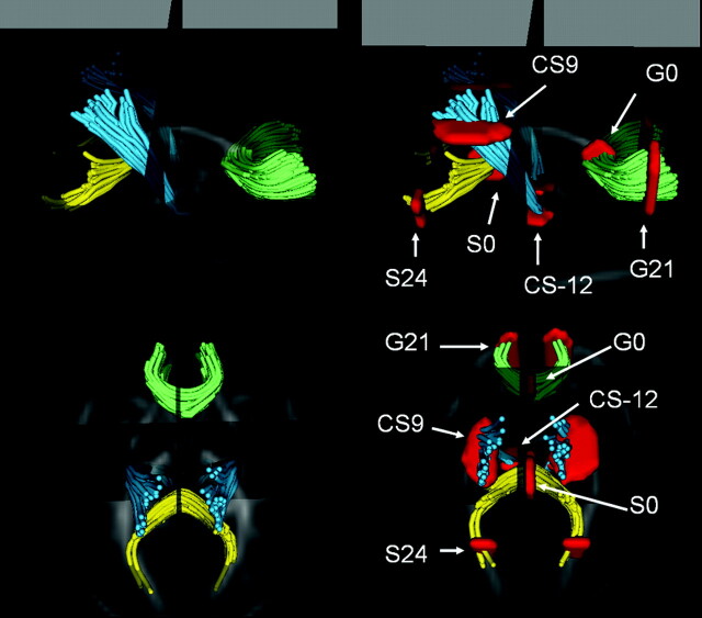
Materials and methods: Isotropic 2 x 2 x 2 mm(3) DTI and structural images were acquired from 47 healthy neonates. DTI and structural images were coregistered and fractional anisotropy (FA), mean diffusivity (MD), and normalized T1-weighted (T1W) and T2-weighted (T2W) signal intensities were determined in central midline and peripheral cortical regions of the white matter tracts of the genu and splenium of the corpus callosum and the central midbrain and peripheral cortical regions of the corticospinal tracts by using quantitative tractography.
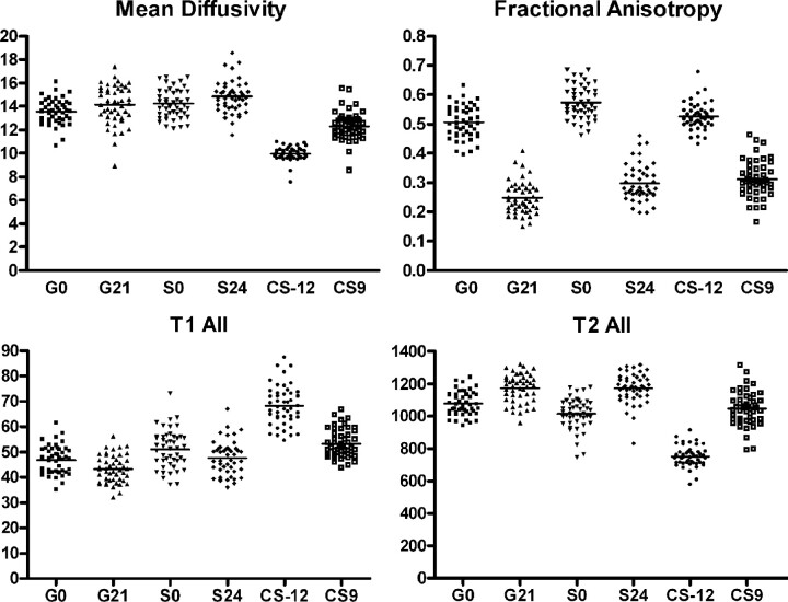
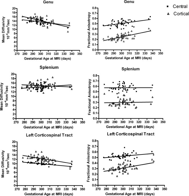
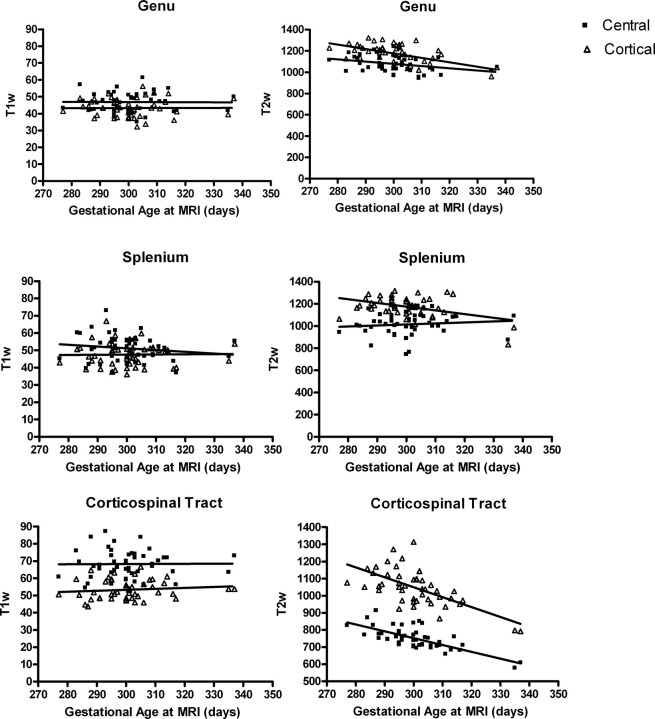
Results: We observed that central regions exhibited lower MD, higher FA values, higher T1W intensity, and lower T2W intensity than peripheral cortical regions. As expected, MD decreased, FA increased, and T2W signal intensity decreased with increasing age in the genu and corticospinal tract, whereas there was no significant change in T1W signal intensity. The central midline region of the splenium fiber tract has a unique pattern, with no change in MD, FA, or T2W signal intensity with age, suggesting different growth trajectory compared with the other tracts. FA seems to be more dependent on tract organization, whereas MD seems to be more sensitive to myelination.
Conclusions: Our novel approach may detect small regional differences and age-related changes in the corpus callosum and corticospinal white matter tracts in unsedated healthy neonates and may be used for future studies of pediatric brain disorders that affect developing white matter.
Figures
Similar articles
-
Diffusion MRI parameters of corpus callosum and corticospinal tract in neonates: Comparison between region-of-interest and whole tract averaged measurements.Eur J Paediatr Neurol. 2018 Sep;22(5):807-813. doi: 10.1016/j.ejpn.2018.05.003. Epub 2018 May 16. Eur J Paediatr Neurol. 2018. PMID: 29804802 Free PMC article.
-
White matter alterations and their associations with motor function in young adults born preterm with very low birth weight.Neuroimage Clin. 2017 Oct 4;17:241-250. doi: 10.1016/j.nicl.2017.10.006. eCollection 2018. Neuroimage Clin. 2017. PMID: 29159041 Free PMC article.
-
White matter microstructural abnormality in children with hydrocephalus detected by probabilistic diffusion tractography.AJNR Am J Neuroradiol. 2013 Dec;34(12):2379-85. doi: 10.3174/ajnr.A3737. Epub 2013 Sep 26. AJNR Am J Neuroradiol. 2013. PMID: 24072621 Free PMC article.
-
The role of diffusion tensor imaging and fractional anisotropy in the evaluation of patients with idiopathic normal pressure hydrocephalus: a literature review.Neurosurg Focus. 2016 Sep;41(3):E12. doi: 10.3171/2016.6.FOCUS16192. Neurosurg Focus. 2016. PMID: 27581308 Review.
-
Application of Diffusion Tensor Imaging (DTI) in the Diagnosis of HIV-Associated Neurocognitive Disorder (HAND): A Meta-Analysis and a System Review.Front Neurol. 2022 Jul 7;13:898191. doi: 10.3389/fneur.2022.898191. eCollection 2022. Front Neurol. 2022. PMID: 35873786 Free PMC article.
Cited by
-
Towards Analysis of Growth Trajectory through Multi-modal Longitudinal MR Imaging.Proc SPIE Int Soc Opt Eng. 2010 Mar 12;7623:76232U-. doi: 10.1117/12.844526. Proc SPIE Int Soc Opt Eng. 2010. PMID: 24353376 Free PMC article.
-
Changes of MR and DTI appearance in early human brain development.Proc SPIE Int Soc Opt Eng. 2010 Mar 12;7623:762324-. doi: 10.1117/12.844912. Proc SPIE Int Soc Opt Eng. 2010. PMID: 24353378 Free PMC article.
-
MULTIVARIATE MODELING OF LONGITUDINAL MRI IN EARLY BRAIN DEVELOPMENT WITH CONFIDENCE MEASURES.Proc IEEE Int Symp Biomed Imaging. 2013:1400-1403. doi: 10.1109/ISBI.2013.6556795. Proc IEEE Int Symp Biomed Imaging. 2013. PMID: 23959506 Free PMC article.
-
Using machine learning to identify air pollution exposure profiles associated with early cognitive skills among U.S. children.Environ Pollut. 2017 Nov;230:730-740. doi: 10.1016/j.envpol.2017.07.023. Epub 2017 Jul 18. Environ Pollut. 2017. PMID: 28732336 Free PMC article.
-
Dynamics of infant white matter maturation from birth to 6 months.bioRxiv [Preprint]. 2025 Feb 13:2025.02.13.638114. doi: 10.1101/2025.02.13.638114. bioRxiv. 2025. PMID: 39990497 Free PMC article. Preprint.
References
-
- Huttenlocher PR, Dabholkar AS. Regional differences in synaptogenesis in human cerebral cortex. J Comp Neurol 1997;387:167–78 - PubMed
-
- Sampaio RC, Truwit CL. Myelination in the developing brain. In: Nelson CA, Luciana M, eds. Handbook of Developmental Cognitive Neuroscience. Cambridge, Mass: MIT Press;2001. :35–44
-
- Kagen J, Herschkowitz N. A Young Mind in a Growing Brain. Mahwah, NJ: Lawrence Erlbaum Associates;2005
-
- Yakovlev PI, Lecours AR. The myelogenetic cycles of regional brain maturation of the brain. In: Mankowski A, editor. Regional Development of the Brain in Early Life. Philadelphia: Davis;1967. :3–69
Publication types
MeSH terms
Grants and funding
LinkOut - more resources
Full Text Sources
Other Literature Sources
Medical
Research Materials