. 2007 Oct 29;204(11):2529-36.
doi: 10.1084/jem.20070462. Epub 2007 Oct 8.

A novel subset of putative stem/progenitor CD34+Oct-4+ cells is the major target for SARS coronavirus in human lung

Affiliations

A novel subset of putative stem/progenitor CD34+Oct-4+ cells is the major target for SARS coronavirus in human lung

Yongxiong Chen et al. J Exp Med. .

Abstract

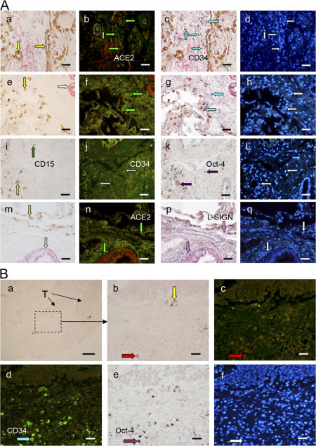
Identification of the nature of severe acute respiratory syndrome (SARS)-infected cells is crucial toward understanding the pathogenesis. Using multicolor colocalization techniques, we previously reported that SARS(+) cells in the lung of fatally infected patients expressed the only known functional receptor, angiotensin-converting enzyme 2, and also a binding receptor, liver/lymph node-specific ICAM-3-grabbing non-integrin (CD209L). In this study, we show that SARS-infected cells also express the stem/progenitor cell markers CD34 and Oct-4, and do not express cytokeratin or surfactant. These putative lung stem/progenitor cells can also be identified in some non-SARS individuals and can be infected by SARS-coronavirus ex vivo. Infection of these cells may contribute to the loss of lung repair capacity that leads to respiratory failure as clinically observed.


Figures

Figure 1.
Figure 1.
SARS+ cells do not express cytokeratin or surfactant in alveoli (a–f) and terminal bronchioles (j–L). SARS lung autopsy samples studied by IHC for surfactant A (brown labeling in a, b, d, and j; some indicated by yellow arrows), for SARS-CoV nucleocapsid antigen (red labeling; some indicated by red arrows in a, b, d, and j; also visible in e and k), and immunofluorescence for cytokeratin (green labeling in e and k; some indicated by white arrows). The black circle in d indicates where cytokeratin+ cells are visualized in e. The yellow circle in e indicates where surfactant+ cells are visualized in d. DAPI counterstaining showing the cellular nuclei is in the right column. Isotype antibody control for IHC and immunofluorescence is in g and h, respectively. Some SARS+ cells were actually “overlapping” with adjacent cytokeratin+ cells (one indicated by a circle in k), which might be mistakenly regarded “colocalized” on two-dimensional photos. Panels of each row represent results of the same section from a patient. Results are representative of six SARS patients who died within 11 d after disease onset. Bar, 20 μm.
Figure 2.
Figure 2.
SARS+ cells in the alveoli express ACE2, CD34, and Oct-4. (a–c) IHC on SARS autopsy lung for CD68 (brown labeling, some indicated by dark blue arrows in a) and for SARS-CoV antigen (red labeling, some indicated by red arrows in a; also visible in b and c), followed by FISH for ACE2 (green labeling in b, some indicated by green arrows and a circle) and ISH for CD34 (purple labeling in c, some indicated by light blue arrows and a circle). Circles in a, b, and c indicate uninfected ACE2+CD34+ cells. (e–g) Isotype antibody control for IHC, ACE2 sense control for FISH, and CD34 sense control for ISH, respectively. (i–k) IHC for SARS-CoV antigen (red labeling, some indicated by red arrows in i; also visible in j and k), followed by FISH for CD34 (green labeling in j, some indicated by light blue arrows and circles), and ISH for Oct-4 (purple labeling in k, some indicated by purple arrows and circles). Circles in j and k indicate uninfected CD34+Oct-4+ cells. Isotype antibody control for IHC, CD34 sense control for FISH, and Oct-4 sense control for ISH are similar to e, f, and g, respectively (not depicted). DAPI counterstaining is in the right column. Panels of each row represent results on the same section of a patient. Results are representative of six SARS patients. Bar, 20 μm.
Figure 3.
Figure 3.
ACE2+CD34+Oct-4+L-SIGN+ cells are present in non-SARS lung and can be infected by SARS CoV ex vivo. (A, a–d) IHC on non-SARS lung samples for surfactant A (brown labeling, some indicated by yellow arrows in a; also visible in c), CD31 (red labeling in a; also visible in b and c), followed by FISH for ACE2 (green labeling in b, some indicated by green arrows) and ISH for CD34 (purple labeling in c, some indicated by light blue arrows). DAPI counterstaining is in d. Circle in b indicates autofluorescence from red blood cells. (e–h) IHC for surfactant A (brown labeling, one indicated by a yellow arrow in e; also visible in g) and for cytokeratin (red labeling, one indicated by a white arrow in e; also visible in f and g), followed by FISH for ACE2 (green labeling in f, some indicated by green arrows) and ISH for CD34 (purple labeling in g, some indicated by light blue arrows). DAPI counterstaining is in h. (i–L) IHC for surfactant A (brown labeling in i, one indicated by a yellow arrow; also visible in k) and for CD15 (red labeling, one indicated by a dark green arrow in i; also visible in j and k), followed by FISH for CD34 (green labeling in j, some indicated by light blue arrows) and ISH for Oct-4 (purple labeling in k, some indicated by purple arrows). DAPI counterstaining is in L. One of the CD34+Oct-4+ cells is juxtaposed to a surfactant+ cell (the yellow arrow in i vs. the lower light blue arrow in j and the lower purple arrow in k). (m–q) IHC for surfactant A (brown labeling, one indicated by a yellow arrow in m; also visible in p) and for cytokeratin (red labeling, one indicated by a white arrow in m; also visible in n and p), followed by FISH for ACE2 (green labeling, some indicated by green arrows in n) and ISH for L-SIGN (purple labeling, some indicated by pink arrows in p). DAPI counterstaining is in q. Some of the ACE2+CD34+Oct4+L-SIGN+ cells are close to surfactant A+ cells (g and k) or cytokeratin+ cells (p). Panels of each row represent results on the same section of a patient. Results are representative of eight patients with lung cancer. Bar, 20 μm. (B) Fresh lung tissues resected from cancer patients were cut into small blocks, exposed to SARS-CoV for 16 h, and studied by IHC for surfactant A (brown labeling in a and b, one indicated by a yellow arrow in b; also visible in e), for SARS antigen (red labeling, indicated by a red arrow in b; also visible in c), followed by FISH for CD34 (green labeling, one indicated by a light blue arrow in d) and ISH for Oct-4 (purple labeling, one indicated by a purple arrow in e). T, tumor. DAPI counterstaining is in f. Results are representative in four of six individuals enrolled. No CD34+ cells could be detected in samples from the other two individuals. Bars: a, 100 μm; b–f, 20 μm.

References

    1. Peiris, J.S., S.T. Lai, L.L. Poon, Y. Guan, L.Y. Yam, W. Lim, J. Nicholls, W.K. Yee, W.W. Yan, M.T. Cheung, et al. 2003. Coronavirus as a possible cause of severe acute respiratory syndrome. Lancet. 361:1319–1325. - PMC - PubMed
    1. Ksiazek, T.G., D. Erdman, C.S. Goldsmith, S.R. Zaki, T. Peret, S. Emery, S. Tong, C. Urbani, J.A. Comer, W. Lim, et al. 2003. A novel coronavirus associated with severe acute respiratory syndrome. N. Engl. J. Med. 348:1953–1966. - PubMed
    1. Drosten, C., S. Gunther, W. Preiser, S. van der Werf, H.R. Brodt, S. Becker, H. Rabenau, M. Panning, L. Kolesnikova, R.A. Fouchier, et al. 2003. Identification of a novel coronavirus in patients with severe acute respiratory syndrome. N. Engl. J. Med. 348:1967–1976. - PubMed
    1. Li, W., M.J. Moore, N. Vasilieva, J. Sui, S.K. Wong, M.A. Berne, M. Somasundaran, J.L. Sullivan, K. Luzuriaga, T.C. Greenough, et al. 2003. Angiotensin-converting enzyme 2 is a functional receptor for the SARS coronavirus. Nature. 426:450–454. - PMC - PubMed
    1. Peiris, J.S., C.M. Chu, V.C. Cheng, K.S. Chan, I.F. Hung, L.L. Poon, K.I. Law, B.S. Tang, T.Y. Hon, C.S. Chan, et al. 2003. Clinical progression and viral load in a community outbreak of coronavirus-associated SARS pneumonia: a prospective study. Lancet. 361:1767–1772. - PMC - PubMed

Publication types

MeSH terms