. 2007 Oct 29;204(11):2561-8.
doi: 10.1084/jem.20070991. Epub 2007 Oct 8.

Development and function of murine B220+CD11c+NK1.1+ cells identify them as a subset of NK cells

Affiliations

Development and function of murine B220+CD11c+NK1.1+ cells identify them as a subset of NK cells

Amanda L Blasius et al. J Exp Med. .

Abstract

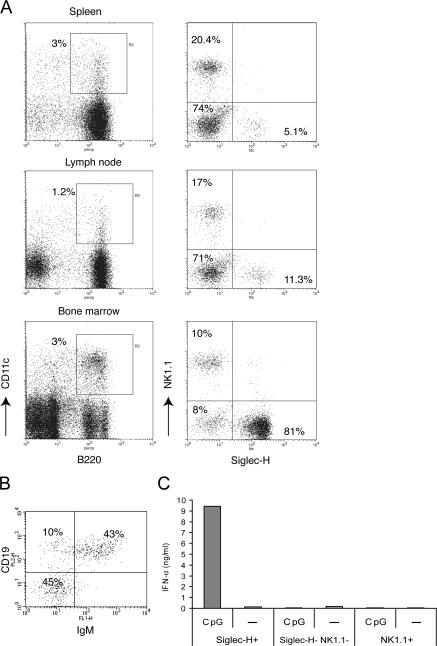
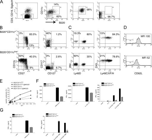
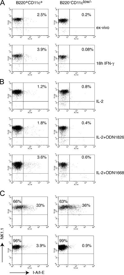
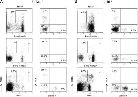
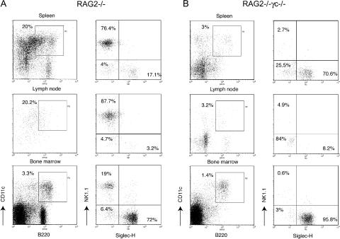
Lymphoid organs contain a B220(+)CD11c(+)NK1.1(+) cell population that was recently characterized as a novel dendritic cell (DC) subset that functionally overlaps with natural killer (NK) cells and plasmacytoid DCs (PDCs). Using Siglec-H and NK1.1 markers, we unambiguously dissected B220(+)CD11c(+) cells and found that PDCs are the only professional interferon (IFN)-alpha-producing cells within this heterogeneous population. In contrast, B220(+)CD11c(+)NK1.1(+) cells are a discrete NK cell subset capable of producing higher levels of IFN-gamma than conventional NK cells. Unlike DCs, only a minute fraction of B220(+)CD11c(+)NK1.1(+) cells in the spleen expressed major histocompatibility complex class II ex vivo or after stimulation with CpG. Consistent with being a NK cell subset, B220(+)CD11c(+)NK1.1(+) cells depended primarily on interleukin 15 and common cytokine receptor gamma chain signaling for their development. In terms of function, expression of distinctive cell surface receptors, and location in lymphoid organs, NK1.1(+)B220(+)CD11c(+) appear to be the murine equivalent of human CD56(bright) NK cells.


Figures

Figure 1.
Figure 1.
Heterogeneity of B220+CD11c+ cells in lymphoid organs. (A) Four-color analysis of spleen, lymph node, and bone marrow cells. B220+CD11c+ double-positive cells (gate R2) were divided into three subsets based on expression of Siglec-H and NK1.1. One subset expresses Siglec-H, a marker of PDCs. A second subset expresses NK1.1. A third subset is Siglec-HNK1.1 and includes mature B cells in the spleen and lymph nodes (not depicted). Percentages of each subset are indicated. Each spleen contained 5–8 × 10⁷ cells. From two femurs of an 8–16-wk-old mouse we recovered 2–4 × 10⁷ bone marrow cells. A Gate (R1) was applied on live cells based on FSC and SSC. FL-1/FL-2 double-positive autofluorescent cellular debris was removed from the analysis by an exclusion gate (R3). (B) The Siglec-HNK1.1 subset displays further heterogeneity in bone marrow. About 40–55% of the cells in different experiments are CD19+IgM+ B cells. However, ∼40–50% of the remaining cells do not express lineage markers. Plots shown in A and B are representative of at least three to four separate experiments with concordant results. (C) Siglec-H+NK1.1, NK1.1+Siglec-H, and Siglec-HNK1.1 were FACS sorted from the spleens of C57BL/6 mice, and identical numbers (105) of cells were stimulated with CpG ODN 2216. After overnight culture, IFN-α secretion was determined in culture supernatants. Results are representative of three independent experiments.
Figure 2.
Figure 2.
B220+CD11c+NK1.1+ cells are enriched in CD27+CD11b+ cells endowed with high IFN-γ secretion capacity. (A) Splenic DX5+ cells were preenriched by DX5 MACS separation. Cells were then stained with a combination of FITC-conjugated anti-CD3, anti-CD19, and anti–Siglec-H antibodies to remove contaminating T cells, B cells, and PDCs (R2). In addition, they were stained with allophycocyanin-conjugated CD11c and PerCP-Cy5.5–conjugated B220. B220+CD11c+ (R3) and B220CD11clow/− (R4) cells were FACS sorted at high purity. One sorting experiment representative of seven is illustrated. (B) Sorted B220+CD11c+ cells (top panels) are highly enriched in CD11b+CD27+ (75–90% in different experiments) and c-kit+ (CD117, 35–45% in different experiments) but not in IL-7Rα+ (CD127) NK cells. One experiment representative of three is shown. (C) B220+CD11c+ cells (top panels) are enriched in Ly49D+ NK cells, whereas other Ly49 family members have a similar distribution between the two subsets. 2B4 is slightly higher in B220+CD11c+ cells. One experiment representative of three is shown. (D) B220+CD11c+ NK cells express more L-selectin than B220CD11clow/− NK cells. (E) B220+CD11c+ NK cells kill YAC cells as efficiently as B220CD11clow/− NK cells. Killing is partially blocked by an anti-NKG2D antibody in both subsets. One of two experiments with identical results is shown. (F) B220+CD11c+ cells produce higher amounts of IFN-γ than B220CD11clow/− NK cells in response to PMA/ionomycin, IL-12 plus IL-18, and CpG ODN1826 and ODN1668. 2 × 10⁴ cells for each condition and subset were assayed in 100 μl of cell culture medium. The supernatants were analyzed after overnight culture. (G) TNF-α was measured after stimulation with PMA/ionomycin and CpG, as described above. No TNF-α secretion was detected upon stimulation with IL-18 plus IL-12. In E and F, one of two experiments with identical results is shown.
Figure 3.
Figure 3.
MHC class II expression in NK cells sorted from the spleen and lymph nodes. (A) Sorted B220+CD11c+ and B220CD11clow/− splenic cell subsets were stained with anti-NK1.1 and anti–I-A/I-E, either ex vivo or after overnight culture with 5 μg/ml IFN-γ. Between 1 and 5% of B220+CD11c+ cells expressed MHC class II in at least six different experiments performed. IFN-γ did not induce a significant up-regulation of MHC class II. One of two experiments with identical outcome is shown. Notably, B220+CD11c+ cells reproducibly expressed slightly higher levels of NK1.1. (B) CpG stimulation slightly increased the percentage of MHC class II+ cells within the B220+CD11c+ cell subset. CpG ODN1668 was more efficient than CpG ODN1826; however, the increment in MHC class II+ cells never exceeded a threefold increase. One of two experiments with similar results is illustrated. (C) Inguinal and laterocervical lymph nodes and spleens of RAG2−/− mice were dissected, and DX5+ cells were enriched by magnetic selection. Cells were stained with B220, CD11c, NK1.1, and MHC class II. Gates were applied on the B220+CD11c+NK1.1+ and B220CD11clow/−NK1.1+ subsets as represented in Fig. 2 A. Although lymph nodes exhibit higher percentages of MHC class II+ NK1.1 cells than spleen, MHC class II expression does not correlate with B220 and/or CD11c. One of three experiments with identical results is shown.
Figure 4.
Figure 4.
B220+CD11c+NK1.1+ cell development is largely FLT3-L independent but requires IL-15. Siglec-H+NK1.1, Siglec-HNK1.1+, and Siglec-HNK1.1 subsets in the spleen, lymph nodes, and bone marrow of FLT3L−/− mice or IL-15–deficient mice were gated within the B220+CD11c+ population as described in Fig. 1. (A) In the absence of FLT3L, Siglec-H+ PDCs are significantly reduced in the spleen and lymph nodes. Although in bone marrow the percentage of Siglec-H+ cells was only partly reduced, and that of NK1.1+ cells was not affected, the entire B220+CD11c+ population was severely reduced (∼10–15-fold). Percentages of each subset are indicated. An FLT3L−/− spleen contained 3.5–7 × 10⁷ cells. From two femurs of an 8–16 wk-old FLT3L−/− mouse we recovered 1.5–3 × 10⁷ bone marrow cells. (B) Lack of IL-15 resulted in a severe decrease of the B220+CD11c+NK1.1+ subset in all the lymphoid organs analyzed. Percentages of each subset are indicated. An IL-15−/− spleen contained 4.5–7.5 × 10⁷ cells. From two femurs of an 8–16-wk-old IL-15−/− mice we recovered 2.5–4 × 10⁷ bone marrow cells. Data presented in this figure were acquired in parallel with data from the B6 WT mice presented in Fig. 1 and are representative of at least four separate experiments with two mice for each genotype.
Figure 5.
Figure 5.
Development of B220+CD11c+NK1.1+ cells requires γc signaling. Siglec-H+NK1.1, Siglec-HNK1.1+, and Siglec-HNK1.1 subsets were evaluated in the spleen, lymph nodes, and bone marrow of RAG2−/− and RAG2−/−γc−/− double-deficient mice. (A) Percentages of Siglec-HNK1.1 cells were strongly reduced in RAG2−/− mice, consistent with their B cell identity. Siglec-H+NK1.1 and Siglec-HNK1.1+ were both present. Percentages of each subset are indicated. Each RAG2−/− spleen contained 1.5–3 × 10⁷ cells. From two femurs of a RAG2−/− mouse we recovered 1.5–4 × 10⁷ bone marrow cells. (B) B220+CD11c+NK1.1+ cells in the spleen, lymph nodes, and bone marrow of RAG2−/−γc−/− mice were dramatically reduced. Each RAG2−/−γc−/− spleen contained 1–2.5 × 10⁷ cells. From two femurs of a RAG2−/−γc−/− mouse we recovered 1–2.5 × 10⁷ bone marrow cells. Data presented in this figure were acquired in parallel with data from the B6 WT mice presented in Fig. 1 and are representative of at least three separate experiments with two mice for each genotype.

Similar articles

Cited by

References

    1. Asselin-Paturel, C., A. Boonstra, M. Dalod, I. Durand, N. Yessaad, C. Dezutter-Dambuyant, A. Vicari, A. O'Garra, C. Biron, F. Briere, and G. Trinchieri. 2001. Mouse type I IFN-producing cells are immature APCs with plasmacytoid morphology. Nat. Immunol. 2:1144–1150. - PubMed
    1. Taieb, J., N. Chaput, C. Menard, L. Apetoh, E. Ullrich, M. Bonmort, M. Pequignot, N. Casares, M. Terme, C. Flament, et al. 2006. A novel dendritic cell subset involved in tumor immunosurveillance. Nat. Med. 12:214–219. - PubMed
    1. Chan, C.W., E. Crafton, H.N. Fan, J. Flook, K. Yoshimura, M. Skarica, D. Brockstedt, T.W. Dubensky, M.F. Stins, L.L. Lanier, et al. 2006. Interferon-producing killer dendritic cells provide a link between innate and adaptive immunity. Nat. Med. 12:207–213. - PubMed
    1. Vremec, D., M. O'Keeffe, H. Hochrein, M. Fuchsberger, I. Caminschi, M. Lahoud, and K. Shortman. 2007. Production of interferons by dendritic cells, plasmacytoid cells, natural killer cells, and interferon-producing killer dendritic cells. Blood. 109:1165–1173. - PubMed
    1. Spits, H., and L.L. Lanier. 2007. Natural killer or dendritic: what's in a name? Immunity. 26:11–16. - PubMed

Publication types