. 2007 Oct 29;204(11):2629-39.
doi: 10.1084/jem.20071262. Epub 2007 Oct 8.

Molecular mechanism of mast cell mediated innate defense against endothelin and snake venom sarafotoxin

Affiliations

Molecular mechanism of mast cell mediated innate defense against endothelin and snake venom sarafotoxin

Lars A Schneider et al. J Exp Med. .

Abstract

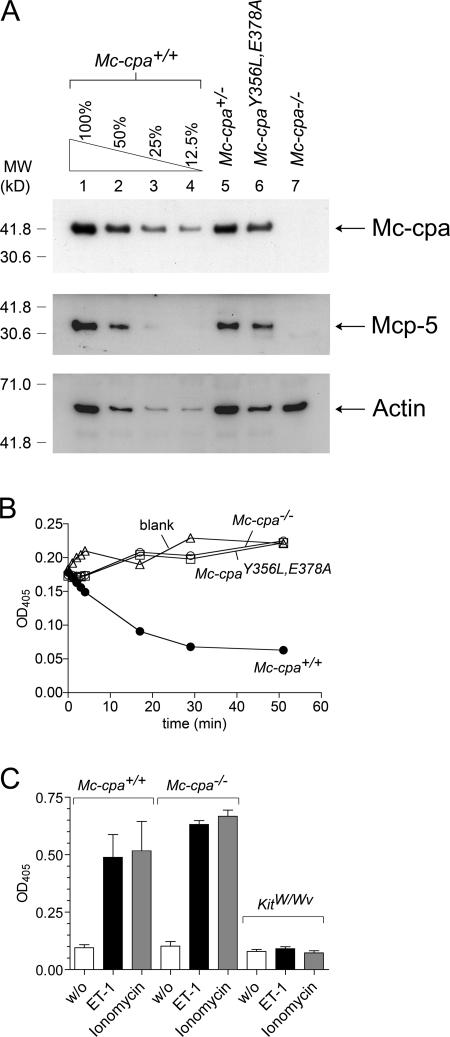
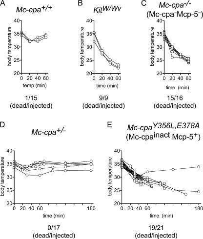
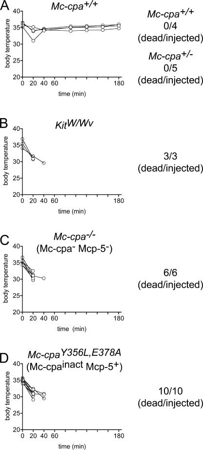
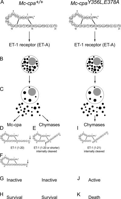
Mast cells are protective against snake venom sarafotoxins that belong to the endothelin (ET) peptide family. The molecular mechanism underlying this recently recognized innate defense pathway is unknown, but secretory granule proteases have been invoked. To specifically disrupt a single protease function without affecting expression of other proteases, we have generated a mouse mutant selectively lacking mast cell carboxypeptidase A (Mc-cpa) activity. Using this mutant, we have now identified Mc-cpa as the essential protective mast cell enzyme. Mass spectrometry of peptide substrates after cleavage by normal or mutant mast cells showed that removal of a single amino acid, the C-terminal tryptophan, from ET and sarafotoxin by Mc-cpa is the principle molecular mechanism underlying this very rapid mast cell response. Mast cell proteases can also cleave ET and sarafotoxin internally, but such "nicking" is not protective because intramolecular disulfide bridges maintain peptide function. We conclude that mast cells attack ET and sarafotoxin exactly at the structure required for toxicity, and hence sarafotoxins could not "evade" Mc-cpa's substrate specificity without loss of toxicity.


Figures

Figure 1.
Figure 1.
Protein expression, carboxypeptidase activity, and ET-1–induced degranulation in mutant mast cells. (A) Lysates of purified peritoneal mast cells from Mc-cpa+/+ (lanes 1–4), Mc-cpa+/− (lane 5), Mc-cpaY356L,E378A (lane 6), and Mc-cpa−/− (lane 7) mice were analyzed by Western blotting for Mc-cpa (top), Mcp-5 (middle), and actin (bottom) expression. Mc-cpa+/+ cell lysates were titrated to estimate the level of Mc-cpa expression in Mc-cpaY356L,E378A mice. 100% in lane 1 corresponds to 14,000 purified peritoneal mast cells. Densitometric measurements of actin expression showed 637, 222, 69, and 46 arbitrary units in lanes 1–4, respectively, and 622, 283, and 601 in lanes 5–7, respectively. Measurements for Mc-cpa expression showed 592, 303, 130, and 70 arbitrary units in lanes 1–4, respectively, and 401, 270, and <1 in lanes 5–7, respectively. Based on these values, we estimate that Mc-cpaY356L,E378A mast cells expressed ∼80% of the Mc-cpa amount expressed in normal mast cells. (B) Lysates of 10,000 peritoneal mast cells from Mc-cpa+/+ (•), Mc-cpa−/− (○), and Mc-cpaY356L,E378A (□) mice, and the blank (without cells) control (▵) were analyzed for carboxypeptidase activity by test substrate (45). Both Mc-cpa−/− and Mc-cpaY356L,E378A mast cells lacked Mc-cpa activity. (C) Mast cell degranulation in response to ET-1 in vitro. PECs from Mc-cpa+/+, Mc-cpa−/−, and KitW/Wv mice were left without stimulus (white bars), treated with ET-1 (black bars), or treated with ionomycin (gray bars). Supernatants were analyzed for β-hexosaminidase release as a measure for degranulation by colorimetric assay (16). The release is mast cell specific, as shown by the absence of β-hexosaminidase release in PECs from KitW/Wv mice. Data summarize the mean ± one SD for three (Mc-cpa+/+ and Mc-cpa−/−) and two (KitW/Wv) independent experiments. Release was significantly different comparing unstimulated versus ET-1–stimulated Mc-cpa+/+ (P = 0.046) and unstimulated versus ET-1–stimulated Mc-cpa−/− (P = 0.001) mast cells, indicating specific degranulation of both genotypes by ET-1. In both genotypes, stimulations by ET-1 and ionomycin were not significantly different. P = 0.5 for Mc-cpa+/+ and P = 0.1 for Mc-cpa−/−.
Figure 2.
Figure 2.
Susceptibility of Mc-cpa−/− and Mc-cpaY356L,E378A mutant mice to ET-1. ET-1 was injected intraperitoneally into Mc-cpa+/+ (A), KitW/Wv (B), Mc-cpa−/− (C), Mc-cpa+/− (D), and Mc-cpaY356L,E378A (E) mice, and the body temperature was tracked by rectal measurements at the indicated time points. Summarized total numbers of dead per injected mice are indicated for each genotype. For clarity, the temperature kinetic is shown for representative mice. Mc-cpa+/+ and Mc-cpa+/− showed a mild and transient temperature drop (A and D), whereas mast cell–deficient KitW/Wv (B), and mice lacking Mc-cpa and Mcp-5 (Mc-cpa−/−; C) succumbed to ET-1 injection within 60 min. Mice lacking only catalytically active Mc-cpa (Mc-cpaY356L,E378A; E) were also largely susceptible to ET-1, but the kinetic of the temperature drop was delayed compared with KitW/Wv or Mc-cpa−/− mice.
Figure 3.
Figure 3.
Degradation of ET-1 by mast cells. (A) ELISA measurements of “residual” ET-1 after incubation of ET-1 with no cells (open bar), or with PECs from Mc-cpa+/+ (solid bar), Mc-cpa−/− (striped bar), or KitW/Wv (shaded bar) mice. Mast cell–containing PECs from Mc-cpa+/+ (P = 0.030 [significant] comparing Mc-cpa+/+ versus KitW/Wv mice), and Mc-cpa−/− (P = 0.037 [significant] comparing Mc-cpa−/− versus KitW/Wv mice) mice abolished ET-1 reactivity by ELISA, suggesting that ET-1 was degraded regardless (P = 0.78 [not significant] comparing Mc-cpa+/+ vs. Mc-cpa−/−) of the mast cell genotype. Data summarize the mean ± one standard deviation for three independent experiments. (B–F) Mass spectrometric analysis of ET-1 degradation (left), and schematic depiction of substrate products (right). ET-1 was left untreated (B) or incubated with ionomycin-stimulated purified peritoneal mast cells from Mc-cpa+/+ (C), Mc-cpa−/− (D), or Mc-cpaY356L,E378A (E and F) mice. C-terminal degradation, evident by the appearance of ET-1 fragments corresponding to the molecular masses of 1–19 and 1–20, was mediated by Mc-cpa+/+ (C), but not by Mc-cpa−/− (D) or Mc-cpaY356L,E378A (E), mast cells. However, after incubation of ET-1 with Mc-cpa−/− (D) or Mc-cpaY356L,E378A (E) mast cells, the molecular mass increased from 2,492 (ET-1; B) to 2,510 daltons (ET-1 plus water; D and E). Hydrolysis of ET-1 was demonstrated after reduction of the samples by the disappearance of the ET-1 1–21 peak, and the appearance of new fragments with molecular masses of 1,639 and 1,786 daltons (see Materials and methods) corresponding to ET-1 1–13 and 1–14, respectively (F). Data are representative of three independent experiments.
Figure 4.
Figure 4.
Removal of tryptophan from the C terminus renders ET-1 nontoxic for mast cell–deficient mice. Synthetic peptides of ET-1 (1–21; A) or of ET-1 minus tryptophan in position 21 (1–20; B) were injected intraperitoneally into mast cell–deficient KitW/Wv mice, and the body temperature was followed by rectal measurements at indicated time points. Mast cell–deficient KitW/Wv mice were resistant to ET-1 (1–20), but succumbed to ET-1 (1–21).
Figure 5.
Figure 5.
Susceptibility of Mc-cpa mutants to S6b. The S6b was injected intraperitoneally into Mc-cpa+/+ (A), Mc-cpa+/− (only numbers shown in A), KitW/Wv (B), Mc-cpa−/− (C), and Mc-cpaY356L,E378A (D) mice, and the body temperature was followed by rectal measurements at the indicated time points. Summarized total numbers of dead per injected mice are indicated for each genotype. The temperature kinetic of Mc-cpa+/− mice (not depicted) was similar to the one from Mc-cpa+/+ mice. All strains except for Mc-cpa+/+ and Mc-cpa+/− mice succumbed to S6b injection within 60 min. The full susceptibility of Mc-cpaY356L,E378A mice demonstrates the essential role of Mc-cpa enzyme activity for survival of snake venom S6b.
Figure 6.
Figure 6.
Degradation of S6b by mast cells. (A–D) Mass spectrometric analyses (left side) of degradation products of S6b. Resulting peptides are schematically depicted on the right side. S6b was left untreated (A) or incubated with ionomycin-stimulated purified mast cells from Mc-cpa+/+ (B) or Mc-cpaY356L,E378A (C and D) mice. C-terminal degradation, which is evident from the appearance of fragments corresponding to the molecular mass of S6b (1–19; 2,266 daltons), was mediated by Mc-cpa+/+ (B), but not by Mc-cpaY356L,E378A (C) mast cells. The peak marked by the asterisk in B does not correspond to a fragment of S6b, and it was not observed in a second experiment. The molecular mass of S6b (1–21) increased after incubation with Mc-cpaY356L,E378A mast cells from 2,565 (1–21; A) to 2,583 daltons (S6b 1–21 plus water; C). This indicated hydrolysis of S6b, which was proven after reduction of the samples by the disappearance of the S6b 1–21 peak, and the appearance of residual fragments with molecular masses of 1,710 and 1,858 daltons (see Materials and methods) corresponding to S6b 1–13 and 1–14, respectively (D). Data are representative for two independent experiments.
Figure 7.
Figure 7.
Molecular mechanism of ET family peptide degradation by Mc-cpa. (A–C) ET-1 and S6b, shown here for ET-1, stimulate mast cells via binding to ET receptors. The receptor type most prominently expressed on peritoneal mast cells is ETA. ETA activation leads to massive degranulation, which is comparable to stimulation via ionomycin, and release of mast cell granule content. These early events are identical in mast cells from Mc-cpa+/+ (A–C; left) and Mc-cpaY356L,E378A (A–C; right) mice. (D–H) Mast cells secrete a set of proteases that include one carboxypeptidase A (Mc-cpa) and several chymases that include Mcp-5. Mc-cpa attacks the C terminus of ETs, and removes amino acids in position 21 (Trp; D) and 20 (Ile; F). These modifications render ETs and related toxins biologically inactive by three orders of magnitude (G; 34), and the mice survive (H). Mc-cpaY356L,E378A mice, which selectively lack active Mc-cpa, are unable to attack the C terminus of ET-1 (I). Mast cell products (possibly Mcp-4) other than Mc-cpa and Mcp-5 attack ET-1 internally by hydrolysis (E and I). Although the molecular structure (46) of position 13–nicked ET-1 is likely altered, ET-1 fragmentation is prevented by the disulfide bridge (1 to 15; E and I), and the molecules retain their toxicity (J and K), unless the C terminus is truncated (E). Mc-cpaY356L,E378A mutant mice are unable to do so, and succumb to exogenous ET-1 and related toxins, thereby demonstrating the essential role of Mc-cpa for this innate immune pathway. Whether or not Mc-cpa is also involved in the degradation of endogenously produced ETs remains to be investigated.

Similar articles

Cited by

References

    1. Echtenacher, B., D.N. Mannel, and L. Hultner. 1996. Critical protective role of mast cells in a model of acute septic peritonitis. Nature. 381:75–77. - PubMed
    1. Malaviya, R., T. Ikeda, E. Ross, and S.N. Abraham. 1996. Mast cell modulation of neutrophil influx and bacterial clearance at sites of infection through TNF-alpha. Nature. 381:77–80. - PubMed
    1. Wort, S.J., and T.W. Evans. 1999. The role of the endothelium in modulating vascular control in sepsis and related conditions. Br. Med. Bull. 55:30–48. - PubMed
    1. Wanecek, M., E. Weitzberg, A. Rudehill, and A. Oldner. 2000. The endothelin system in septic and endotoxin shock. Eur. J. Pharmacol. 407:1–15. - PubMed
    1. Magder, S., and P. Cernacek. 2003. Role of endothelins in septic, cardiogenic, and hemorrhagic shock. Can. J. Physiol. Pharmacol. 81:635–643. - PubMed

Publication types