. 2007 Oct 8;179(1):75-86.
doi: 10.1083/jcb.200704166.

Two regulatory steps of ER-stress sensor Ire1 involving its cluster formation and interaction with unfolded proteins

Affiliations

Two regulatory steps of ER-stress sensor Ire1 involving its cluster formation and interaction with unfolded proteins

Yukio Kimata et al. J Cell Biol. .

Abstract

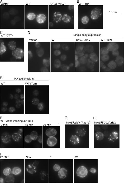
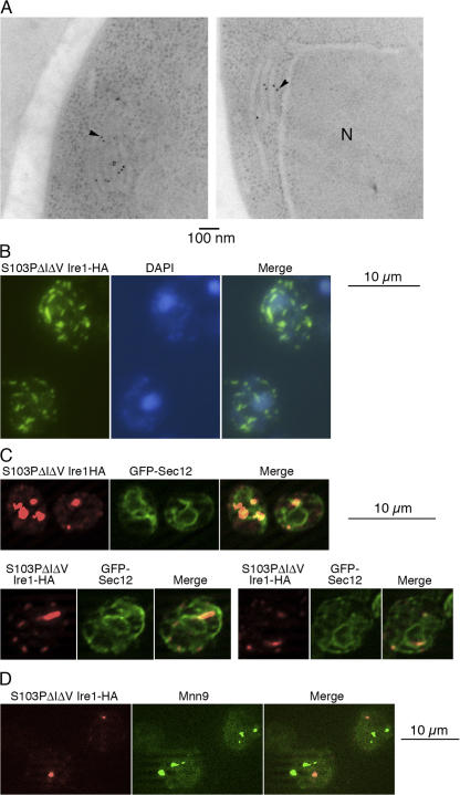
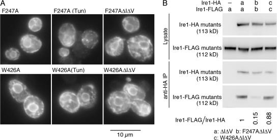
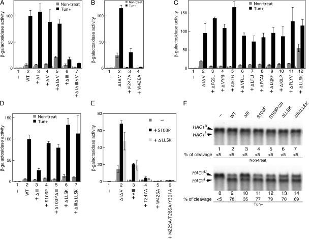
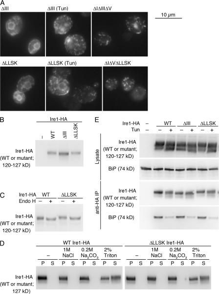
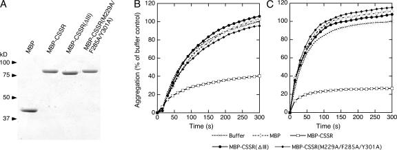
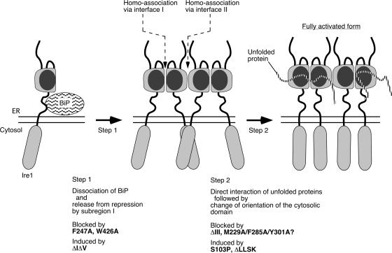
Chaperone protein BiP binds to Ire1 and dissociates in response to endoplasmic reticulum (ER) stress. However, it remains unclear how the signal transducer Ire1 senses ER stress and is subsequently activated. The crystal structure of the core stress-sensing region (CSSR) of yeast Ire1 luminal domain led to the controversial suggestion that the molecule can bind to unfolded proteins. We demonstrate that, upon ER stress, Ire1 clusters and actually interacts with unfolded proteins. Ire1 mutations that affect these phenomena reveal that Ire1 is activated via two steps, both of which are ER stress regulated, albeit in different ways. In the first step, BiP dissociation from Ire1 leads to its cluster formation. In the second step, direct interaction of unfolded proteins with the CSSR orients the cytosolic effector domains of clustered Ire1 molecules.


Figures

Figure 1.
Figure 1.
Structure of yeast Ire1 and mutations used in this study. Structure of the luminal domain according to Kimata et al. (2004), Oikawa et al. (2005), and Credle et al. (2005) is illustrated. Subregions I (aa 32–111), III (aa 243–272), and V (aa 455–524) are loosely folded, whereas subregions II (aa 112–242) and IV (aa 273–454) form the tightly folded CSSR. The position of a hydrophobic segment (aa 527–570) that is deduced to be the transmembrane domain is also shown. The dashed lines indicate positions of amino acid residues deleted in ΔI (aa 32–91), ΔIII (aa 253–272), and ΔV (aa 463–524) mutants, respectively. The positions of another deletion mutation, 567LLSK570, and point mutations S103, F247, and W246 are also indicated. Double circles respectively represent M229, F285, and Y301, which are simultaneously replaced by Ala in Figs. 5 E and 7. Because we now assign the initiation methionine according to the data from the Saccharomyces genome database (http://www.yeastgenome.org/), the amino acid numbers of Ire1 in the present paper differ by 7 aa from those in two of our previous papers (Kimata et al., 2004; Oikawa et al., 2005).
Figure 2.
Figure 2.
Cluster formation of Ire1. Excepting E, Ire1-HA variants were expressed from wild type (WT) or mutant versions of pRS315-IRE1-HA (D; centromeric plasmid) or pRS423-IRE1-HA (A–C and E–I; 2-μm plasmid) in yeast strain KMY1015 (ire1Δ; D), YKY1004 (ire1Δ hac1Δ; G), or KMY1516 (ire1Δ; A–C, E and F, and H and I) and detected by anti-HA immunofluorescent staining. For vector control not carrying the Ire1-HA gene, cells carrying pRS423 (A) or pRS315 (D) were used. (E) An Ire1-HA gene knockin strain YKY2005 was examined. Where indicated, Tun (2 μg/ml final concentration) or DTT (10 mM final concentration) was added into cultures 1 h before harvest. (F) Cells treated with 10 mM DTT for 30 min were washed with medium and further incubated for the indicated times. Cells were observed by a conventional fluorescent microscope, and exposure times for image acquisition were 2 s for D and E and 1 s for other panels.
Figure 3.
Figure 3.
Cellular localization of Ire1 clusters as examined by electron microscopy and double fluorescent staining. YKY1004 (ire1Δ hac1Δ; A and C) or KMY1516 (ire1Δ; B and D) cells carrying the S103PΔIΔV mutant version of pRS423-Ire1-HA were examined. (C) The cells also contained a GFP-Sec12 expression plasmid as described in the Materials and methods. (A) Ultrathin sections of the cells were immunogold stained with anti-HA antibody and observed with a transmission electron microscope. In each panel, the arrowhead points to one of the gold particles (N, nucleus). (B) The cells were doubly stained with anti-HA antibody (FITC) and DAPI and observed by a conventional fluorescent microscope. (C and D) Cells were doubly stained with anti-HA antibody (Cy5) and anti-GFP (C) or anti-Mnn9 (D) antibody (FITC). (C) Cy5 image in the top panels is brighter than those in the bottom panels, partly because of difference in exposure times for image acquisition.
Figure 4.
Figure 4.
Mutations that impair cluster formation. (A) KMY1516 (ire1Δ) cells carrying the indicated mutant version of pRS423-IRE1-HA were subjected to anti-HA immunofluorescent staining. Cells were observed by a conventional fluorescent microscope, and exposure times for image acquisition were 1 s for all panels. When indicated, Tun (2 μg/ml final concentration) was added into cultures 1 h before harvest. (B) Diploid cells KMY1015 × KMY1520 (ire1Δ/ire1Δ) coexpressing Ire1-HA and Ire1-FLAG carrying the indicated mutations from mutant versions of pRS315-IRE1-HA and pRS426-IRE1-FLAG, respectively, were lysed and subjected to anti-HA immunoprecipitation. The lysates and IPs were then analyzed by anti-HA or anti-FLAG Western blotting to detect the indicated proteins. For vector control (−), cells carried empty vector pRS315. The ratios of Ire1-Flag to Ire1-HA signal in anti-HA IP (Ire1-Flag/Ire1-HA) were normalized to that of the ΔIΔV mutant and are presented. Multiple independent clones were examined to obtain standard deviations < 20% of the means.
Figure 5.
Figure 5.
Activity of Ire1 mutants. (A–E) Cellular β-galactosidase activity of KMY1015 (ire1Δ) cells carrying pRS315-IRE1-HA (wild type [WT] or mutant) and UPRE-lacZ reporter plasmid pCZY1 was assayed and is presented as the mean and standard deviation of three independent transformants, which has been normalized to the Tun + wild-type Ire1-HA control (set at 100) in all panels. Column 1 in each panel is vector control, in which the cells carried an empty vector pRS315. The value from wild-type or ΔIΔV Ire1-HA cells is presented in column 2. In column 3 and the subsequent columns, the mutations indicated on the x axis were introduced into WT (A and D) or ΔIΔV (B, C, and E) Ire1-HA. (C) The results from aa deletion scanning from aa 531–534 (ΔFGSL) to aa 567–570 (ΔLLSK) are presented in columns 2 and 3. The result from the N-terminal mutation (deletion of aa 527–530) was not reproducible (not depicted). (E) S103P, ΔLLSK, or no (−) mutation was additionally introduced as indicated. (F) Total RNA prepared from KMY1516 (ire1Δ) cells expressing untagged Ire1 variants from the WT or mutant versions of pRS313-IRE1 was analyzed by Northern blot detection of the HAC1 mRNA variants. For vector control (lanes 1 and 8), cells carried an empty vector pRS313. The percentage of HAC1u mRNA cleavage was estimated as described in Kimata et al. (2003). For Tun+ samples, Tun was added into cultures at 2 μg/ml final concentration 4 (A–D) or 1 h (F) before harvest.
Figure 6.
Figure 6.
Cluster formation, localization, cellular expression, or BiP dissociation is not significantly altered by either the ΔIII or the ΔLLSK mutation. (A) KMY1516 (ire1Δ) cells carrying the indicated mutant version of pRS423-IRE1-HA were subjected to anti-HA immunofluorescent staining. Cells were observed by a conventional fluorescent microscope, and exposure times for image acquisition were 1 s for all panels. (B and C) Lysates from KMY1015 (ire1Δ) cells carrying pRS315-IRE1-HA (wild type [WT] or mutant) were analyzed by anti-HA Western blotting. (C) Cell lysate was treated with Endo H where indicated. (D) Detergent-free crude cell lysate containing 0.7 M sorbitol was prepared from KMY1516 cells carrying pRS423-IRE1-HA (WT or ΔLLSK) and incubated with the indicated reagents (final concentration) on ice for 10 min. The samples were then centrifuged at 100,000 g for 30 min, and pellet (P) and supernatant (S) fractions prepared from the same amount of samples were analyzed by anti-HA Western blotting. (E) Lysates from KMY1516 cells carrying pRS423-IRE1-HA (WT or mutant) were subjected to anti-HA immunoprecipitation, and the lysates and the IPs were analyzed by anti-HA or anti-BiP Western blotting to detect the indicated proteins. When indicated in A and E, Tun was added at 2-μg/ml final concentration into cultures 1 h before harvest. For vector control (−), cells carried an empty vector, pRS315 (B) or pRS423 (E).
Figure 7.
Figure 7.
Direct interaction of CSSR with unfolded proteins as monitored by inhibition of in vitro aggregation. (A) Bacterially expressed MBP-CSSR, its mutant version, or unfused MBP were purified; run on 10% SDS-PAGE gel (1 μg of protein per lane); and stained with Coomassie blue. (B and C) At time 0, 25 μM of luciferase (B) or 50 μM of citrate synthase (C) in guanidine HCl–denaturing solution was 50- (for luciferase) or 66-fold (for citrate synthase) diluted into assay buffer containing MBP-CSSR, its mutant version, unfused MBP (2 μM each for luciferase or 0.5 μM each for citrate synthase), or buffer only. Turbidity of the sample mixtures was monitored, normalized against the maximal value of the buffer sample, and presented as aggregation.
Figure 8.
Figure 8.
Our current model for the mechanism by which ER stress activates Ire1. Our view about effects of the Ire1 mutations is also indicated. See text for details.

Similar articles

Cited by

References

    1. Bertolotti, A., Y. Zhang, L.M. Hendershot, H.P. Harding, and D. Ron. 2000. Dynamic interaction of BiP and ER stress transducers in the unfolded-protein response. Nat. Cell Biol. 2:326–332. - PubMed
    1. Christianson, T.W., R.S. Sikorski, M. Dante, J.H. Shero, and P. Hieter. 1992. Multifunctional yeast high-copy-number shuttle vectors. Gene. 110:119–122. - PubMed
    1. Cox, J.S., and P. Walter. 1996. A novel mechanism for regulating activity of a transcription factor that controls the unfolded protein response. Cell. 87:391–404. - PubMed
    1. Cox, J.S., C.E. Shamu, and P. Walter. 1993. Transcriptional induction of genes encoding endoplasmic reticulum resident proteins requires a transmembrane protein kinase. Cell. 73:1197–1206. - PubMed
    1. Craven, R.A., M. Egerton, and C.J. Stirling. 1996. A novel Hsp70 of the yeast ER lumen is required for the efficient translocation of several protein precursors. EMBO J. 15:2640–2650. - PMC - PubMed

Publication types

MeSH terms