. 2007 Oct 8;179(1):117-28.
doi: 10.1083/jcb.200706196.

Regulatory interactions between two actin nucleators, Spire and Cappuccino

Affiliations

Regulatory interactions between two actin nucleators, Spire and Cappuccino

Margot E Quinlan et al. J Cell Biol. .

Abstract

Spire and Cappuccino are actin nucleation factors that are required to establish the polarity of Drosophila melanogaster oocytes. Their mutant phenotypes are nearly identical, and the proteins interact biochemically. We find that the interaction between Spire and Cappuccino family proteins is conserved across metazoan phyla and is mediated by binding of the formin homology 2 (FH2) domain from Cappuccino (or its mammalian homologue formin-2) to the kinase noncatalytic C-lobe domain (KIND) from Spire. In vitro, the KIND domain is a monomeric folded domain. Two KIND monomers bind each FH2 dimer with nanomolar affinity and strongly inhibit actin nucleation by the FH2 domain. In contrast, formation of the Spire-Cappuccino complex enhances actin nucleation by Spire. In Drosophila oocytes, Spire localizes to the cortex early in oogenesis and disappears around stage 10b, coincident with the onset of cytoplasmic streaming.


Figures

Figure 1.
Figure 1.
Localization of Spir in Drosophila oocytes. (A–C) Ovaries were dissected from wild-type flies and processed according to Robinson and Cooley (1997). Spir is detected at the actin-rich oocyte cortex during midoogenesis (green, anti-Spir; red, actin detected with rhodamine-phalloidin). Examples of stage 9 (A) and stage 6 (B) oocytes are shown. Posterior is to the right. For comparison with spir1 flies, see Fig. S1. (C) Spir is no longer at the cortex by stage 10b. (D) Spir and Capu interact in vivo. Spir coimmunoprecipitates with Capu from Drosophila ovary lysates. 1% of input is shown. Spir did not precipitate with beads alone or beads bound to nonspecific IgG (not depicted). The three boxes in each row are from the same exposure, moved for presentation. For more information, see Fig. S1 (available at http://www.jcb.org/cgi/content/full/jcb.200706196/DC1). Bars, 10 μm.
Figure 2.
Figure 2.
Interaction of Spir and Capu is mediated by the KIND and FH2 domains. (A) Domain organization of Spir and Capu. (top) The central region of Spir proteins contains a cluster of four actin-binding WH2 motifs, which nucleate actin. The C-terminal part consists of a modified FYVE zinc finger (mFYVE), which targets the protein to intracellular membranes. The adjacent Spir box (S-box) is similar to motifs found in proteins that bind Rab-3a and may also play a role in subcellular localization. The KIND domain is a novel motif that may function as a protein–protein interaction module. (bottom) Capu, a formin, contains a proline-rich region, the formin homology 1 domain (FH1), and a C-terminal formin homology 2 domain (FH2) that dimerizes and nucleates actin. (B–E) NIH 3T3 cells transfected with individual expression vectors (B) or cotransfected with two expression vectors (C–E) encoding the indicated proteins. EGFP fusion proteins are green, and the myc-tagged counterpart is localized by immunofluorescence using anti-myc antibodies (red). (B) When expressed alone, full-length Capu and Capu-FH2 are diffuse throughout the cell. Spir is punctate as previously described (Kerkhoff et al., 2001). (C) When cotransfected with Spir, the localization of Capu shifts to a punctate pattern coinciding with Spir. (D) The Spir-KIND domain and Capu-FH2 domain are sufficient for colocalization. The KIND domain is driven to membranes by a CAAX box. Capu-FH2 is found concentrated at these same structures. (E) These interactions are conserved in mammalian proteins. Here, we show the colocalization of Spir-1–KIND and Fmn2-FH2 (Fig. S2 C, available at http://www.jcb.org/cgi/content/full/jcb.200706196/DC1). Insets are magnified (2.3 times) images of the boxed areas. (F) Coimmunoprecipitation of EGFP-Capu-FH2 and myc-Spir-NT from NIH 3T3 cells expressing these constructs with a myc antibody. Bars, 10 μm.
Figure 3.
Figure 3.
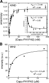
Binding affinities of direct interactions between Spir and Capu. (A) Polarization anisotropy of 10 nM AlexaFluor488-labeled KIND in the presence of the Capu-FH1FH2 domain. By fitting the concentration-dependent change in anisotropy to a quadratic binding curve, we determined a dissociation equilibrium constant of 1 ± 2 nM. (inset) Competition of fluorescently labeled KIND with unlabeled KIND. We mixed 10 nM AlexaFluor488-labeled KIND and 50 nM Capu-FH1FH2 with varying concentrations of unlabeled KIND. Fitting the decrease in anisotropy to a competition binding curve (see Materials and methods) yields a dissociation equilibrium constant of 5 ± 3 nM. Error bars represent SD. (B) The affinity of 20 nM AlexaFluor488-labeled WH2 was measured by fitting a quadratic binding curve to concentration-dependent intensity changes induced by adding Capu-FH1FH2. The Kd is 2.4 ± 0.9 μM. Error bars are smaller than the symbols.
Figure 4.
Figure 4.
Two KIND domains bind each Capu dimer. (A and B) We determined that KIND domains from Drosophila Spir and human Spir-1 are monomeric using equilibrium centrifugation. We measured the solution molecular weights of purified KIND (A) and Spir-1–KIND (B) as described in Materials and methods. We spun samples to equilibrium at 10,000 (open circles), 14,000 (closed circles), and 20,000 rpm (open squares). Symbols are data points; lines represent the best fit to a single species model. Residuals for each dataset are shown below. In both cases, the apparent molecular weight by centrifugation was slightly higher than the predicted monomer molecular weight, suggesting a possible weak tendency to self-associate. By fitting the ultracentrifugation data with a monomer-dimer equilibrium model, we placed lower bounds on the dissociation equilibrium constants for dimerization. For KIND, the Kd for dimerization is at least 92 μM, and, for Spir-1–KIND, it is >530 μM. Neither dataset was well fit by a single-species model with the molecular weight of a KIND homodimer. (C) The KIND domain is highly asymmetric. We measured the sedimentation coefficient, Stokes radius, and aspect ratio of KIND by velocity sedimentation. Both oblate and prolate ellipsoids with an aspect ratio of 1:8 fit the data. The molecular weight measurement agrees with that found by equilibrium sedimentation (44.7 vs. 44.6 kD). Because this value is larger than expected, we confirmed that the actual molecular mass is 37.7 kD with mass spectometry. Velocity sedimentation analysis of Spir-1–KIND indicates a 1:8 aspect ratio as well (not depicted). (D) We measured the solution molecular weight of three molar ratios of AlexaFluor488-labeled Capu-FH1FH2 and KIND (1 [Capu dimer]:1 [KIND], 1:2, and 1:4). We spun samples to equilibrium at 5,000 (open circles), 7,000 (closed circles), and 14,000 rpm (open squares) and measured protein concentration as a function of radius by absorbance at 500 nm to track the AlexaFluor488-labeled protein. The apparent molecular mass of the single species is 225 kD, which is very close to the predicted mass of 223.6 kD (the sum of one Capu-FH1FH2 dimer [134 kD] plus two KIND molecules [Mapp = 2 × 44.6 kD]). We found that Capu-FH1FH2 alone is unstable under the same conditions (not depicted). Therefore, in the case in which there was excess Capu-FH1FH2 (1:1), we believe that unbound Capu-FH1FH2 precipitated, leaving only the complex to be detected. Conditions: buffer, 50 mM KCl, 10 mM Hepes, pH 7, 1 mM TCEP, and 0.01% sodium azide; temperature, 24°C.
Figure 5.
Figure 5.
Interaction between Spir and Capu affects actin nucleation. (A) The Spir-KIND domain inhibits actin nucleation by Capu family formins. We induced polymerization by mixing pyrene-labeled actin with Mg2+, K+, and Capu-FH1FH2. The addition of KIND to Capu-FH1FH2 before mixing with actin potently inhibited nucleation activity in a dose-dependent manner. Protein concentrations were as follows: actin, 4 μM (5% pyrene labeled); Capu-FH1FH2, 10 nM; KIND, as indicated. (B) Plot of nucleation rates (calculation shown in Fig. S3 D, available at http://www.jcb.org/cgi/content/full/jcb.200706196/DC1) versus concentration of KIND added. Data were fit with a quadratic binding curve. The inhibition constants for Capu-FH2 (Ki = 5 ± 1 nM), Capu-FH1FH2 (Ki = 10 ± 2 nM), and NTSpir[A*B*C*D*] (Ki = 6 ± 6 nM) are similar, indicating that the effect of KIND is independent of the Capu-FH1 domain and Spir-WH2 cluster. (C) Capu enhances actin nucleation by Spir. We mixed several concentrations of a nucleation-incompetent mutant of Capu-FH1FH2 (Capu-FH1FH2(I706A)) with the N-terminal half of Spir (NTSpir), which contains the KIND domain and four WH2 domains (only three concentrations are shown for clarity). Nucleation activity of NTSpir was increased by Capu-FH1FH2(I706A) until the proteins were approximately equimolar, and then activity decreased. (inset) Capu-FH1FH2(I706A) does not enhance the activity of the WH2 cluster alone, indicating that an interaction between the KIND domain and the formin is necessary. Protein concentrations were as follows: actin, 4 μM (5% pyrene labeled), NTSpir and WH2, 250 nM; Capu-FH1FH2(I706A), as indicated. (D) Capu and Spir affect each other's actin-nucleation activity. 100 nM Capu-FH1FH2 was mixed with a range of NTSpir concentrations. The activity is not the sum of the individual components. Baseline activities are shown for comparison: 100 nM Capu-FH1FH2 (red) and 250 nM NTSpir (green). (inset) Expanded view of the early time shows a lag when Spir and Capu are mixed that is absent for Capu alone.
Figure 6.
Figure 6.
The KIND domain, microtubules, and actin bind Capu-FH1FH2 competitively. (A) Gels of cosedimentation assays show that the majority of the Capu-FH1FH2 cosediments when 0.5 μM Capu-FH1FH2 and 2 μM of stabilized microtubules are combined and centrifuged (left-most lanes of each gel). The addition of increasing amounts of KIND (0.0625, 0.125, 0.25, …16 μM) leads to the dissociation of Capu-FH1FH2 from microtubules, resulting in decreasing amounts of the formin in the pellet. (B) Gels of cosedimentation assays show that the majority of the Capu-FH1FH2 cosediments when 0.5 μM Capu-FH1FH2 and 2 μM of stabilized actin filaments are combined and centrifuged (left-most lanes of each gel). The addition of increasing amounts of KIND (0.0625, 0.125, 0.25, …16 μM) leads to the dissociation of Capu-FH1FH2 from actin, resulting in decreasing amounts of formin in the pellet. (C) Gels were quantified with Sypro-Red staining. Circles are Capu-FH1FH2 in the supernatant; squares are from the pellet. It takes ∼2.5 μM KIND to compete about half of the Capu-FH1FH2 away from microtubules because Capu-FH1FH2–microtubule interaction is very high affinity, as determined by fitting the data to a competition equation (Kd = 0.5 ± 0.1 nM). Only ∼0.5 μM KIND is required to compete about half of the Capu-FH1FH2 away for actin, indicating that the affinity of Capu-FH1FH2 for actin is lower than that for microtubules, although still tight (Kd = 7 ± 1 nM). Only the data that meet the assumptions (solid symbols) were used for the fit (see Materials and methods). (D) Actin cross-linking assay. 2 μM of filamentous actin plus 0.5 μM Capu-FH1FH2 or α-actinin were centrifuged at 16,000 g for 5 min. The supernatants and pellets were separated and analyzed by SDS-PAGE. Lines are for ease of comparison. The samples were all run on the same gel. Actin is found in the pellet in the presence of Capu-FH1FH2 and α-actinin, a known actin cross-linker.

Similar articles

Cited by

References

    1. Ciccarelli, F.D., P. Bork, and E. Kerkhoff. 2003. The KIND module: a putative signalling domain evolved from the C lobe of the protein kinase fold. Trends Biochem. Sci. 28:349–352. - PubMed
    1. Clark, I., E. Giniger, H. Ruohola-Baker, L.Y. Jan, and Y.N. Jan. 1994. Transient posterior localization of a kinesin fusion protein reflects anteroposterior polarity of the Drosophila oocyte. Curr. Biol. 4:289–300. - PubMed
    1. Cooper, J.A., S.B. Walker, and T.D. Pollard. 1983. Pyrene actin: documentation of the validity of a sensitive assay for actin polymerization. J. Muscle Res. Cell Motil. 4:253–262. - PubMed
    1. Emmons, S., H. Phan, J. Calley, W. Chen, B. James, and L. Manseau. 1995. Cappuccino, a Drosophila maternal effect gene required for polarity of the egg and embryo, is related to the vertebrate limb deformity locus. Genes Dev. 9:2482–2494. - PubMed
    1. Harris, E.S., F. Li, and H.N. Higgs. 2004. The mouse formin, FRLalpha, slows actin filament barbed end elongation, competes with capping protein, accelerates polymerization from monomers, and severs filaments. J. Biol. Chem. 279:20076–20087. - PubMed

Publication types

MeSH terms