

Three-dimensional gray matter atrophy mapping in mild cognitive impairment and mild Alzheimer disease
- PMID: 17923632
- PMCID: PMC3197839
- DOI: 10.1001/archneur.64.10.1489
Three-dimensional gray matter atrophy mapping in mild cognitive impairment and mild Alzheimer disease
Abstract
Background: Alzheimer disease (AD) is the most common form of dementia worldwide. Mild cognitive impairment (MCI) is the recent terminology for patients with cognitive deficiencies in the absence of functional decline. Most patients with MCI harbor the pathologic changes of AD and demonstrate transition to dementia at a rate of 10% to 15% per year. Patients with AD and MCI experience progressive brain atrophy.
Objective: To analyze the structural magnetic resonance imaging data for 24 patients with amnestic MCI and 25 patients with mild AD using an advanced 3-dimensional cortical mapping technique.
Design: Cross-sectional cohort design. Patients/
Methods: We analyzed the structural magnetic resonance imaging data of 24 amnestic MCI (mean MMSE, 28.1; SD, 1.7) and 25 mild AD patients (all MMSE scores, >18; mean MMSE, 23.7; SD, 2.9) using an advanced 3-dimensional cortical mapping technique.
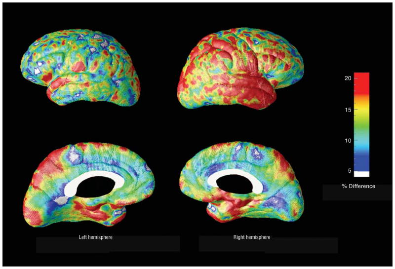
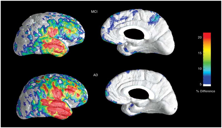
Results: We observed significantly greater cortical atrophy in patients with mild AD. The entorhinal cortex, right more than left lateral temporal cortex, right parietal cortex, and bilateral precuneus showed 15% more atrophy and the remainder of the cortex primarily exhibited 10% to 15% more atrophy in patients with mild AD than in patients with amnestic MCI.
Conclusion: There are striking cortical differences between mild AD and the immediately preceding cognitive state of amnestic MCI. Cortical areas affected earlier in the disease process are more severely affected than those that are affected late. Our method may prove to be a reliable in vivo disease-tracking technique that can also be used for evaluating disease-modifying therapies in the future.
Figures


References
-
- Jicha GA, Parisi JE, Dickson DW, et al. Neuropathologic outcome of mild cognitive impairment following progression to clinical dementia. Arch Neurol. 2006;63(5):674–681. - PubMed
-
- Duyckaerts C, Dickson DW. Neuropathology of Alzheimer’s disease. In: Dickson DW, editor. Neurodegeneration: The Molecular Pathology of Dementia and Movement Disorders. Basel, Switzerland: ISN Neuropath Press; 2003. pp. 47–65.
-
- Mesulam MM. Aging, Alzheimer’s disease and dementia. In: Mesulam MM, editor. Principles of Behavioral and Cognitive Neurology. 2. Oxford, England: Oxford University Press; 2000. pp. 439–510.
-
- Braak H, Braak E. Frequency of stages of Alzheimer-related lesions in different age categories. Neurobiol Aging. 1997;18(4):351–357. - PubMed
-
- Braak H, Del Tredici K, Schultz C, Braak E. Vulnerability of select neuronal types to Alzheimer’s disease. Ann N Y Acad Sci. 2000;924:53–61. - PubMed
Publication types
MeSH terms
Grants and funding
LinkOut - more resources
Full Text Sources
Medical
