. 2007 Dec;27(24):8510-21.
doi: 10.1128/MCB.01615-07. Epub 2007 Oct 8.

T-bet's ability to regulate individual target genes requires the conserved T-box domain to recruit histone methyltransferase activity and a separate family member-specific transactivation domain

Affiliations

T-bet's ability to regulate individual target genes requires the conserved T-box domain to recruit histone methyltransferase activity and a separate family member-specific transactivation domain

Megan D Lewis et al. Mol Cell Biol. 2007 Dec.

Abstract

Appropriate cellular differentiation and specification rely upon the ability of key developmental transcription factors to precisely establish gene expression patterns. These transcription factors often regulate epigenetic events. However, it has been unclear whether this is the only role that they play in functionally regulating developmental gene expression pathways or whether they also participate in downstream transactivation events at the same promoter. The T-box transcription factor family is important in cellular specification events in many developmental systems, and determining the molecular mechanisms by which this family regulates gene expression networks warrants attention. Here, we examine the mechanism by which T-bet, a critical T-box protein in the immune system, influences transcription. T-bet is both necessary and sufficient to induce permissive histone H3-K4 dimethyl modifications at the CXCR3 and IFN-gamma promoters. A T-bet structure-function analysis revealed that the conserved T-box domain, with a small C-terminal portion, is required for recruiting histone methyltransferase activity to promoters. Interestingly, this function is conserved in the T-box family and is necessary, but not sufficient, to induce transcription, with an independent transactivation activity also required. The requirement for two separable functional activities may ultimately contribute to the stringent role for T-box proteins in establishing specific developmental gene expression pathways.


Figures

FIG. 1.
FIG. 1.
T-bet is required and sufficient for the induction of the permissive H3-K4 dimethyl modification at the CXCR3 and IFN-γ promoters. (A) Shown are representative results from a ChIP experiment examining the H3-K4 dimethyl (H3K4me2) and trimethyl (H3K4me3) modifications at various promoters in CD4+ T cells isolated from either wild-type (WT) or T-bet−/− (knockout [KO]) mice and cultured under Th1-skewing conditions. A ChIP assay was performed with antibodies specific to the histone H3-K4Me2 and H3-K4Me3 modifications or a nonspecific immunoglobulin G (IgG) control as indicated below the graphs. Quantitative PCR analysis was performed with promoter-specific primers as indicated above each graph. The y axis of each graph represents the level of specific-antibody precipitation relative to the total-input control for each cell type. (B) EL4 T cells were transfected with either a control pcDNA3.1 (lanes 1 to 4) or a T-bet (lanes 5 to 8) expression vector. Cells were cross-linked and a ChIP analysis was performed using antibodies specific to histone H3-K4Me3 (lanes 1 and 5) or H3-K4Me2 (lanes 2 and 6) or a nonspecific IgG antibody control (lanes 4 and 8). An aliquot of the total chromatin input is also shown (lanes 3 and 7). Promoter-specific primers were utilized in the PCR portion of the assay, as indicated to the left of the gel images.
FIG. 2.
FIG. 2.
Schematic representation of the T-bet deletion constructs. All T-bet expression constructs were cloned into the pcDNA3.1 vector with a C-terminal V5 epitope tag for monitoring protein expression levels. In all cases, the T-box DNA binding domain, indicated in black, was contained within the constructs to allow for binding to the endogenous T-bet target genes. The initial N-terminal and final C-terminal amino acids for each mutant are indicated.
FIG. 3.
FIG. 3.
Transactivation potential is contained within both the N- and C-terminal domains of T-bet. EL4 T cells were transfected with either the control pcDNA3.1 expression vector or the T-bet C-terminal (A and B) or N-terminal (C and D) deletion constructs as indicated. Aliquots of the transfected samples were processed for RNA and quantitative RT-PCR analyses (A and C) and the evaluation of protein expression by Western analysis utilizing a V5 antibody (αV5) to examine construct expression or a glyceraldehyde-3-phosphate dehydrogenase antibody (αGapdh) as a loading control (B and D). The quantitative RT-PCR analysis was performed with gene-specific primers to monitor the influence of the wild-type and mutant T-bet constructs on the expression of the endogenous target genes indicated at the bottom of the graph. The y axis indicates the level of expression of each deletion mutant relative to that of the wild-type T-bet 1-530 transfection after the normalization of values for all samples to beta-actin as a control. Results for the C-terminal (A and B) and N-terminal (C and D) deletion constructs from a representative experiment monitoring both target gene expression and the mutant protein expression levels in transfected cells are shown.
FIG. 4.
FIG. 4.
T-bet's N- and C-terminal domains are dispensable for initiating the H3-K4 dimethyl (H3K4me2) modification at the CXCR3 promoter. (A) Shown are results from representative ChIP experiments examining the status of the H3-K4 dimethyl modification in response to T-bet deletion mutant overexpression. EL4 T cells were transfected with the control pcDNA3.1 vector (lanes 1 to 4) or one of the following T-bet expression constructs as indicated above the gel images: T-bet 1-530 (lanes 5 to 8), T-bet 1-468 (lanes 9 to 12), T-bet 1-402 (lanes 13 to 16), T-bet 1-331 (lanes 17 to 20), or T-bet 120-530 (lanes 21 to 24). Transfected samples were cross-linked, and ChIP assays were performed with an antibody specific to either H3-K4 trimethyl (H3K4me3; lanes 1, 5, 9, 13, 17, and 21), H3-K4 dimethyl (lanes 2, 6, 10, 14, 18, and 22), or a nonspecific IgG control (lanes 4, 8, 12, 16, 20, and 24). An aliquot of the total input chromatin from each transfection is also shown (lanes 3, 7, 11, 15, 19, and 23). Primers specific to the promoters of CXCR3, IFN-γ, JMJD1A, or IL-4 were utilized as indicated to the left of the gel images. (B) A quantitative PCR analysis of the samples from panel A was performed as described in the legend to Fig. 1A.
FIG. 5.
FIG. 5.
The loss of T-bet's N- and C-terminal domains results in a complete absence of transactivation potential. T-bet deletion constructs that had the N terminus deleted in conjunction with the progressive deletion of the C-terminal domain were utilized. (A and B) EL4 T cells were transfected with the T-bet mutant constructs as indicated in the legends to Fig. 3 and 4. (A) The ability of the mutant constructs to activate endogenous target gene expression was monitored by quantitative RT-PCR. (B) A Western analysis examining the levels of expression of the constructs in the transfected cells from panel A is shown. αV5, V5 antibody; αGAPDH, glyceraldehyde-3-phosphate dehydrogenase antibody.
FIG. 6.
FIG. 6.
Localization of the domain required for functional recruitment of methyltransferase activity to the CXCR3 and IFN-γ promoters. (A) The ability of the double-truncation mutant proteins to induce the H3-K4 dimethyl (H3K4me2) modification was monitored by ChIP as described in the legend to Fig. 4. EL4 T cells were transfected with a pcDNA3.1 control vector (lanes 1 to 5), T-bet 120-468 (lanes 6 to 9), T-bet 120-402 (lanes 10 to 13), or T-bet 120-331 (lanes 14 to 17). The transfected samples were precipitated with antibodies specific to H3-K4 trimethyl (H3K4me3; lanes 1, 6, 10, and 14), H3-K4 dimethyl (lanes 2, 7, 11, and 15), H3-K9 acetyl (H3AcK9) (lane 3), or an IgG control (lanes 5, 9, 13, and 17). An aliquot of the total input chromatin is also shown (lanes 4, 8, 12, and 16). (B) Shown is the quantitative PCR analysis of the samples described in the legend to panel A. The construct symbol key is the same as that in Fig. 5. (C) A ChIP assay was performed to determine if the double-truncation mutant proteins retain the ability to bind to the IFN-γ promoter. EL4 T cells were transfected with either a pcDNA3.1 control (lanes 1 to 3), T-bet 120-468 (lanes 4 to 6), T-bet 120-402 (lanes 7 to 9), T-bet 120-331 (lanes 10 to 12), or T-bet 1-530 (lanes 13 to 15). The transfected samples were processed for standard ChIP analysis and precipitated with either the V5 epitope tag antibody (lanes 1, 4, 7, 10, and 13) or an IgG control (lanes 3, 6, 9, 12, and 15). An aliquot of the input chromatin from each transfection is also shown (lanes 2, 5, 8, 11, and 14).
FIG. 7.
FIG. 7.
The ability to recruit histone methyltransferase activity to target genes is a conserved function for the T-box family. (A) Brachyury (Brach.) is able to associate with the T-bet target genes. Shown is a ChIP analysis performed with the human 721 B-cell line. The chromatin was precipitated with an antibody specific to either Brachyury (lane 1), T-bet (lane 2), or a nonspecific IgG control (lane 4). The total input chromatin is also shown (lane 3), and the promoter-specific primers utilized in the PCR are indicated to the left of the gel image. (B) EL4 T cells were transfected with either a pcDNA3.1 control vector (lanes 1 to 4), Tbx6 (lanes 5 to 8), Eomes (lanes 9 to 12), or Brachyury (lanes 13 to 16) as indicated. The ability of the T-box family members to induce the H3-K4 dimethyl (H3K4me2) modification was monitored by ChIP. Transfected samples were precipitated with an antibody to H3-K4 trimethyl (H3K4me3; lanes 1, 5, 9, and 13), H3-K4 dimethyl (lanes 2, 6, 10, and 14), or a nonspecific IgG control (lanes 4, 8, 12, and 16), and the analysis of an aliquot of the total input chromatin is also shown (lanes 3, 7, 11, and 15). Promoter-specific primers were used in the PCR portion of the assay as indicated to the left of the gel image. (C and D) The ability of these T-box family members to regulate endogenous target gene expression was examined by quantitative RT-PCR (C), and a Western analysis to monitor protein expression levels was performed as indicated in the legend to Fig. 3 (D).
FIG. 8.
FIG. 8.
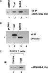
T-bet can associate with histone methyltransferase activity. A co-IP experiment was performed, and histone methyltransferase activity was monitored. (A) EL4 T cells were transfected with either a pcDNA 3.1 control vector (lane 1), T-bet 1-530 (lane 2), or SET7/9 (lane 3). The T-bet 1-530 and SET7/9 constructs contain a C-terminal V5 epitope tag. Whole-cell extracts were made from the individual samples and precipitated with a V5 antibody (lanes 1 to 3). A methyltransferase activity assay was performed with the precipitated samples, which were subsequently processed for Western analysis to monitor H3-K4 dimethyl-specific methyltransferase activity. An amount of the recombinant histone H3 equal to that used in the methyltransferase reaction was also run as a control (−; lane 4). The membrane was probed with an antibody specific to histone H3-K4 dimethyl (αH3K4Me2) as shown. (B) The membrane was then stripped and reprobed with a V5-specific antibody (αV5) to confirm the precipitation of the transfected constructs. The minor band indicated by the asterisk represents cross-reactivity with the heavy chain. (C) A co-IP experiment to monitor methyltransferase activity was performed as described in panel A. 721 B cells were stimulated with phorbol 12-myristate 13-acetate and ionomycin (P/I). Nuclear extracts were precipitated with either a control Cdk6 antibody (lane 1) or a T-bet-specific antibody (lane 2). The co-IP samples were subjected to a methyltransferase activity assay, and the Western analysis was performed with an H3-K4 dimethyl-specific antibody.

Similar articles

Cited by

References

    1. Afkarin, M., J. R. Sedy, J. Yang, N. G. Jacobson, N. Cereb, S. Y. Yang, T. L. Murphy, and K. M. Murphy. 2002. T-bet is a STAT1-induced regulator of IL-12R expression in naive CD4+ T cells. Nat. Immunol. 3:549-557. - PubMed
    1. Agarwal, S., and A. Rao. 1998. Modulation of chromatin structure regulates cytokine gene expression during T cell differentiation. Immunity 9:765-775. - PubMed
    1. Ansel, K. M., I. Djuretic, B. Tanasa, and A. Rao. 2006. Regulation of Th2 differentiation and IL4 locus accessibility. Annu. Rev. Immunol. 24:607-656. - PubMed
    1. Avni, O., D. Lee, F. Macian, S. J. Szabo, L. H. Glimcher, and A. Rao. 2002. T(H) cell differentiation is accompanied by dynamic changes in histone acetylation of cytokine genes. Nat. Immunol. 3:643-651. - PubMed
    1. Baala, L., S. Briault, H. C. Etchevers, F. Laumonnier, A. Natiq, J. Amiel, N. Boddaert, C. Picard, A. Sbiti, A. Asermouh, T. Attie-Bitach, F. Encha-Razavi, A. Munnich, A. Sefiani, and S. Lyonnet. 2007. Homozygous silencing of T-box transcription factor EOMES leads to microcephaly with polymicrogyria and corpus callosum agenesis. Nat. Genet. 39:454-456. - PubMed

Publication types

MeSH terms

LinkOut - more resources