. 2007 Dec;27(23):8164-77.
doi: 10.1128/MCB.00555-07. Epub 2007 Oct 8.

Differential control of Wnt target genes involves epigenetic mechanisms and selective promoter occupancy by T-cell factors

Affiliations

Differential control of Wnt target genes involves epigenetic mechanisms and selective promoter occupancy by T-cell factors

Simon Wöhrle et al. Mol Cell Biol. 2007 Dec.

Abstract

Canonical Wnt signaling and its nuclear effectors, beta-catenin and the family of T-cell factor (TCF) DNA-binding proteins, belong to the small number of regulatory systems which are repeatedly used for context-dependent control of distinct genetic programs. The apparent ability to elicit a large variety of transcriptional responses necessitates that beta-catenin and TCFs distinguish precisely between genes to be activated and genes to remain silent in a specific context. How this is achieved is unclear. Here, we examined patterns of Wnt target gene activation and promoter occupancy by TCFs in different mouse cell culture models. Remarkably, within a given cell type only Wnt-responsive promoters are bound by specific subsets of TCFs, whereas nonresponsive Wnt target promoters remain unoccupied. Wnt-responsive, TCF-bound states correlate with DNA hypomethylation, histone H3 hyperacetylation, and H3K4 trimethylation. Inactive, nonresponsive promoter chromatin shows DNA hypermethylation, is devoid of active histone marks, and additionally can show repressive H3K27 trimethylation. Furthermore, chromatin structural states appear to be independent of Wnt pathway activity. Apparently, cell-type-specific regulation of Wnt target genes comprises multilayered control systems. These involve epigenetic modifications of promoter chromatin and differential promoter occupancy by functionally distinct TCF proteins, which together determine susceptibility to Wnt signaling.


Figures

FIG. 1.
FIG. 1.
Schematic representation of regulatory regions of Wnt target genes. Shown are parts of the murine Axin2, Cdx1, and T/Bra gene loci. Thin lines represent DNA, TBEs are indicated by red bars and numbered, exons are highlighted in green, and CpG islands are in black. Blue arrowheads mark the location of primer pairs used for genomic bisulfite sequencing. White arrowheads indicate primer pairs used for the analyses of chromatin immunoprecipitates. Numbers refer to nucleotide positions relative to the transcriptional start sites (arrows, +1). For each gene, DNA fragments used to drive the expression of a luciferase reporter gene (yellow bar) are displayed and nucleotide coordinates are shown. In each case, reporter gene expression driven by the promoter fragments used recapitulates important aspects of Axin2, Cdx1, and T/Bra embryonic expression patterns in transgenic animals and mutational inactivation of TCF binding elements contained within the promoter regions abrogates transgene expression in cognate T/Bra, Cdx1, and Axin2 expression domains (7, 48, 81).
FIG. 2.
FIG. 2.
Wnt target genes are differentially activated in neural, myogenic, and embryonic cell lines. (A) Wnt target gene activation in C17.2 neural stem-cell like cells, C2C12 myogenic cells, and E14 embryonic stem cells. Total RNA was isolated from cells treated with 25 μM (C17.2, C2C12) or 5 μM (E14) SB-216763 (lanes 1 to 3) and 200 ng/ml recombinant Wnt3a (lanes 5 to 10) as indicated. Activation of the Wnt target genes Axin2, Cdx1, and T/Bra was analyzed by RT-PCR. Amplification of GAPDH was used to control sample integrity and loading. Control reactions received no template (no templ.), cDNA samples which were mock processed in the absence of RT (−RT), or plasmid DNA or a pretested cDNA as the template (pos. control). n.a.: not analyzed. (B) Quantitative real-time PCR of Wnt target gene activation. C17.2, C2C12, and E14 cells were treated with recombinant Wnt3a for the times indicated. Activation of the Wnt target genes Axin2, Cdx1, and T/Bra is shown as induction (n-fold) compared to transcript levels in untreated cells. Data given are the average and SEM from at least three experiments.
FIG. 3.
FIG. 3.
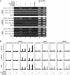
Promoters of Wnt-inducible target genes are occupied by endogenous TCF4 irrespective of pathway activity. (A) ChIP analysis for TCF4 in C17.2, C2C12, or E14 cells. Cells were stimulated for 24 h with 25 μM (C17.2, C2C12) or 5 μM (E14) SB-216763 or with DMSO as the solvent control. Formaldehyde-fixed chromatin was precipitated using 2 μg of a TCF4-specific antibody or 2 μg of a nonspecific goat IgG as control. Precipitated DNA was analyzed by PCR using primer sets indicated in Fig. 1 with close proximity to TCF-binding elements in the Axin2, Cdx1, and T/Bra promoters. Primers within the coding region of the GAPDH gene, which is not regulated by TCFs, were used to monitor nonspecific precipitation. Control PCRs received no template (no templ.) or genomic DNA as the positive control. (B) Quantitative real-time PCR of ChIP DNA from C17.2, C2C12, and E14 cells as described above. For each gene, precipitated DNA was analyzed using multiple primer sets. Numbers refer to nucleotide positions of the amplicons relative to the transcriptional start site as indicated in Fig. 1. Bars represent relative enrichment compared to that of the nonspecific isotype control (relative enrichment of 1). Data given are the average and SEM from at least three experiments. α-, anti-.
FIG. 4.
FIG. 4.
Promoter occupancy by different TCF family members is limited to Wnt-responsive target genes. ChIP analysis was performed in C17.2, C2C12, or E14 cells stably transfected with expression vectors for HA-tagged TCFs. Chromatin was precipitated using 2 μg of a rat anti-HA (α-HA) antibody or equal amounts of the corresponding isotype control. Precipitated DNA was analyzed by quantitative real-time PCR using primer sets within the Axin2, Cdx1, and T/Bra promoter regions. Nucleotide positions of the amplicons relative to the transcriptional start site are indicated (Fig. 1). Bars represent relative enrichment compared to that of the nonspecific isotype control (relative enrichment of 1). Data given are the average and SEM from at least four experiments.
FIG. 5.
FIG. 5.
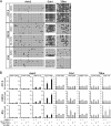
Differential Wnt inducibility correlates with distinct epigenetic states of the corresponding target genes. (A) Analysis of DNA methylation patterns at the Axin2, Cdx1, and T/Bra promoters. Genomic DNA was isolated from C17.2, C2C12, or E14 cells after treatment for 24 h with 25 μM (C17.2, C2C12) or 5 μM (E14) SB-216763 or with DMSO as the solvent control. Bisulfite-treated DNA fragments of the promoter regions were PCR amplified with primer pairs shown in Fig. 1, subcloned, and analyzed by sequencing. Each line represents the results from a single sequence and each circle denotes a CpG dinucleotide (filled circles, methylated CpGs; open circles, nonmethylated CpGs). (B) ChIP analysis for acetylated histone H3 in C17.2, C2C12, or E14 cells. Formaldehyde-fixed chromatin was prepared after treatment with 25 μM (C17.2, C2C12) or 5 μM (E14) SB-216763 or with DMSO as the solvent control for 24 h. Chromatin was precipitated with 2 μg of an anti-acetylated histone H3 (α-H3ac) antibody or equal amounts of a nonspecific rabbit isotype control. ChIP DNA was analyzed by quantitative real-time PCR with primer pairs within the Axin2, Cdx1, and T/Bra promoter regions. Nucleotide positions of the amplicons relative to the transcriptional start site are indicated (Fig. 1). Bars represent relative enrichment compared to that of the nonspecific isotype control (relative enrichment of 1). Data given are the average and SEM from at least three experiments.
FIG. 6.
FIG. 6.
Wnt responsiveness is paralleled by the cell-type-specific occurrence of methylated H3K4 at target gene promoters. ChIP analysis for methylated histone H3K4 in C17.2, C2C12, or E14 cells. Formaldehyde-fixed chromatin was prepared after treatment with 25 μM (C17.2, C2C12) or 5 μM (E14) SB-216763 or with DMSO as the solvent control for 24 h. Chromatin was precipitated with 2 μg of an antibody recognizing trimethylated histone H3K4 (α-H3K4me3) or equal amounts of a nonspecific rabbit isotype control. ChIP DNA was analyzed by quantitative real-time PCR with primer pairs within the Axin2, Cdx1, and T/Bra promoter regions. Nucleotide positions of the amplicons relative to the transcriptional start site are indicated (Fig. 1). Bars represent relative enrichment compared to that of the nonspecific isotype control (relative enrichment of 1). Data given are the average and SEM from at least three experiments.
FIG. 7.
FIG. 7.
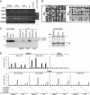
Nonresponsive Wnt target genes remain refractory to Wnt induction upon inhibition of histone deacetylases or DNA methyltransferases. (A) TSA increases the basal expression level but does not restore the Wnt inducibility of the T/Bra gene. C17.2 cells were treated for 24 h with 0.5 μM TSA or 5 μM Aza in combination with 50 ng/ml Wnt3a purified from conditioned medium (lanes 1 to 6) as indicated. Total RNA was isolated, and the activation of the Wnt target genes Axin2, Cdx1, and T/Bra was analyzed by RT-PCR. Amplification of GAPDH was used to control sample integrity and loading. Control reactions were done as described for Fig. 2A. (B) DNA methylation analysis of the T/Bra promoter in C17.2 cells upon TSA and Aza treatment. Bisulfite-treated DNA was amplified by PCR with primers indicated in Fig. 1 and analyzed as described before (Fig. 5). (C) Treatment with TSA increases levels of acetylated histone H3. Immunoprecipitations with anti-acetylated H3 (α-acH3) antibodies were performed with nuclear lysates from C17.2 cells treated for 24 h with 0.5 μM TSA, 5 μM Aza, or DMSO as the solvent control. A fraction of the nuclear (nucl.) lysates (2.5%) and the immunoprecipitates were analyzed by immunoblotting with anti-acetylated H3 antibodies. Coomassie brilliant blue staining of nuclear lysates separated by SDS-PAGE was used to confirm equal loading. (D) Inhibition of histone deacetylases does not alter the epigenetic states and TCF promoter occupancy of Wnt target genes. ChIP analysis for TCF4, acetylated histone H3, and methylated histone H3K27 in TSA-treated C17.2 cells. Formaldehyde-fixed chromatin was prepared from C17.2 cells after treatment with 1 μM TSA or DMSO as the solvent control for 24 h. Chromatin was precipitated using 2 μg of a TCF4-specific antibody, antibodies to acetylated histone H3 (2 μg) or trimethylated histone H3K27 (α-H3K27me3) (4 μg), or equivalent amounts of the corresponding isotype controls. Precipitated DNA was analyzed by quantitative real-time PCR using primer sets within the promoter regions of the Axin2, Cdx1, and T/Bra genes. Nucleotide positions of the amplicons relative to the transcriptional start site are indicated (Fig. 1). Bars represent relative enrichment compared to that of the nonspecific isotype controls (relative enrichment of 1). Data given are the average and SEM from at least three experiments.

Similar articles

Cited by

References

    1. Akiyama, H., J. P. Lyons, Y. Mori-Akiyama, X. Yang, R. Zhang, Z. Zhang, J. M. Deng, M. M. Taketo, T. Nakamura, R. R. Behringer, P. D. McCrea, and B. de Crombrugghe. 2004. Interactions between Sox9 and beta-catenin control chondrocyte differentiation. Genes Dev. 18:1072-1087. - PMC - PubMed
    1. Aoki, M., A. Hecht, U. Kruse, R. Kemler, and P. K. Vogt. 1999. Nuclear endpoint of Wnt signaling: neoplastic transformation induced by transactivating lymphoid-enhancing factor 1. Proc. Natl. Acad. Sci. USA 96:139-144. - PMC - PubMed
    1. Arce, L., N. N. Yokoyama, and M. L. Waterman. 2006. Diversity of LEF/TCF action in development and disease. Oncogene 25:7492-7504. - PubMed
    1. Arias, A. M., and P. Hayward. 2006. Filtering transcriptional noise during development: concepts and mechanisms. Nat. Rev. Genet. 7:34-44. - PubMed
    1. Arnold, S. J., J. Stappert, A. Bauer, A. Kispert, B. G. Herrmann, and R. Kemler. 2000. Brachyury is a target gene of the Wnt/beta-catenin signaling pathway. Mech. Dev. 91:249-258. - PubMed

Publication types

MeSH terms

LinkOut - more resources