. 2007 Dec;27(24):8522-32.
doi: 10.1128/MCB.01007-07. Epub 2007 Oct 8.

Transcription alters chromosomal locations of cohesin in Saccharomyces cerevisiae

Affiliations

Transcription alters chromosomal locations of cohesin in Saccharomyces cerevisiae

Christoph Bausch et al. Mol Cell Biol. 2007 Dec.

Abstract

In eukaryotic cells, cohesion between sister chromatids allows chromosomes to biorient on the metaphase plate and holds them together until they separate into daughter cells during mitosis. Cohesion is mediated by the cohesin protein complex. Although the association of this complex with particular regions of the genome is highly reproducible, it is unclear what distinguishes a chromosomal region for cohesin association. Since one of the primary locations of cohesin is intergenic regions between converging transcription units, we explored the relationship between transcription and cohesin localization. Chromatin immunoprecipitation followed by hybridization to a microarray (ChIP chip) indicated that transcript elongation into cohesin association sites results in the local disassociation of cohesin. Once transcription is halted, cohesin can reassociate with its original sites, independent of DNA replication and the cohesin loading factor Scc2, although cohesin association with chromosomes in G2/M is not functional for cohesion. A computer program was developed to systematically identify differences between two ChIP chip data sets. Our results are consistent with a model for cohesin association in which (i) a portion of cohesin can be dynamically loaded and unloaded to accommodate transcription and (ii) the cohesin complex has preferences for features of chromatin that are a reflection of the local transcriptional status. Taken together, our results suggest that cohesion may be degraded by transcription.


Figures

FIG. 1.
FIG. 1.
Alterations of Mcd1-18Myc location during amino acid starvation. Strain 1827-22D was grown in YPD at 30°C and arrested with nocodazole. Following arrest, one-third of the culture was harvested and two-thirds of the culture was transferred to medium lacking amino acids and containing nocodazole. After 30 min, one-third of the culture was harvested and one-third was transferred back to YPD with nocodazole for an additional 30 min. Arrest was maintained throughout the experiment, as verified by FACS analysis. An aliquot of each culture was harvested for gene expression analysis by being frozen in liquid nitrogen (values are indicated above each ORF) and for ChIP chip analysis by formaldehyde cross-linking. The log2 values of the red/green ratios for the ChIP results (y axis) are shown for YPD (black), 30 min of amino acid starvation (red), and return to YPD for 30 min following amino acid starvation (gray). A diagram of each locus is shown on the x axis of each graph, and the Saccharomyces Genome Database (SGD) coordinates are given in kb. The x-fold change in gene expression from YPD to amino acid starvation is indicated for each feature; numbers of >1 indicate an increase in gene expression during amino acid starvation, and numbers of <1 indicate a decrease in gene expression during amino acid starvation. Genes carried on the Watson strand are shown in red, and genes carried on the Crick strand are shown in blue. Gray arrows are “dubious” ORFs, according to SGD. Pink indicates a long terminal repeat, and green indicates a tRNA. CEN8 is indicated by a star. (A) MET17; (B) CIT2; (C) SUL1; (D) RPL2B; (E) RPL14B; (F) ADE17.
FIG. 2.
FIG. 2.
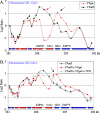
Galactose-induced transcription at GAL2 affects cohesin localization. ChIP chip analysis was performed for Mcd1-18Myc in strain 1827-22D under medium conditions that either induced or repressed transcription from the GAL2 locus. Cell cycle arrests were confirmed in all samples by FACS analysis. The midpoint of each feature is used to represent the log2 of the median red/green ratio (y axis). A depiction of the locus is shown along with coordinates, in kb, for chromosome XII from SGD. (A) A culture grown in YPraff was arrested in G1 with alpha factor. Following arrest, half of the culture was released into YPgal with nocodazole (red line), and half of the culture was released into YPraff with nocodazole (black line). (B) A culture grown in YPraff was arrested in G2/M with nocodazole and collected for ChIP (black line). Two-thirds of the culture was transferred to YPgal medium and nocodazole for 1 h and collected for ChIP (red line). Half of this culture was transferred to YPD with nocodazole for 1 h and collected for ChIP (gray line).
FIG. 3.
FIG. 3.
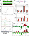
Galactose-induced Mcd1-6HA associates with GAL2 when induced in G2/M. Strain 1827-11B, which contains Mcd1-18Myc under the control of the endogenous Mcd1 promoter and an ectopic copy of Mcd1-6HA under the control of the GAL1-10 promoter, was grown in YPD to mid-log phase and arrested with nocodazole. Following arrest, cells were transferred to YPgal medium with nocodazole (t = 0 h). Samples were taken every hour for Western analysis, poly(A) mRNA isolation, and FACS analysis. At 6 h, the culture was harvested for ChIP chip analysis. (A) Western blot for GAL1-10-Mcd1-6HA and Mcd1-18Myc. Thirty micrograms of protein was loaded in each lane. (B) mRNA expression of GAL2, measured by microarray analysis. (C) FACS analysis. (D) Diagram of PCR products at GAL2 locus monitored by real-time PCR. (E) Real-time PCR performed in triplicate at time zero and at 6 h for Mcd1-18Myc-associated DNA at GAL2 as well as CEN3 and FMP32, a cohesin arm site where surrounding transcription is not affected by the switch to galactose medium. (F) Real-time PCR of GAL1-10-Mcd1-6HA-associated DNA. (G) ChIP chip results for Mcd1-18Myc and Mcd1-6HA. This region was monitored on our microarrays by both PCR products and 70-mer oligonucleotides. The width of each product on the microarray is represented by the width of the bar, with the narrowest bars representing the oligonucleotides. The height represents the log2 value of the median red/green ratio (y axis). The GAL2 ORF is indicated by vertical dashed lines.
FIG. 4.
FIG. 4.
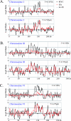
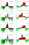
Galactose-induced Mcd1-6HA associates with the same genomic locations as Mcd1-18Myc when induced in G2/M. The ChIP patterns of GAL1-10-Mcd1-6HA (red) and Mcd1-18Myc (black) at time zero and following 6 h of induction in galactose were plotted using Peakfinder (13). Data from a ChIP performed without antibody are shown in gray. Vertical gray lines indicate 50-kb intervals. (A) Chromosome I at 0 and 6 h. (B) Chromosome III at 0 and 6 h. (C) Chromosome VI at 0 and 6 h.
FIG. 5.
FIG. 5.
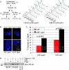
Galactose-induced Mcd1-6HA that associates with chromosomes in G2/M is not functional for cohesion. Strains containing either a chromosome arm marked with GFP (CBY601) or a telomere marked with GFP (JMH303) were grown in YPraff at the permissive temperature of 23°C for mcd1/scc1-173 and arrested with nocodazole (NZ). Following arrest, galactose was added to half of the culture for 3 h, while arrest was maintained. Cultures were shifted to the nonpermissive temperature of 37°C by adding an equal volume of medium at 53°C. (A) Schematic of experiment and FACS profiles confirming the arrest throughout the course of the experiment. The slight peak to the left of the 1N peak in raffinose sample 6 at 60 min is due to cellular debris. (B) Images of strains CBY601 and JMH303 showing either one or two GFP spots and DNA stained with DAPI. (C) Quantitation of the percent increase in two GFP spots following 1 h at 37°C. The numbers of cells for which GFP spots were counted prior to temperature shift (n0) and following temperature shift (n60) are shown. Prior to the temperature shift, there was no significant difference in the numbers of cells with one GFP spot versus two GFP spots in the YPraff culture compared to the YPraff+gal cultures. (D) Strain SEN111 was grown in YPD and arrested with nocodazole for 2 hours prior to the transfer of half of the culture to YPgal with nocodazole. Cultures were then incubated in YPD or YPgal with nocodazole for 5 hours. As a control, the same strain was grown overnight in YPgal and then arrested with nocodazole. FACS analysis verified all arrests. Immunoprecipitation from whole-cell extract (WCE) was performed with anti-HA antibody, anti-FLAG antibody, or no antibody. The precipitated material was subjected to sodium dodecyl sulfate-polyacrylamide gel electrophoresis, transferred to a nitrocellulose membrane, and probed with anti-FLAG antibody.
FIG. 6.
FIG. 6.
Scc2 is not required for loading or disruption of Smc1-6HA in G2/M. Strain K8250, which includes a temperature-sensitive mutation in Scc2 and Smc1-6HA, was grown in YPD at 23°C. Following metaphase arrest for 1.5 h, the culture was split in half and incubated in either YPD at the permissive temperature of 23°C or YPD at the nonpermissive temperature of 33°C for 30 min while maintaining the arrest. Each of these two cultures was then resuspended in YPD with nocodazole or medium lacking amino acids (no AA) and containing nocodazole at their respective temperatures for 1 h. Cells were collected for ChIP. We used microarrays that contained both PCR products and closely spaced oligonucleotide probes for several loci of interest for this experiment. Each vertical bar indicates the log2 ratio (y axis) for a probe on the array, and the width indicates its length. SGD coordinates are shown in kb on the x axis. Probes corresponding to the MET17 ORF (A) and the RPL2B ORF (B) are indicated with blue vertical lines and red arrowheads.

References

    1. Antoniacci, L. M., and R. V. Skibbens. 2006. Sister-chromatid telomere cohesion is nonredundant and resists both spindle forces and telomere motility. Curr. Biol. 16:902-906. - PubMed
    1. Bhalla, N., S. Biggins, and A. W. Murray. 2002. Mutation of YCS4, a budding yeast condensin subunit, affects mitotic and nonmitotic chromosome behavior. Mol. Biol. Cell 13:632-645. - PMC - PubMed
    1. Blat, Y., and N. Kleckner. 1999. Cohesins bind to preferential sites along yeast chromosome III, with differential regulation along arms versus the centric region. Cell 98:249-259. - PubMed
    1. Bohlander, S. K., R. Espinosa III, M. M. Le Beau, J. D. Rowley, and M. O. Diaz. 1992. A method for the rapid sequence-independent amplification of microdissected chromosomal material. Genomics 13:1322-1324. - PubMed
    1. Ciosk, R., M. Shirayama, A. Shevchenko, T. Tanaka, A. Toth, and K. Nasmyth. 2000. Cohesin's binding to chromosomes depends on a separate complex consisting of Scc2 and Scc4 proteins. Mol. Cell 5:243-254. - PubMed

Publication types

MeSH terms