

Review
doi: 10.3346/jkms.2007.22.S.S139.
Anaplastic ganglioglioma in a middle-aged woman: a case report with a review of the literature
Affiliations
- PMID: 17923741
- PMCID: PMC2694381
- DOI: 10.3346/jkms.2007.22.S.S139
Item in Clipboard
Review
Anaplastic ganglioglioma in a middle-aged woman: a case report with a review of the literature
J Korean Med Sci.
2007 Sep.
Abstract
We report a case of anaplastic ganglioglioma. A 45-yr-old woman was admitted with a 5-month history of headache and dizziness, both of which progressed slowly. Preoperative magnetic resonance imaging revealed a strong enhancing mass in the left frontal lobe extending to the cingulate gyrus. Adjuvant radiation therapy and chemotherapy were given after gross total resection of the tumor. Histological and immunohistochemical studies showed an anaplastic ganglioglioma. Gangliogliomas of the central nervous system are rather uncommon tumors, and anaplastic ones are extremely rare. The pertinent literature regarding gangliogliomas is reviewed.
Figures
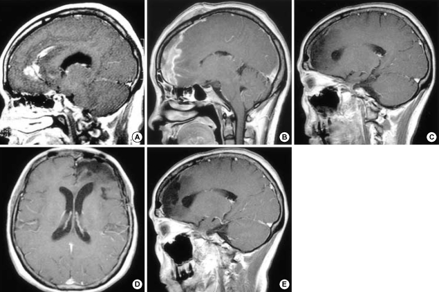
Preoperative MRI showing an irregular strong enhancing mass in the left frontal lobe extending to the cingulate gyrus (A). MRI on the 12th postoperative day showing multiple irregularly shaped enhancing lesions along the resection margin of the left frontal lobe and cingulate gyrus (B). After chemotherapy and radiation therapy, MRI 5 months postoperatively revealed that the irregular enhancing lesions in the resected margin of the left frontal lobe and cingulate gyrus had disappeared (C). And, the last follow-up MRI at 35 months postoperatively shows no evidence of recurrence (D, E).
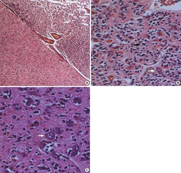
Photomicrographs showing the histopathology of the ganglioglioma in our case. (A) The tumor cells infiltrated the gray matter and subarachnoid space. The tumor cells in the gray matter consisted of neoplastic ganglion cells and glial cells. Those in the subarachnoid space (asterisk) are mainly glial cells and show increased cellularity, nuclear atypism, and occasional mitosis (hematoxylin and eosin, original magnification, ×40). (B) Atypical glial cells (arrow) and scattered ganglion cells (white arrow) constitute the ganglioglioma. Note the increased cellularity and nuclear atypism of the glial cells (hematoxylin and eosin, original magnification, ×400). (C) Neoplastic ganglion cells (arrow) are clustered abnormally and lack orientation and polarity. Most have large nuclei with prominent nucleoli (hematoxylin and eosin, original magnification, ×400).
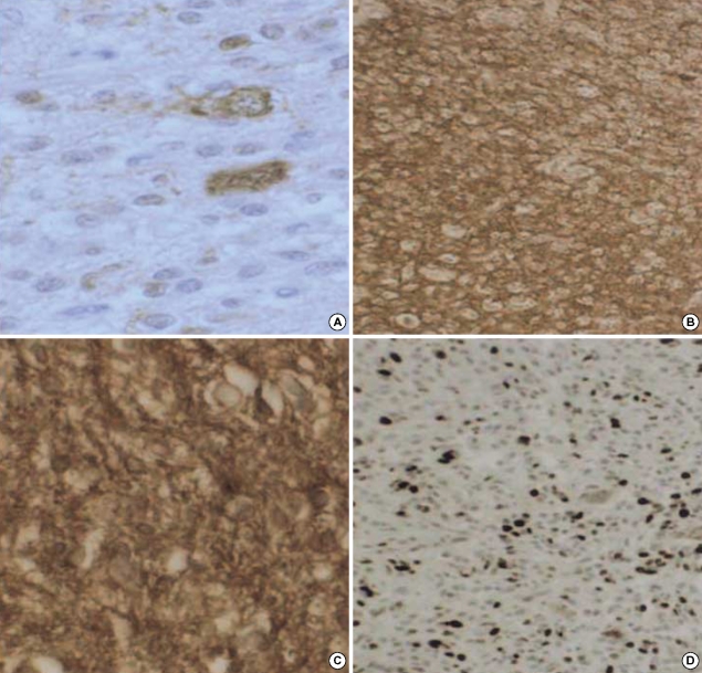
Immunostaining features suggestive of anaplastic gangliogliomas. Neoplastic ganglion cells are positive for NSE (A) and synaptophysin (B). (C) Glial components are positive for GFAP, and scattered ganglion cells are negative. (D) The Ki-67 LI is high (20%) in the region of anaplastic transformation.
Similar articles
-
Cerebellopontine angle gangliogliomas: Report of two cases.Neurochirurgie. 2016 Oct;62(5):266-270. doi: 10.1016/j.neuchi.2016.07.001. Epub 2016 Oct 19. Neurochirurgie. 2016. PMID: 27771109
-
Pediatric primary anaplastic ganglioglioma with malignant neuronal component.Turk J Pediatr. 2018;60(1):102-106. doi: 10.24953/turkjped.2018.01.017. Turk J Pediatr. 2018. PMID: 30102489
-
[Malignant progression of an anaplastic ganglioglioma into a glioblastoma multiforme--report on two cases and review of the literature].Khirurgiia (Sofiia). 2009;(2-3):69-74. Khirurgiia (Sofiia). 2009. PMID: 20506796 Bulgarian.
-
Anaplastic ganglioglioma: case report and review of the literature.Br J Neurosurg. 1998 Jun;12(3):277-80. doi: 10.1080/02688699845159. Br J Neurosurg. 1998. PMID: 11013696 Review.
-
Gangliogliomas: a clinicopathological study of 25 cases and review of the literature.J Neurooncol. 1993 Aug;17(2):139-54. doi: 10.1007/BF01050216. J Neurooncol. 1993. PMID: 8145057 Review.
Cited by
-
Malignant ganglioglioma: case report and review of literature.J Neurooncol. 2011 Jan;101(2):311-8. doi: 10.1007/s11060-010-0248-z. Epub 2010 Jun 4. J Neurooncol. 2011. PMID: 20524041 Review.
-
Epilepsy due to a temporal ganglioglioma and its subsequent malignant transformation into a primitive neuroectodermal tumor.Surg Neurol Int. 2012;3:79. doi: 10.4103/2152-7806.98511. Epub 2012 Jul 14. Surg Neurol Int. 2012. PMID: 22937479 Free PMC article.
-
Practical aspects regarding the histopathological grading and anaplastic transformation of gangliogliomas - a literature review.Rom J Morphol Embryol. 2021 Apr-Jun;62(2):385-393. doi: 10.47162/RJME.62.2.04. Rom J Morphol Embryol. 2021. PMID: 35024726 Free PMC article. Review.
-
Outcome and prognostic features in anaplastic ganglioglioma: analysis of cases from the SEER database.J Neurooncol. 2011 Dec;105(3):539-45. doi: 10.1007/s11060-011-0615-4. Epub 2011 May 29. J Neurooncol. 2011. PMID: 21626070
-
Impact of surgery, adjuvant treatment, and other prognostic factors in the management of anaplastic ganglioglioma.Childs Nerv Syst. 2018 Jun;34(6):1207-1213. doi: 10.1007/s00381-018-3780-3. Epub 2018 Mar 29. Childs Nerv Syst. 2018. PMID: 29594461
References
-
- Russell DS, Rubinstein LJ. Pathology of tumors of the nervous system. 5th Ed. Baltimore, USA: Williams & Wilkins; 1989. pp. 289–307.
-
- Haddad SF, Moore SA, Menezes AH, VanGilder JC. Ganglioglioma: 13 years of experience. Neurosurgery. 1992;31:171–178. - PubMed
-
- Hall WA, Yunis EJ, Albright AL. Anaplastic ganglioglioma in an infant: case report and review of the literature. Neurosurgery. 1986;19:1016–1020. - PubMed
-
- Kitano M, Takayama S, Nagao T, Yoshimura O. Malignant ganglioglioma of the spinal cord. Acta Pathol Jpn. 1987;37:1009–1018. - PubMed
-
- Lang FF, Epstein FJ, Ransohoff J, Allen JC, Wisoff J, Abbott IR, Miller DC. Central nervous system gangliogliomas. Part 2: clinical outcome. J Neurosurg. 1993;79:867–873. - PubMed
Publication types
MeSH terms
LinkOut - more resources
Full Text Sources
Medical
