

Pituitary carcinoma with mandibular metastasis: a case report
- PMID: 17923742
- PMCID: PMC2694386
- DOI: 10.3346/jkms.2007.22.S.S145
Pituitary carcinoma with mandibular metastasis: a case report
Abstract
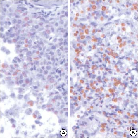
Pituitary carcinomas are rare primary adenohypophyseal tumors with cerebrospinal or extracranial metastasis. The present case, the first report of the disease in Korea, involved a 36-yr-old woman who presented with a 3-week history of headache. Brain magnetic resonance imaging revealed a 2.5-cm sellar and suprasellar mass showing heterogeneous enhancement with suspicious invasion of both cavernous sinuses. The patient underwent gross-total resection. The tumor cells were composed of polygonal cells singly or in variable-sized nests. The nuclei were large and round with prominent nucleoli. The cytoplasms was acidophilic and granular. Marked pleomorphism and frequent mitoses (3 per 10 HPFs) were found. By immunohistochemistry, tumor cells were strongly positive for prolactin, but negative for ACTH and GH. Additional immunostainings for cytokeratin, vimentin, and glial fibrillary acidic protein (GFAP) were negative. After the surgery, the patient received radiotherapy because of the atypical histologic features. The prolactin level fell from 123.17 ng/mL to 5.17 ng/mL after surgery. Nine months after the initial diagnosis, the patient died from mandibular metastasis associated with the pituitary carcinoma.
Figures


Similar articles
-
What causes a prolactinoma to be aggressive or to become a pituitary carcinoma?Hormones (Athens). 2012 Oct-Dec;11(4):477-82. doi: 10.14310/horm.2002.1380. Hormones (Athens). 2012. PMID: 23422771
-
Prolactin-producing pituitary adenoma with atypical spindle cell morphology: a case report.World J Surg Oncol. 2015 Jul 31;13:229. doi: 10.1186/s12957-015-0655-x. World J Surg Oncol. 2015. PMID: 26228535 Free PMC article.
-
Malignant prolactinoma: is metastasis a must? Clinico-pathologic and immunohistochemical study of a case.J Neurosurg Sci. 2004 Mar;48(1):37-41. J Neurosurg Sci. 2004. PMID: 15257264
-
[Pituitary carcinoma. Anatomic and clinical features of cases reported in literature].Neurochirurgie. 2007 Aug;53(4):284-8. doi: 10.1016/j.neuchi.2007.01.001. Neurochirurgie. 2007. PMID: 17524431 Review. French.
-
Prolactin-secreting carcinoma of the pituitary: clinicopathological and immunohistochemical study of a case with intracranial and intraspinal dissemination.Br J Neurosurg. 1997 Aug;11(4):350-5. doi: 10.1080/02688699746177. Br J Neurosurg. 1997. PMID: 9337937 Review.
Cited by
-
Corticotrophic pituitary carcinoma with cervical metastases: case series and literature review.Pituitary. 2018 Jun;21(3):290-301. doi: 10.1007/s11102-018-0872-8. Pituitary. 2018. PMID: 29404894 Review.
-
FDG-PET/CT findings of a metastatic pituitary tumor.Cancer Imaging. 2010 Mar 18;10(1):114-6. doi: 10.1102/1470-7330.2010.0015. Cancer Imaging. 2010. PMID: 20299302 Free PMC article.
-
Orbital metastasis of pituitary growth hormone secreting carcinoma causing lateral gaze palsy.Surg Neurol Int. 2013 Apr 18;4:59. doi: 10.4103/2152-7806.110658. Print 2013. Surg Neurol Int. 2013. PMID: 23646269 Free PMC article.
-
Clinical features of pituitary carcinoma: analysis based on a case report and literature review.Front Endocrinol (Lausanne). 2024 Oct 31;15:1440247. doi: 10.3389/fendo.2024.1440247. eCollection 2024. Front Endocrinol (Lausanne). 2024. PMID: 39544231 Free PMC article. Review.
References
-
- Pernicone PJ, Scheithauer BW, Sebo TJ, Kovacs KT, Horvath E, Young WF, Jr, Lloyd RV, Davis DH, Guthrie BL, Schoene WC. Pituitary carcinoma: a clinicopathologic study of 15 cases. Cancer. 1997;79:804–812. - PubMed
-
- Cairns H, Russel DS. Intracranial and spinal metastasis in gliomas of the brain. Brain. 1931;54:377–420.
-
- Scheithauer BW, Kovacs KT, Horvath E, Roncaroli F, Ezzat S, Asa SL, Lloid RV, Nose V, Watwon RE, Jr, Lindell EP. Pituitary carcinoma. In: DeLellis RA, Lloyd RV, Hritz PU, Eng C, editors. Pathology & Genetics Tumours of Endocrine Organs. Lyon, France: International Agency for Research on Cancer (IARC) Press; 2004. pp. 36–39.
-
- Long MA, Colquhoun IR. Case report: multiple intra-cranial metastases from a prolactin-secreting pituitary tumour. Clin Radiol. 1994;49:356–358. - PubMed
-
- Bayindir C, Balak N, Gazioglu N. Prolactin-secreting carcinoma of the pituitary: clinicopathological and immunohistochemical study of a case with intracranial and intraspinal dissemination. Br J Neurosurg. 1997;11:350–355. - PubMed
Publication types
MeSH terms
Substances
LinkOut - more resources
Full Text Sources
Medical
Miscellaneous
